. 2024 Oct 9;22(1):482.
doi: 10.1186/s12964-024-01855-9.

TMEM132A regulates Wnt/β-catenin signaling through stabilizing LRP6 during mouse embryonic development

Affiliations

TMEM132A regulates Wnt/β-catenin signaling through stabilizing LRP6 during mouse embryonic development

Shin Ae Oh et al. Cell Commun Signal. .

Abstract

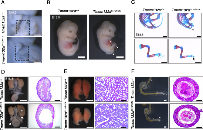
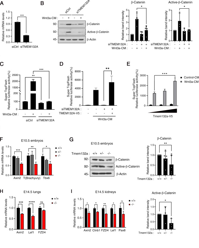
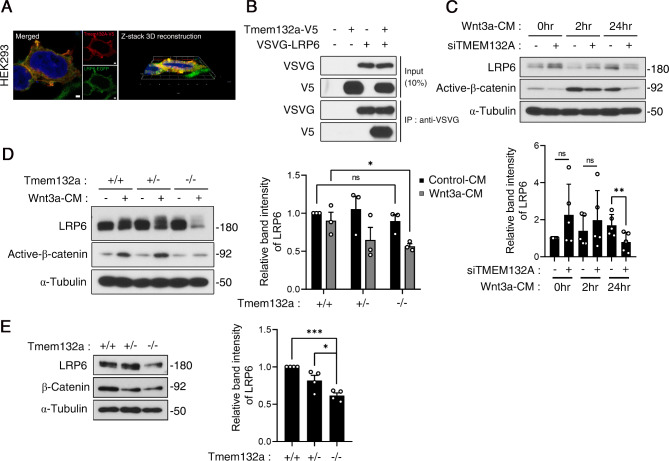
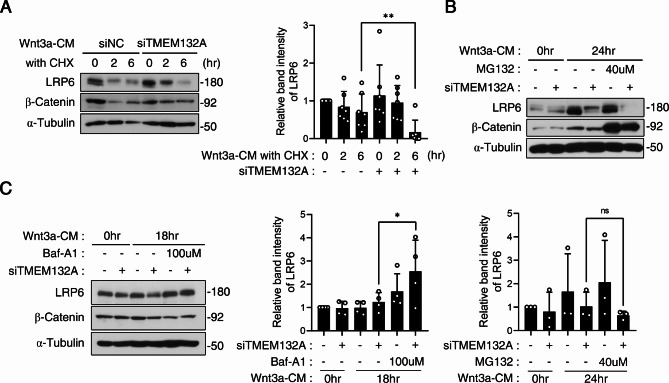
The Wnt/β-catenin signaling pathway is crucial for embryonic development and adult tissue homeostasis. Dysregulation of Wnt signaling is linked to various developmental anomalies and diseases, notably cancer. Although numerous regulators of the Wnt signaling pathway have been identified, their precise function during mouse embryo development remains unclear. Here, we revealed that TMEM132A is a crucial regulator of canonical Wnt/β-catenin signaling in mouse development. Mouse embryos lacking Tmem132a displayed a range of malformations, including open spina bifida, caudal truncation, syndactyly, and renal defects, similar to the phenotypes of Wnt/β-catenin mutants. Tmem132a knockdown in cultured cells suppressed canonical Wnt/β-catenin signaling. In developing mice, loss of Tmem132a also led to diminished Wnt/β-catenin signaling. Mechanistically, we showed that TMEM132A interacts with the Wnt co-receptor LRP6, thereby stabilizing it and preventing its lysosomal degradation. These findings shed light on a novel role for TMEM132A in regulating LRP6 stability and canonical Wnt/β-catenin signaling during mouse embryo development. This study provides valuable insights into the molecular intricacies of the Wnt signaling pathway. Further research may deepen our understanding of Wnt pathway regulation and offer its potential therapeutic applications.

Keywords: LRP6; Lysosomal degradation; Mouse development; TMEM132A; Wnt/β-catenin signaling.


Conflict of interest statement

The authors declare no competing interests.

Figures

Fig. 1
Fig. 1
TMEM132A is crucial for mouse embryonic development. A. Abnormal formation of posterior neural tube in Tmem132atm1a/tm1a mice at embryonic day 10.5 (E10.5). The dashed box indicates incomplete elongation and disclosure of the caudal neural tube. Scale bars: 2 mm. B. Morphological features of E13.5 mutant mouse embryos. The mutants displayed restrained overall posterior development, characterized by spina bifida (asterisk) and underdeveloped fore and hind limb buds (arrowheads). Scale bars: 2 mm. C. Alizarin red/Alcian blue staining of E18.5 embryos revealed truncation of the body axis (asterisk) and forelimb digit loss (arrow) in Tmem132atm1a mutants. Scale bars: 2 mm. D-F. Comparison of the morphological and histological features of key organs between E18.5 control and mutant mice. Tissues were fixed in 4% paraformaldehyde overnight, processed for paraffin embedding, and sectioned at 5 μm. (D) Loss of TMEM132A caused hydronephrosis, characterized by hypoplastic and/or renal pelvis dilation (asterisk) and a urinary tract filled with fluid (arrows). Hematoxylin and eosin staining of kidney sections provided a detailed view of renal cysts. Scale bars: 2 mm, 200 μm. (E) Tmem132atm1a mutants with hypoplastic and less vascularized lungs. Hematoxylin and eosin staining revealed suppressed alveolar formation in the mutants. Scale bars: 2 mm and 200 μm, respectively. (F). The mutant mice also exhibited a shortened colon with an obstructed anus (asterisk) but normal histology. The dashed line indicates where the tissue sections were obtained. Scale bars: 2 mm and 100 μm
Fig. 2
Fig. 2
TMEM132A regulates Wnt/β-catenin signaling. A. qRT‒PCR was used to determine the efficiency of TMEM132A knockdown in HEK293 cells. HEK293 cells were transfected with either negative control siRNA or human TMEM132A-siRNA for 30 h and lysed with TRIzol reagent for qRT‒PCR. The expression levels of the genes were normalized to that of GAPDH. The data are presented as the mean ± SEM. Statistical significance was determined by two-sided unpaired Student’s t tests in GraphPad Prism. ***P < 0.001. B. TMEM132A-knockdown (KD) and control HEK293 cells were treated with Wnt3a-CM overnight to observe changes in the response to Wnt3a ligands, which was analyzed by western blotting. Knockdown of TMEM132A led to decreased Wnt signaling, as shown by reduced β-catenin and active β-catenin levels. Band intensities were quantified by normalization to β-actin (n = 3). The data are presented as the mean ± SEM. Statistical significance was assessed by two-sided unpaired Student’s t tests in GraphPad Prism. *P < 0.05. C. Dual-luciferase assays were used to examine the transcriptional Wnt activity in TMEM132A-KD STF-HEK293 cells. HEK293 cells stably expressing the SuperTopflash reporter were transfected with TMEM132A-siRNA or control siRNA and treated with Wnt3a-CM. The data are presented as the mean ± SEM. Statistical significance was assessed by two-sided unpaired Student’s t tests. **P < 0.01; ***P < 0.001. D. Overexpression of TMEM132A rescues inhibited Wnt signaling in TMEM132A-KD cells. STF-HEK293 cells were transfected with siRNA and/or the TMEM132A-V5 expression vector along with Renilla vectors. The data are presented as the mean ± SEM (n = 3). Statistical significance was assessed by two-sided unpaired Student’s t tests in GraphPad Prism. **P < 0.01. E. TMEM132A overexpression increases Wnt signaling activity in a dose-dependent manner in STF-HEK cells. The data are presented as the mean ± SEM. Statistical significance was assessed by Tukey’s multiple comparisons test in GraphPad Prism. *P < 0.05. F. mRNA expression analysis of downstream Wnt/β-catenin target genes in control and Tmem132Atm1a mutant mouse embryos at E10.5 revealed decreased Wnt signaling activity in mutants. Total RNA was extracted from whole mouse embryos and analyzed by qRT-PCR (n = 5). The expression levels of the genes were normalized to GAPDH. Data are presented as the mean ± SEM. Statistical significance was assessed by two-sided unpaired Student’s t tests. *P < 0.05; **P < 0.01; and ***P < 0.001. G. Western blotting analysis of protein lysates from whole E10.5 embryos showed reduced Wnt signaling in mutants. Band intensities were normalized to β-actin (n = 4). Data are presented as the mean ± SEM. Statistical significance was assessed by two-sided unpaired Student’s t tests. **P < 0.01. H, I. Decreased expression of Wnt/β-catenin signaling components in organs of E14.5 Tmem132a mutant mice. qRT-PCR analysis of E14.5 lungs (H) and kidneys (I) revealed downregulation of Wnt/β-catenin signaling comonents in Tmem132a mutant organs compared to controls. Total RNA was extracted and analysed by qRT‒PCR. The expression levels of the genes were normalized to GAPDH. Data are presented as the mean ± SEM. Statistical significance was determined by Dunnett’s multiple comparisons test. *P < 0.05; **P < 0.01; and ***P < 0.001
Fig. 3
Fig. 3
TMEM132A regulates Wnt/β-catenin signaling through LRP6. A. TMEM132A partially co-localized with LRP6, as indicated by the merged signal (yellow). HEK293 cells co-transfected with Tmem132a-V5 and LRP6-EGFP plasmids were immunostained with V5 (red) and imaged by confocal microscopy. Nuclei were stained with DAPI. 3D reconstruction of a confocal z-stack showing colocalization of TMEM132A and LRP6. Scale bar: 2 μm. B. Co-IP showing the interaction of TMEM132A with LRP6 in HEK293 cells. TMEM132A-V5 and VSVG-LRP6 were overexpressed in HEK293 cells and pulled down by a VSVG antibody in a co-IP experiment. C. Western blot analysis showed decreased protein levels of LRP6 and active β-catenin in TMEM132A-KD cells upon chronic stimulation with Wnt3a. HEK293 cells with TMEM132A-siRNA-mediated knockdown were treated with Wnt3a-CM for the indicated times and harvested for western blot analysis. Total protein levels were analyzed by western blotting, and relative band intensities were normalized to those of α-tubulin. The data are presented as the mean ± SEM (n = 5). Statistical significance was assessed by Tukey’s multiple comparisons test in GraphPad Prism. **P < 0.01. D. The protein expression of LRP6 decreased in Wnt3a-treated Tmem132a mutant MEFs compared with control MEFs. Tmem132a wild-type, heterozygous, and mutant MEFs were treated with Wnt3a-CM overnight and analyzed by western blotting. Total protein levels were analyzed by western blotting, and relative band intensities were normalized to those of α-tubulin. The data are presented as the mean ± SEM (n = 3). Statistical significance was assessed by Dunnett’s multiple comparisons test in GraphPad Prism. *P < 0.05. E. The LRP6 protein level decreased in E10.5 Tmem132a mutant mouse embryos. Total protein lysate was obtained from E10.5 Tmem132atm1a embryos and analyzed by western blotting. Relative band intensities were normalized to those of α-tubulin. Total protein levels were analyzed by western blotting, and relative band intensities were normalized to those of α-tubulin. The data are presented as the mean ± SEM (n = 4). Statistical significance was assessed by Tukey’s multiple comparisons test in GraphPad Prism. ***P < 0.001
Fig. 4
Fig. 4
TMEM132A regulates LRP6 stability by blocking the autophagy degradation pathway. A. Western blotting showed decreased protein levels of LRP6 and active β-catenin in TMEM132A-KD cells upon chronic stimulation with Wnt3a. Transfection of HEK293 cells with control or TMEM132A-siRNA was performed, and the cells were treated with cycloheximide (35 µM) in Wnt3a-CM for the indicated times. Total protein levels were analyzed by western blotting, and relative band intensities were normalized to those of α-tubulin. The data are presented as the mean ± SEM (n = 7). Statistical significance was assessed by two-sided unpaired Student’s t tests in GraphPad Prism. **P < 0.01. B. Decreased protein levels of LRP6 were observed upon treatment with MG132 (40 µM) (n = 4). Total protein levels were analyzed by western blotting, and relative band intensities were normalized to those of α-tubulin. The data are presented as the mean ± SEM. Statistical significance was assessed by two-sided unpaired Student’s t tests in GraphPad Prism. C. Maintenance of LRP6 protein expression upon treatment with bafilomycin A1 (100 µM). Total protein levels were analyzed by western blotting, and relative band intensities were normalized to those of α-tubulin. The data are presented as the mean ± SEM (n = 3). Statistical significance was assessed by Tukey’s multiple comparisons test in GraphPad Prism. *P < 0.05

References

    1. Miller RK, McCrea PD. Wnt to build a tube: contributions of wnt signaling to epithelial tubulogenesis. Dev Dyn. 2010;239:77–93. 10.1002/dvdy.22059. - PMC - PubMed
    1. Zhao T, Gan Q, Stokes A, Lassiter RN, Wang Y, Chan J, Han JX, Pleasure DE, Epstein JA, Zhou CJ. β-catenin regulates Pax3 and Cdx2 for caudal neural tube closure and elongation. Development. 2014;141:148–57. 10.1242/dev.101550. - PMC - PubMed
    1. Wang Y, Song L, Zhou CJ. The canonical Wnt/β-catenin signaling pathway regulates Fgf signaling for early facial development. Dev Biol. 2011;349:250–60. 10.1016/j.ydbio.2010.11.004. - PubMed
    1. Ybot-Gonzalez P, Savery D, Gerrelli D, Signore M, Mitchell CE, Faux CH, Greene ND, Copp AJ. Convergent extension, planar-cell-polarity signalling and initiation of mouse neural tube closure. Development. 2007;134:789–99. 10.1242/dev.000380. - PMC - PubMed
    1. Wallingford JB. Planar cell polarity, ciliogenesis and neural tube defects. Hum Mol Genet. 2006;15:R227–34. 10.1093/hmg/ddl216. - PubMed

Substances

LinkOut - more resources