. 2024 Oct 9;21(1):253.
doi: 10.1186/s12985-024-02514-8.

Upregulation of keratin 15 is required for varicella-zoster virus replication in keratinocytes and is attenuated in the live attenuated vOka vaccine strain

Affiliations

Upregulation of keratin 15 is required for varicella-zoster virus replication in keratinocytes and is attenuated in the live attenuated vOka vaccine strain

Cristina Tommasi et al. Virol J. .

Abstract

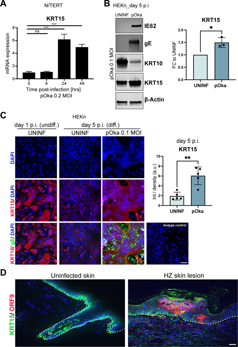
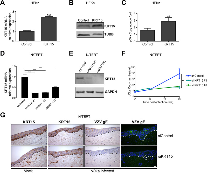
Varicella-zoster virus (VZV) is the etiological agent of chickenpox and shingles, diseases characterised by epidermal virus replication in skin and mucosa and the formation of blisters. We have previously shown that VZV infection has a profound effect on keratinocyte differentiation, altering the normal pattern of epidermal gene expression. In particular, VZV infection reduces expression of suprabasal keratins 1 and 10 and desmosomal proteins, disrupting epidermal structure to promote expression of a blistering phenotype. Here, we extend these findings to show that VZV infection upregulates the expression of keratin 15 (KRT15), a marker expressed by basal epidermal keratinocytes and hair follicles stem cells. We demonstrate that KRT15 is essential for VZV replication in the skin, since downregulation of KRT15 inhibits VZV replication in keratinocytes, while KRT15 exogenous overexpression supports viral replication. Importantly, our data show that VZV upregulation of KRT15 depends on the expression of the VZV immediate early gene ORF62. ORF62 is the only regulatory gene that is mutated in the live attenuated VZV vaccine and contains four of the five fixed mutations present in the VZV Oka vaccine. Our data indicate that the mutated vaccine ORF62 is not capable of upregulating KRT15, suggesting that this may contribute to the vaccine attenuation in skin. Taken together our data present a novel association between VZV and KRT15, which may open a new therapeutic window for a topical targeting of VZV replication in the skin via modulation of KRT15.

Keywords: IE62; KRT15; VZV; vOka vaccine.


Conflict of interest statement

The authors declare no competing interests.

Figures

Fig. 1
Fig. 1
VZV infection increases KRT15 expression. (A) N/TERT keratinocytes were infected with cell-free VZV parental strain pOka at an MOI of 0.2 and induced to differentiate at day 1 post-infection. Infected N/TERTs were then tested at the indicated time points for KRT15 mRNA expression by quantitative reverse transcription PCR (qRT-PCR). Tubulin beta (TUBB) levels were used for normalization for RNA signal input. n = 3 independent experiments. Error bars: SD. (B) HEKn keratinocytes were infected with pOka at an MOI of 0.1 for 5 days. HEKn were induced to differentiate at day 3 post-infection and left in culture for 2 further days before being collected. Protein levels were analysed by Western blotting. KRT15 densitometry was quantified and normalised to β-actin in n = 3 independent experiments and presented as fold change (FC) to UNINF. Error bars: SD. (C) KRT15 expression as visualised by immunofluorescence in HEKn keratinocytes infected with pOka at an MOI of 0.1 for 5 days. HEKn were induced to differentiate at day 3 post-infection and left in culture for 2 further days before being fixed. Infection with pOka is indicated by the expression of the VZV gE protein, as well as by the presence of syncytia structures displayed in the DAPI channel. Images shown are representative of n = 2 independent experiments. Graph shows average quantification of integrated density of KRT15 signal relative to number of nuclei in 5 fields of view for each of the UNINF and pOka conditions at day 5 p.i. Error bars: SD. (D) KRT15 and ORF9 protein expression as visualised by immunofluorescence in uninfected skin (displaying hair follicle, HF) and in vesicular skin lesion in herpes zoster (HZ). Images are representative of n = 4 patients skin samples analysed per condition. Dotted lines mark the epidermal-dermal junction. Scale bar, 50 μm. Statistical significance was calculated by one-way ANOVA and two-tailed t test ( and denoted by *P < 0.05, **P < 0.01,***P < 0.001. ns, not significant; hrs, hours; UNINF, uninfected; p.i., post infection; undiff, undifferentiated; diff, differentiated; SD, standard deviation
Fig. 2
Fig. 2
VZV immediate early gene ORF62 induces KRT15 upregulation. (A) PAA-treated or control-treated N/TERT keratinocytes were harvested at the indicated time points (hrs post-infection) and analysed for KRT15 mRNA levels by qRT-PCR. KRT15 mRNA levels were normalised to TUBB levels. Error bars: SD (n = 3 experiments). (B) KRT15 protein levels detected by Western blotting were analysed in N/TERT keratinocytes transduced to overexpress the VZV transcriptional regulatory proteins; ORF61, ORF62 or ORF63. (C) KRT15 mRNA levels were analysed by qRT-PCR in N/TERT keratinocytes overexpressing ORF62 as compared to control. KRT15 mRNA levels were normalised to TUBB levels. Error bars: SD (n = 3 experiments). Statistical significance was calculated by one-way ANOVA in (A) and two-tailed t test in (C) and denoted by **P < 0.01, ***P < 0.001. SD, standard deviation
Fig. 3
Fig. 3
KRT15 upregulation supports VZV replication. (A, B) Transduced HEKn overexpressing the KRT15 gene were analysed for KRT15 (A) mRNA by qRT-PCR and (B) protein levels by Western Blotting. Graph in (A) is average quantification of n = 3 biological replicates ± SD. (C) KRT15-overexpressing HEKn were then infected with VZV pOka and analysed by qPCR for VZV genome copy number and normalized per cell. Error bars: SD (n = 3 independent experiments). (D, E) N/TERT keratinocytes were infected with lentivirus expressing KRT15 specific short hairpin RNAs (shRNAs). 72 h post-infection cells were analysed for KRT15 (D) mRNA by qRT-PCR and (E) protein levels by Western Blotting. Graph in (D) is average quantification of n = 3 biological replicates ± SD. (F) Cells were harvested at the indicated time points post-VZV (pOka) infection and analysed for VZV genome copy number per cells using qPCR. Error bars: SD (n = 3 independent experiments). (G) Organotypic rafts of control and KRT15 knockdown (by means of siRNA) N/TERT keratinocytes were mock-infected or infected with VZV pOka nine days post-lifting at the air-liquid interface. Five days post-infection, the rafts were fixed and stained using antibodies for KRT15 or the viral protein VZV gE. Dotted lines mark the epidermal-dermal junction. Data is representative of biological triplicates and representative of results obtained in n = 2 independent experiments. Statistical significance was calculated by two-tailed t test (A) and one-way ANOVA (D and F) and denoted by *P < 0.05, **P < 0.01, ***P < 0.001. Scale bar, 50 μm. SD, standard deviation
Fig. 4
Fig. 4
The mutations in ORF62 in the vaccine strain vOka prevent KRT15 upregulation. (A) HEKn cells were infected either with pOka or with vOka at MOI 0.1 and analysed 5 days post-infection for gE expression by means of Fast Red staining. Graphs represent average quantification of n = 3 biological replicates for surface area gE positive cells ± SD. (B) HEKn cells were infected either with pOka or with vOka and analysed 5 days post-infection for gE protein expression by Western Blotting. Data representative of n = 2 independent experiments. (C) HEKn cells were infected either with pOka or with vOka and analysed 5 days post-infection for IE62 protein expression by Western Blotting. Data representative of n = 2 independent experiments. (D) HEKn cells were infected either with pOka or with vOka and analysed 5 days post-infection for IE62 mRNA expression by qRT-PCR. Data representative of n = 2 independent experiments. (E) KRT15 expression levels analysed by Western blotting in N/TERT keratinocytes after infection with increasing (as indicated by a triangle) MOI of the parental (pOka) and vaccine (vOka) VZV strains, with the highest MOI being 0.2. Data representative of n = 2 independent experiments. (F) HEKn cells were infected either with pOka at MOI 0.1 or with vOka at MOI 0.1 or 0.2 and analysed 5 days post-infection for KRT15 expression. VZV infection is indicated by staining for IE62 protein. Graph shows average quantification of integrated density of KRT15 signal in 4 fields of view for each of the UNINF and vOka conditions and 3 fields of view for the pOka condition. Error bars: SD. Data representative of n = 2 independent experiments. (G) (a) Schematic of the 5 regions of IE62. High homology regions 2 & 4 are highlighted. (b) Known functional/ protein interaction domains mapped to IE62 and their aminoacid co-ordinates. SSED = high serine-acidic tract. NLS = nuclear import signal. (c) Mapped vaccine SNPs (with reference to VZV Dumas) are indicated by short vertical lines. Those in orange are near-fixed, and other SNP coding changes (in blue) are indicated. The percentage of vaccine allele SNP in Varivax (Merck) and Varilix (GSK) are indicated. (d) The SNP mutations present in vOka-ID5-M9 are indicated. (H) HEKn cells were infected either with pOka or with vOka-ID5-M9 and analysed 5 days post-infection for pan VZV expression by means of Fast Red staining. Data representative of n = 3 biological replicates. (I) KRT15 protein levels from HEKn cells transfected with WT and mutant ORF62 were analysed by Western blotting. Data representative of n = 2 independent experiments. (J) HEKn cells expressing the pOka or vOka version of the viral ORF62 were infected by the vOka strain and analysed for VZV copy number per cell by qPCR (n = 3 independent experiments). Statistical significance was calculated by two-tailed t test (A) and one-way ANOVA (F and J) and denoted by *P < 0.05, **P < 0.01. Scale bar, 100 μm. ns, not significant; SD, standard deviation

References

    1. Arvin AM. Varicella-Zoster virus. Clin Microbiol Rev. 1996;9:361–81. - PMC - PubMed
    1. Gershon AA, Breuer J, Cohen JI, Cohrs RJ, Gershon MD, Gilden D, et al. Varicella Zoster virus infection. Nat Rev Dis Primers. 2015;1:1–18. - PMC - PubMed
    1. Moffat JF, Stein MD, Kaneshima H, Arvin AM. Tropism of varicella-zoster virus for human CD4 + and CD8 + T lymphocytes and epidermal cells in SCID-hu mice. J Virol. 1995;69:5236–42. - PMC - PubMed
    1. Moffat JF, Zerboni L, Kinchington PR, Grose C, Kaneshima H, Arvin AM. Attenuation of the vaccine Oka strain of varicella-zoster virus and role of glycoprotein C in alphaherpesvirus virulence demonstrated in the SCID-hu mouse. J Virol. 1998;72:965–74. - PMC - PubMed
    1. Candi E, Schmidt R, Melino G. The cornified envelope: a model of cell death in the skin. Nat Rev Mol Cell Biol. 2005;6:328–40. - PubMed

Publication types

MeSH terms

LinkOut - more resources