. 2024 Oct 10;12(1):159.
doi: 10.1186/s40478-024-01870-4.

Crucial role of Aquaporin-4 extended isoform in brain water Homeostasis and Amyloid-β clearance: implications for Edema and neurodegenerative diseases

Affiliations

Crucial role of Aquaporin-4 extended isoform in brain water Homeostasis and Amyloid-β clearance: implications for Edema and neurodegenerative diseases

Pasqua Abbrescia et al. Acta Neuropathol Commun. .

Abstract

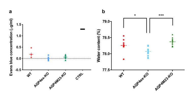
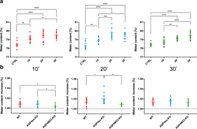
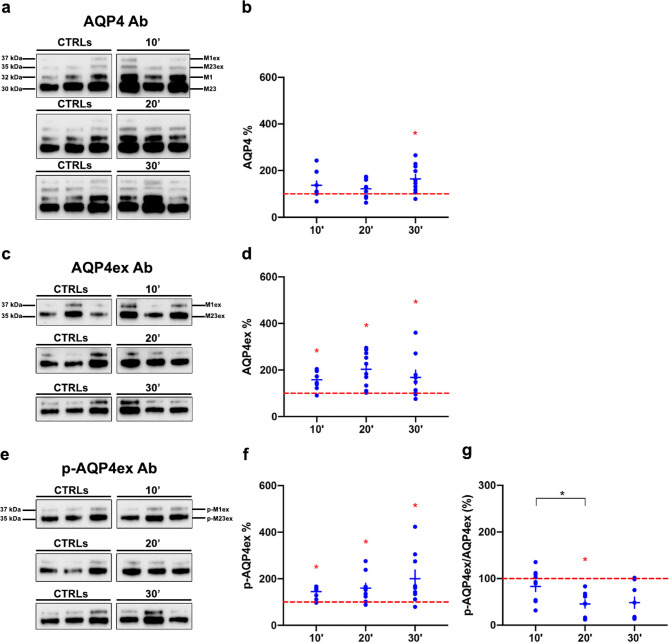
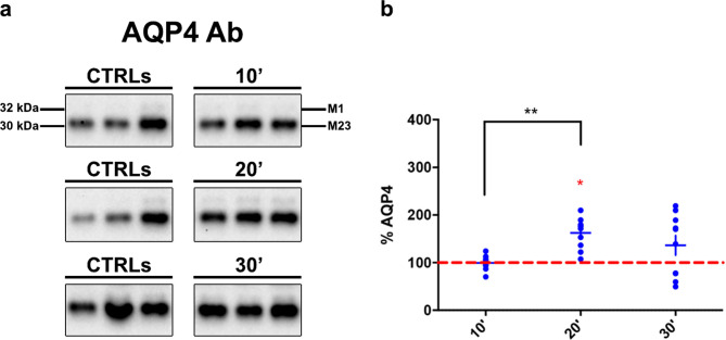
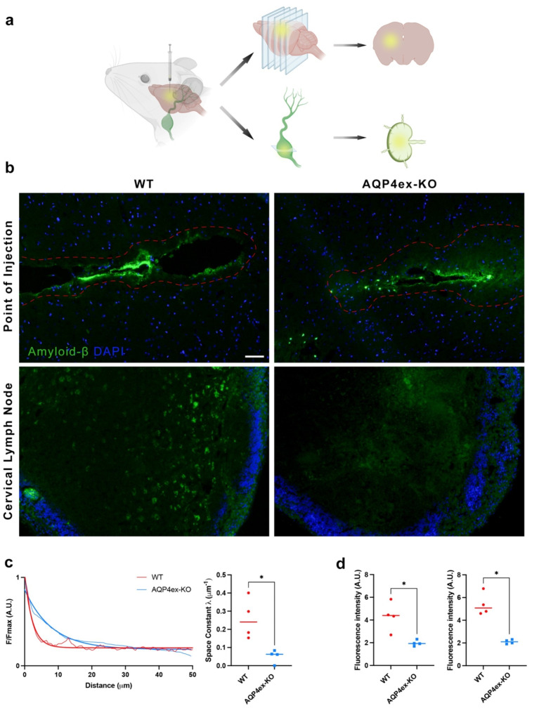
The water channel aquaporin-4 (AQP4) is crucial for water balance in the mammalian brain. AQP4 has two main canonical isoforms, M23, which forms supramolecular structures called Orthogonal Arrays of Particles (OAP) and M1, which does not, along with two extended isoforms (M23ex and M1ex). This study examines these isoforms' roles, particularly AQP4ex, which influences water channel activity and localization at the blood-brain barrier. Using mice lacking both AQP4ex isoforms (AQP4ex-KO) and lacking both AQP4M23 isoforms (OAP-null) mice, we explored brain water dynamics under osmotic stress induced by an acute water intoxication (AWI) model. AQP4ex-KO mice had lower basal brain water content than WT and OAP-null mice. During AWI, brain water content increased rapidly in WT and AQP4ex-KO mice, but was delayed in OAP-null mice. AQP4ex-KO mice had the highest water content increase at 20 min. Immunoblot analysis showed stable total AQP4 in WT mice initially, with increases at 30 min. AQP4ex and its phosphorylated form (p-AQP4ex) levels rose quickly, but the p-AQP4ex/AQP4ex ratio dropped at 20 min. AQP4ex-KO mice showed a compensatory rise in canonical AQP4 at 20 min post-AWI. These findings highlight the important role of AQP4ex in water content dynamics in both normal and pathological states. To evaluate brain waste clearance, amyloid-β (Aβ) removal was assessed using a fluorescent Aβ intra-parenchyma injection model. AQP4ex-KO mice demonstrated markedly impaired Aβ clearance, with extended diffusion distances and reduced fluorescence in cervical lymph nodes, indicating inefficient drainage from the brain parenchyma. Mechanistically, the polarization of AQP4 at astrocytic endfeet is essential for efficient clearance flow, aiding interstitial fluid movement into the CSF and lymphatic system. In AQP4ex-KO mice, disrupted polarization forces reliance on slower, passive diffusion for solute clearance, significantly reducing Aβ removal efficiency and altering extracellular space dynamics. Our results underscore the importance of AQP4ex in both brain water homeostasis and solute clearance, particularly Aβ. These findings highlight AQP4ex as a potential therapeutic target for enhancing waste clearance mechanisms in the brain, which could have significant implications for treating brain edema and neurodegenerative diseases like Alzheimer's.

Keywords: AQP4ex; Amyloid-β clearance; Aquaporin-4; Brain edema; Glymphatic system; Neurodegenerative diseases.


Conflict of interest statement

The authors declare that they have no competing interests.

Figures

Fig. 1
Fig. 1
Characterization of blood vessel permeability and brain basal water content. (a) Aligned dot plot showing quantitation of Evans Blue extravasation in WT (red), AQP4ex-KO (blue) and OAP-null (green) mice brain after intracaudal injection of dye. No differences among genotypes are observed (n = 5 for each analyzed group). The CTRL value is from a non-perfused animal. (b) Basal brain water content expressed in percentage. Aligned dot plot shows significantly reduced brain water content in AQP4ex-KO brain compared to WT and OAP-null. One-way ANOVA, Tukey’s multiple comparisons test, *p < 0.05, ***p < 0.0001; data are expressed as means ± SEM
Fig. 2
Fig. 2
Evaluation of brain water content after acute water intoxication at different time points. (a) Brain water measurements expressed as percentage after 10’, 20’ and 30’ of water intoxication in WT (red), AQP4ex-KO (blue) and OAP-null (green) mice. (b) Comparison of brain water content between different genotypes at each time point of AWI. Data were expressed as fold increase compared to basal water content for each genotype, set at 1.00 and indicated as black dashed line. WT and AQP4ex-KO showed higher water increase compared to OAP-null initially, but AQP4ex-KO had greater water accumulation at 20 min. Data are expressed as means ± SEM. (*p < 0.05, **p < 0.01, ***p < 0.001, ****p < 0.0001, One-Way ANOVA with post hoc comparison via a Bonferroni multiple comparison) n = 14, except for (a) AQP4ex-KO 20’ (n = 13), AQP4M23-KO 10’ and 20’ (n = 12); (b) AQP4M23-KO 10’ (n = 12), AQP4ex-KO 20’ (n = 13), AQP4M23-KO 20’ (n = 12)
Fig. 3
Fig. 3
AQP4 isoforms protein expression in WT mice after different time points of AWI. (a, c, e) Immunoblot results of AQP4, AQP4ex and p-AQP4ex expression in three different untreated (CTRLs) and treated mice at 10, 20 and 30 minuses post-AWI. Western blot experiments are representative and the samples shown belong to the same electrophoretic run. (b, d, f) Results of the densitometric analysis shown as the percentage variation compared to baseline (red dashed line) of AQP4, AQP4ex and P-AQP4ex respectively (Red asterisk indicates the Student’s t-test significant differences for the comparison with basal level, while the black one indicates the comparison between different time points). (g) Ratio between p-AQP4ex and AQP4ex showing that only half of additional AQP4ex was phosphorylated at 20 min. Data are expressed as means ± SEM (*p < 0.05; One-Way ANOVA n = 10)
Fig. 4
Fig. 4
AQP4 canonical isoform expression in AQP4ex-KO mice post-AWI. (a) Typical immunoblot of M1 and M23 isoforms detection in three untreated and treated mice; Western blot experiments are representative and the samples shown belong to the same electrophoretic run. (b) Results of the densitometric aanalysis shown as the percentage variation compared to baseline (red dashed line) of AQP4. Note that after 20 min of AWI canonical isoforms significantly increased. (Red asterisk indicates the Student’s t-test significant differences for the comparison with basal level, while black one the comparison between different time points, *p < 0.05, **p < 0.01, n = 10)
Fig. 5
Fig. 5
Amyloid-β drainage from brain parenchyma. (a) Schematic model of the protocol used to evaluate the cleaance of Aβ in the central nervous system of mice. Amyloid-β (1–42) Hylexa Fluor-488 was stereotaxically injected into the striatum and six hours post-injection, the fluorescence of Aβ were assessed after cryosection of brains and lymphnodes. (b) Brain section that exhibits the maximum fluorescence intensity among all sections at the injection site was analyzed together with lymph nodes sections. The red dotted line indicates the region where fluorescence levels were detectable. Cell nuclei were stained with DAPI (in blue). Note that the fluorescence intensity of amyloid-β in WT appears lowers than AQP4ex-KO at the point of injection while the ipsilateral cervical lymph nodes have markedly higher levels of fluorescence in WT mice compared to AQP4ex-KO mice. Scale bar 50 μm. (c) Aβ fluorescence values plotted as a function of distance from the injection site in the striatum. Quantification of fluorescent signal quantified using the space constant λ (µm⁻¹). (d) Quantification of Aβ in cortical (left) and medullar (right) regions of ipsilateral lymph node of WT and AQP4ex-KO mice (n = 4). Unpaired t-test, *p < 0.05

References

    1. Verkman AS, Binder DK, Bloch O, Auguste K, Papadopoulos MC (2006) Three distinct roles of Aquaporin-4 in brain function revealed by knockout mice. Biochim Biophys Acta 1758(8):1085–1093. 10.1016/j.bbamem.2006.02.018 - DOI - PubMed
    1. Manley GT, Fujimura M, Ma T, Noshita N, Filiz F, Bollen AW, Chan P, Verkman AS (2000) Aquaporin-4 deletion in mice reduces Brain Edema after Acute Water Intoxication and ischemic stroke. Nat Med 6(2):159–163. 10.1038/72256 - DOI - PubMed
    1. Papadopoulos MC, Manley GT, Krishna S, Verkman AS (2004) Aquaporin-4 facilitates reabsorption of excess fluid in Vasogenic Brain Edema. FASEB J 18(11):1291–1293. 10.1096/fj.04-1723fje - DOI - PubMed
    1. Iliff JJ, Wang M, Liao Y, Plogg BA, Peng W, Gundersen GA, Benveniste H, Vates GE, Deane R, Goldman SA et al (2012) A paravascular pathway facilitates CSF Flow through the Brain Parenchyma and the Clearance of Interstitial Solutes, including amyloid β. Sci Transl Med 4(147):147ra111. 10.1126/scitranslmed.3003748 - DOI - PMC - PubMed
    1. Hablitz LM, Plá V, Giannetto M, Vinitsky HS, Stæger FF, Metcalfe T, Nguyen R, Benrais A, Nedergaard M (2020) Circadian control of Brain Glymphatic and Lymphatic Fluid Flow. Nat Commun 11(1):4411. 10.1038/s41467-020-18115-2 - DOI - PMC - PubMed

Publication types

MeSH terms

LinkOut - more resources