. 2024 Dec;51(12):1677-1687.
doi: 10.1111/jcpe.14070. Epub 2024 Oct 9.

Wide Restorative Emergence Angle Increases Marginal Bone Loss and Impairs Integrity of the Junctional Epithelium of the Implant Supracrestal Complex: A Preclinical Study

Affiliations

Wide Restorative Emergence Angle Increases Marginal Bone Loss and Impairs Integrity of the Junctional Epithelium of the Implant Supracrestal Complex: A Preclinical Study

Franz J Strauss et al. J Clin Periodontol. 2024 Dec.

Abstract

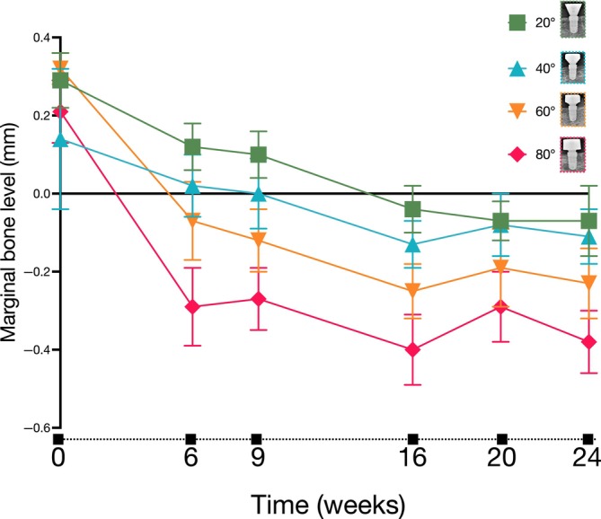
Aim: To assess the influence of the emergence angle on marginal bone loss (MBL) and supracrestal soft tissue around dental implants.

Materials and methods: In six mongrel dogs, the mandibular premolars and molars were extracted. After 3 months of healing, four dental implants were placed in each hemimandible. The implants were randomly allocated to receive one of four customized healing abutments, each with a different value of the restorative emergence angle: 20°, 40°, 60° or 80°. Intra-oral radiographs were taken after placing the healing abutments and at 6, 9, 16 and 24 weeks of follow-up. Then, micro-CT and undecalcified histology and synchrotron were performed. MBL over time was analysed with generalized estimating equations (GEEs) and adjusted for baseline soft-tissue thickness.

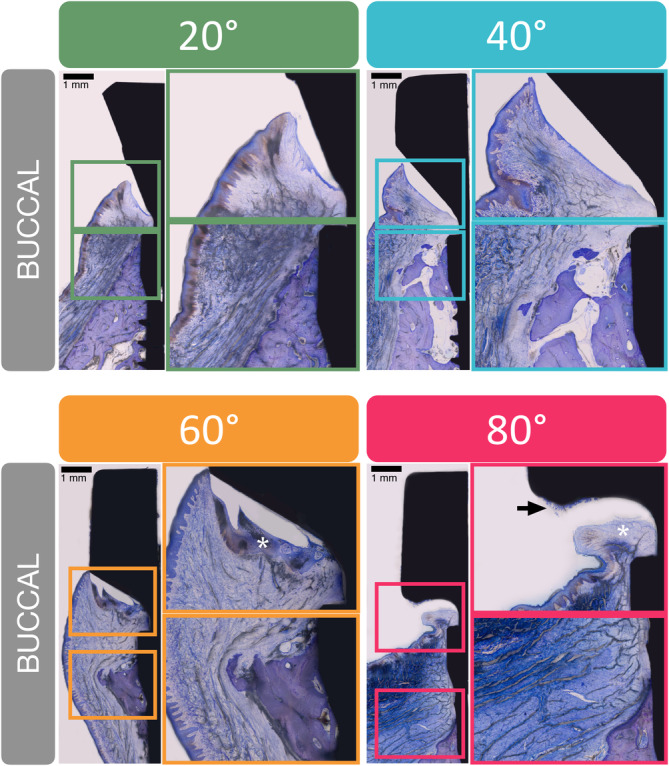
Results: From implant placement to 24 weeks, GEE modelling showed that the MBL at mesial and distal sites consistently increased over time, indicating MBL in all groups (p < 0.001). The model indicated that MBL varied significantly across the different restorative angles (angle effect, p < 0.001), with 80° showing the greatest bone loss. Micro-CT, histology and synchrotron confirmed the corresponding trends and showed that wide restorative angles (60° and 80°) impaired the integrity of the junctional epithelium of the supracrestal tissue.

Conclusions: A wide restorative angle increases MBL and impairs the integrity of the junctional epithelium of the implant supracrestal complex.

Keywords: CAD/CAM; dental implant; dental implant‐abutment design; emergence profile; histology; prosthodontics; restorative angle; titanium abutments.


Conflict of interest statement

The authors declare no conflicts of interest.

Figures

FIGURE 1
FIGURE 1
Line chart indicating the mean marginal bone level at each time points for the different angle groups. Error bars indicate standard error. Differences were tested using a linear model under the generalized estimating equations with angle, time and their interaction and adjusted for soft tissue thickness at baseline (implant placement). Bonferroni correction was applied for the multiple comparisons.
FIGURE 2
FIGURE 2
First bone‐to‐implant contact (fBIC) values in the different groups at different sites (mesial/distal and buccal) based on micro‐CT analysis. Differences were tested using the Kruskal–Wallis test with the Dunn's test for multiple comparison adjustments.
FIGURE 3
FIGURE 3
First bone‐to‐implant contact (fBIC) values at the different groups based on histomorphometric analysis (scale bars indicate 1 mm). Differences were tested using the Kruskal–Wallis test.
FIGURE 4
FIGURE 4
Histological images of the buccal soft tissue surrounding the customized abutments for each group. For each group, a representative image and two magnifications focused on the supracrestal soft tissue and crestal bone at buccal sites are shown (scale bars indicate 1 mm). The white asterisk shows the disorganized or lacking junctional epithelium. The black arrow indicates the presence of bacterial biofilm against the abutment surface.
FIGURE 5
FIGURE 5
Distance from inflammatory infiltrate to fBIC. The distance between the apical termination of the thickest part of the inflammatory infiltrate (red) and the fBIC (purple) was measured (yellow arrow). One line parallel to the implant axis was drawn from the apical termination of the thickest part of the inflammatory infiltrate (identified by the accumulation of immune cells). This line intersected another line drawn perpendicular to the implant axis at the level of the fBIC (purple). Differences were tested using the one‐way ANOVA test with the Dunnet test for multiple comparison adjustments.

Similar articles

Cited by

References

    1. Abrahamsson, I. , Berglundh T., and Lindhe J.. 1998. “Soft Tissue Response to Plaque Formation at Different Implant Systems. A Comparative Study in the Dog.” Clinical Oral Implants Research 9, no. 2: 73–79. 10.1034/j.1600-0501.1998.090202.x. - DOI - PubMed
    1. Academy of Prosthodontics . 2023. “The Glossary of Prosthodontic Terms 2023: Tenth Edition.” The Journal of Prosthetic Dentistry 130, no. 4 Suppl 1: e7–e126. 10.1016/j.prosdent.2023.03.002. - DOI - PubMed
    1. Albrektsson, T. , Zarb G., Worthington P., and Eriksson A. R.. 1986. “The Long‐Term Efficacy of Currently Used Dental Implants: A Review and Proposed Criteria of Success.” The International Journal of Oral & Maxillofacial Implants 1, no. 1: 11–25. - PubMed
    1. Belser, U. C. , Buser D., Hess D., Schmid B., Bernard J. P., and Lang N. P.. 1998. “Aesthetic Implant Restorations in Partially Edentulous Patients—A Critical Appraisal.” Periodontology 2000 2000, no. 17: 132–150. 10.1111/j.1600-0757.1998.tb00131.x. - DOI - PubMed
    1. Berglundh, T. , Abrahamsson I., Welander M., Lang N. P., and Lindhe J.. 2007. “Morphogenesis of the Peri‐Implant Mucosa: An Experimental Study in Dogs.” Clinical Oral Implants Research 18, no. 1: 1–8. 10.1111/j.1600-0501.2006.01380.x. - DOI - PubMed

Publication types