. 2024 Oct 1;17(10):dmm050566.
doi: 10.1242/dmm.050566. Epub 2024 Oct 23.

RNA sequencing reveals molecular mechanisms of endometriosis lesion development in mice

Affiliations

RNA sequencing reveals molecular mechanisms of endometriosis lesion development in mice

Kavita Panir et al. Dis Model Mech. .

Abstract

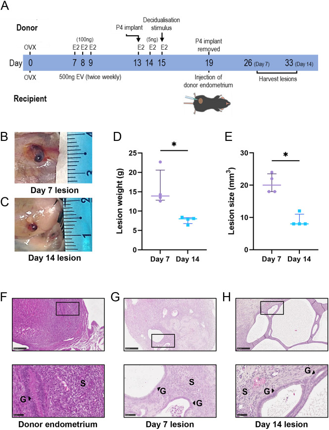
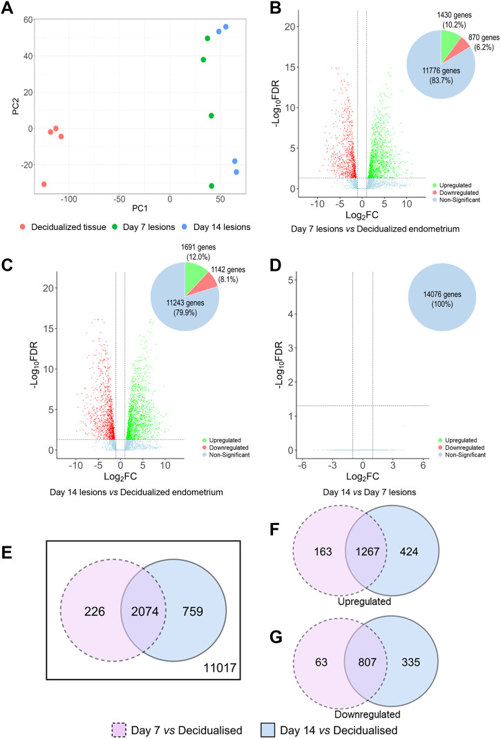
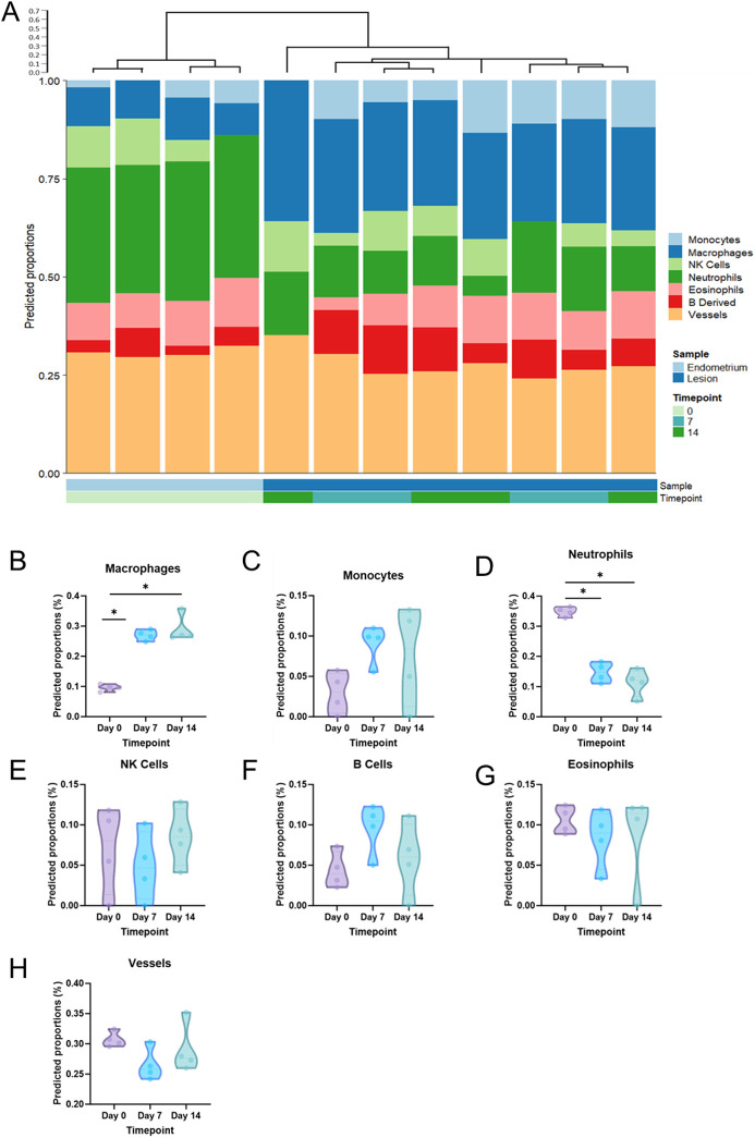
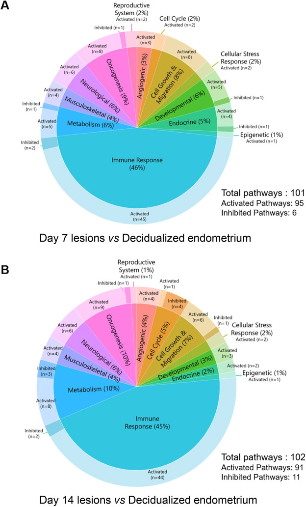
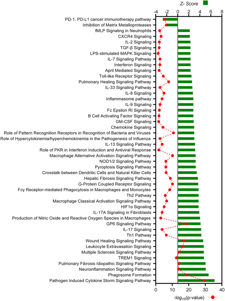
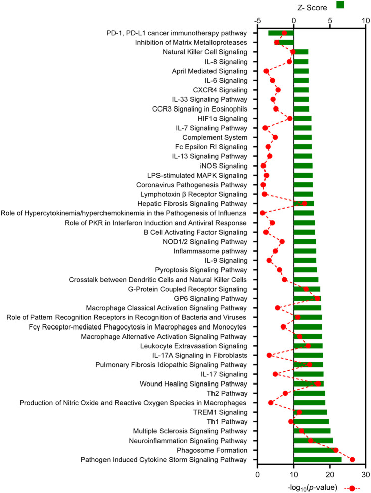
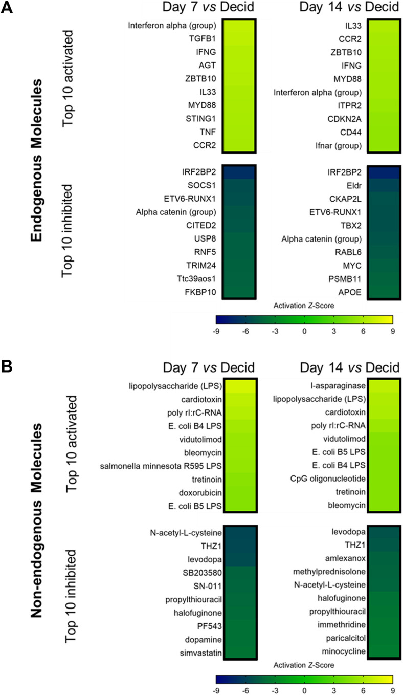
Understanding of molecular mechanisms contributing to the pathophysiology of endometriosis, and upstream drivers of lesion formation, remains limited. Using a C57Bl/6 mouse model in which decidualized endometrial tissue is injected subcutaneously in the abdomen of recipient mice, we generated a comprehensive profile of gene expression in decidualized endometrial tissue (n=4), and in endometriosis-like lesions at Day 7 (n=4) and Day 14 (n=4) of formation. High-throughput mRNA sequencing allowed identification of genes and pathways involved in the initiation and progression of endometriosis-like lesions. We observed distinct patterns of gene expression with substantial differences between the lesions and the decidualized endometrium that remained stable across the two lesion timepoints, and showed similarity to transcriptional changes implicated in human endometriosis lesion formation. Pathway enrichment analysis revealed several immune and inflammatory response-associated canonical pathways, multiple potential upstream regulators, and involvement of genes not previously implicated in endometriosis pathogenesis, including IRF2BP2 and ZBTB10, suggesting novel roles in disease progression. Collectively, the provided data will be a useful resource to inform research on the molecular mechanisms contributing to endometriosis-like lesion development in this mouse model.

Keywords: Endometriosis; Immune regulation; Mouse model; RNA Sequencing.


Conflict of interest statement

Competing interests The authors declare no competing or financial interests.

Figures

Fig. 1.
Fig. 1.
Development of endometriosis lesions in mice. (A) A modified version of a menstrual mouse model of endometriosis was used to establish subcutaneous endometriosis-like lesions in recipient mice. E2, beta-estradiol; EV, estradiol valerate; OVX, ovariectomy surgery; P4, progesterone. (B,C) Representative images of lesions at Day (D)7 (B) and D14 (C). (D,E) Lesions were excised, weighed (D) and measured (E). (F-H) Representative photomicrographs of Hematoxylin and Eosin (H&E)-stained sections from donor decidualized endometrium (F), D7 lesions (G) and D14 lesions (H). Top row: 10× magnification; bottom row: 40× magnification. Scale bars: 50 μm. G, glands; S, stroma. Data are presented as median±interquartile range, with each symbol representative of a single lesion in one mouse used for RNA sequencing (RNA-seq). Statistical analysis was performed using the Mann–Whitney test (*P<0.05).
Fig. 2.
Fig. 2.
Comparison of the gene expression profile in donor decidualized endometrium, and D7 and D14 endometriosis-like lesions. Differentially expressed genes (DEGs) were identified using limma/voom and edgeR with a cut-off of false discovery rate-adjusted P-value (FDR)≤0.05 and log2fold change (log2FC)≥+1 or ≤−1. (A) Principal component (PC) analysis of RNA-seq results visualizing the gene expression pattern of individual samples used in this analysis. (B-D) Volcano plots comparing log10FC and −log10(FDR) among detected genes between D7 lesions versus decidualized endometrium (B), D14 lesions versus decidualized endometrium (C), and D14 lesions versus D7 lesions (D). (E) Venn diagram displays the distribution and overlap of DEGs (both upregulated and downregulated) between D7 lesion versus decidualized endometrium and D14 lesion versus decidualized endometrium. (F,G) Additional Venn diagrams were generated to determine the number of upregulated (F) and downregulated (G) DEGs during lesion development compared to decidualized endometrial tissue. A complete list of DEGs can be found in Tables S1-S3.
Fig. 3.
Fig. 3.
Cellular deconvolution of RNA-seq data. (A) Cellular deconvolution with Euclidean clustering was performed for each of the 12 tissue samples. (B-H) Predicted proportions of macrophages (B), monocytes (C), neutrophils (D), natural killer (NK) cells (E), B cells (F), eosinophils (G) and vessels (H) were compared across the three timepoints (D0, decidualized endometrium; D7, Day 7 lesions; D14, Day 14 lesions). Effect of tissue status was analyzed by Mann–Whitney test (*P<0.05).
Fig. 4.
Fig. 4.
Classification of canonical pathways identified by Ingenuity Pathway Analysis (IPA). (A,B) Canonical pathways identified in D7 endometriosis-like lesions versus decidualized endometrium (A), and D14 endometriosis-like lesions versus decidualized endometrium (B). The full list of canonical pathways is available in Tables S4 and S5. All pathways shown have a Z-activation score of ≥+2 or ≤−2, and corresponding P-value of ≤0.05.
Fig. 5.
Fig. 5.
All canonical pathways associated with immune responses in D7 endometriosis-like lesions versus decidualized endometrium identified by IPA. Canonical pathways predicted to be associated with immunological response are listed, with Z-score shown on the top axis and −log10(P-value) on the bottom axis. All pathways shown have a Z-activation score of ≥+2 or ≤−2, and corresponding P-value of ≤0.05. The full list of canonical pathways is available in Table S4.
Fig. 6.
Fig. 6.
All canonical pathways associated with immune responses in D14 endometriosis-like lesions versus decidualized endometrium identified by IPA. Canonical pathways predicted to be associated with immunological response are listed, with Z-score shown on the top axis and −log10(P-value) on the bottom axis. All pathways shown have a Z-activation score of ≥+2 or ≤−2, and corresponding P-value of ≤0.05. The full list of canonical pathways is available in Table S5.
Fig. 7.
Fig. 7.
Molecules predicted to be upstream regulators of differentially expressed genes in Day 7 and Day 14 endometriosis-like lesions versus decidualized endometrium identified by IPA. (A) Heatmaps of the top 10 activated and top 10 inhibited endogenous upstream regulators are shown for D7 lesion versus decidualized endometrium and D14 lesion versus decidualized endometrium. (B) Heatmaps of the top 10 activated and top 10 inhibited non-endogenous upstream regulators are shown for D7 lesion versus decidualized endometrium and D14 lesion versus decidualized endometrium. All upstream regulators have a Z-activation score of ≥+2 or ≤−2, and corresponding P-value of ≤0.05. The full list of upstream regulators is available in Tables S6 and S7.

References

    1. Acién, P., Quereda, F., Campos, A., Gomez-Torres, M.-J., Velasco, I. and Gutierrez, M. (2002). Use of intraperitoneal interferon alpha-2b therapy after conservative surgery for endometriosis and postoperative medical treatment with depot gonadotropin-releasing hormone analog: a randomized clinical trial. Fertil. Steril. 78, 705-711. 10.1016/S0015-0282(02)03330-7 - DOI - PubMed
    1. Ali, A. F. M., Fateen, B., Ezzet, A., Badawy, H., Ramadan, A. and El-Tobge, A. (2000). Laparoscopic intraperitoneal injection of human interferon-α2b in the treatment of pelvic endometriosis: a new modality. Obstet. Gynecol. 95 4, Suppl. 1, S47-S48. 10.1016/S0029-7844(00)00684-0 - DOI
    1. Anastasi, E., Scaramuzzino, S., Viscardi, M. F., Viggiani, V., Piccioni, M. G., Cacciamani, L., Merlino, L., Angeloni, A., Muzii, L. and Porpora, M. G. (2023). Efficacy of N-acetylcysteine on endometriosis-related pain, size reduction of ovarian endometriomas, and fertility outcomes. Int. J. Environ. Res. Public Health 20, 4686. 10.3390/ijerph20064686 - DOI - PMC - PubMed
    1. Aznaurova, Y. B., Zhumataev, M. B., Roberts, T. K., Aliper, A. M. and Zhavoronkov, A. A. (2014). Molecular aspects of development and regulation of endometriosis. Reprod. Biol. Endocrinol. 12, 1-25. 10.1186/1477-7827-12-50 - DOI - PMC - PubMed
    1. Bacci, M., Capobianco, A., Monno, A., Cottone, L., Di Puppo, F., Camisa, B., Mariani, M., Brignole, C., Ponzoni, M., Ferrari, S.et al. (2009). Macrophages are alternatively activated in patients with endometriosis and required for growth and vascularization of lesions in a mouse model of disease. Am. J. Pathol. 175, 547-556. 10.2353/ajpath.2009.081011 - DOI - PMC - PubMed