. 2024 Nov 27;52(21):13368-13385.
doi: 10.1093/nar/gkae875.

Human selenocysteine synthase, SEPSECS, has evolved to optimize binding of a tRNA-based substrate

Affiliations

Human selenocysteine synthase, SEPSECS, has evolved to optimize binding of a tRNA-based substrate

Anupama K Puppala et al. Nucleic Acids Res. .

Abstract

The evolution of the genetic code to incorporate selenocysteine (Sec) enabled the development of a selenoproteome in all domains of life. O-phosphoseryl-tRNASec selenium transferase (SepSecS) catalyzes the terminal reaction of Sec synthesis on tRNASec in archaea and eukaryotes. Despite harboring four equivalent active sites, human SEPSECS binds no more than two tRNASec molecules. Though, the basis for this asymmetry remains poorly understood. In humans, an acidic, C-terminal, α-helical extension precludes additional tRNA-binding events in two of the enzyme monomers, stabilizing the SEPSECS•tRNASec complex. However, the existence of a helix exclusively in vertebrates raised questions about the evolution of the tRNA-binding mechanism in SEPSECS and the origin of its C-terminal extension. Herein, using a comparative structural and phylogenetic analysis, we show that the tRNA-binding motifs in SEPSECS are poorly conserved across species. Consequently, in contrast to mammalian SEPSECS, the archaeal ortholog cannot bind unacylated tRNASec and requires an aminoacyl group. Moreover, the C-terminal α-helix 16 is a mammalian innovation, and its absence causes aggregation of the SEPSECS•tRNASec complex at low tRNA concentrations. Altogether, we propose SEPSECS evolved a tRNASec binding mechanism as a crucial functional and structural feature, allowing for additional levels of regulation of Sec and selenoprotein synthesis.


Figures

Graphical Abstract
Graphical Abstract
Figure 1.
Figure 1.
Helix α16 of human SEPSECS utilizes negatively charged surfaces to engage the tRNASec binding pocket and entrance into the active site. Electrostatic potential maps of the α16 faces (PDBID 7MDL) show acidic residues along α16 comprise a negatively charged (red surfaces) face (A) and a more neutral face when rotated 180° along the y-axis (B). (C) Human holo SEPSECS (PDBID 7L1T) harbors large positively charged surfaces (blue) that constitute the tRNA binding pockets and active sites. (D) α16 nestles into the tRNA binding pocket of SEPSECS in a manner that orients the acidic face of α16 toward the tRNA binding pocket. As illustrated by the bar, surfaces are contoured from −5 (red) to +5 (blue) kT/e based on the potential of the solvent accessible surface.
Figure 2.
Figure 2.
Δ470 SEPSECS maintains catalytic competency and forms higher-orders species at low concentrations of human tRNASec. (A) EtBr staining shows the change in migration of human tRNASec (htRNASec) in a titration series upon binding to either WT or Δ470 SEPSECS. Binding of htRNASec to WT or Δ470 SEPSECS formed soluble complex, as indicated by the ‘higher’ and ‘lower’ EtBr-staining bands with reduced electrophoretic mobility. However, binding to Δ470 SEPSECS induced a higher-order complex at low substrate concentrations, as indicated by staining within the well. The higher-order species was more prevalent with only one tRNA per tetramer (lane 11) and only minimally present when 2 tRNAs were provided per tetramer (Lane 12). (B) Coomassie staining of the same gel confirms the presence of protein in the complex. lane 1—free tRNASec, lane 2—WT, lane 3—4:1 WT: tRNASec, lane 4—4:2 WT: tRNASec, lane 5—4:4 WT: tRNASec, lane 6—4:6 WT: tRNASec, lane 7—4:8 WT: tRNASec, lane 8—empty, lane 9—free tRNASec, lane 10—Δ470, lane 11—4:1 Δ470: tRNASec, lane 12—4:2 Δ470: tRNASec, lane 13—4:4 Δ470: tRNASec, lane 14—4:6 Δ470: tRNASec, lane 15—4:8 Δ470: tRNASec. (C) Only co-expression of human SEPSECS and Methanocaldococcus jannaschii PSTK allows ΔselA JS2(DE3) cells to synthesize formate dehydrogenase and reduce benzyl viologen (BV) to its purple form. Δ470 SEPSECS exhibits similar levels of BV reduction as the WT enzyme throughout the dilution series. Experiment was performed in duplicate with one representative result shown.
Figure 3.
Figure 3.
MALS traces reveal large, polydisperse species of the Δ470 SEPSECS binary complex. Light scattering traces of WT SEPSECS (blue) reveal a single, monodisperse species with a horizontal molar mass distribution across the peak that corresponds to the SEPSECS•tRNASec binary complex. Analogous Δ470 SEPSECS samples (red traces) possess an aggregate peak (earlier eluting peak), and polydispersity within the sample precludes accurate molecular weight determination of the Δ470 SEPSECS species. MALS traces at SEPSECS•tRNASec molar ratios of (A) 4:1, (B) 4:2, (C) 4:4, (D) 4:6, (E) 4:8.
Figure 4.
Figure 4.
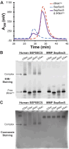
M. maripaludis SepSecS does not form a stable complex with unacylated tRNASec. (A) Size-exclusion chromatograms showed no observable binary complex peak upon mixing MMP SepSecS and MMP tRNASec. Instead, two peaks that coincide with the protein and MMP tRNASec alone, respectively, are observed. The EMSA gel analyzing binding of human SEPSECS and archaeal SepSecS to several tRNASec species, using either EtBr (B) or Coomassie staining (C). When mixed with a molar equivalent of tRNA, human SEPSECS formed a stable complex with human (hSec), MMP (aSec) and E. coli (bSec) tRNASec. The smear observed in the presence of human tRNASer (hSer) indicated nonspecific binding. MMP SepSecS did not interact with any of the unacylated tRNASec species.
Figure 5.
Figure 5.
Phylogenetic reconstruction of SepSecS evolution. 874 SepSecS orthologs spanning from archaea to eukarya were used to reconstruct the evolutionary history of SepSecS, rooted at M. kandleri. Archaeal orthologs are colored in red, while the eukaryotic orthologs are divided into eight groups: protozoans (dark blue), fungi (black), invertebrates (dark green), fishes (yellow), amphibians (light green), mammals (brown), reptiles (light blue), and birds (pink). The cladogram illustrates the divergence of eukaryotic orthologs from archaeal orthologs, as well as the divergence of mammals from other vertebrates. An alternate view of the phylogeny with support values and branch lengths is available as online supporting materials in Supplementary Figure S14.
Figure 6.
Figure 6.
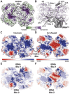
The enzyme surface including the tRNA-binding motifs are weakly conserved. (A) Consurf-generated evolutionary conservation grades mapped onto a surface representation of human holo SEPSECS (PDBID 7L1T). The active sites are strongly conserved (purple), while the enzyme surface has more variability (green). (B) Conservation of α1, α9, α14 and α15 which comprise two tRNA-binding pockets across each face of the human enzyme, with human tRNASec (PDBID 7MDL) modeled onto Site 2. (C) The electrostatic potential map of human SEPSECS (PDBID 7L1T) largely features a positively charged solvent-exposed surface (blue). (D) Apart from the positively charged active sites, archaeal SepSecS (PDBID 2Z67) exhibits a more negatively charged surface (red), particularly across the center of the tetramer. (E) Helices α1, α9, α14 and α15 comprise the positively charged tRNA binding pocket (tRNA Site 1 and 2). (F) The comparable helices in archaeal SepSecS generate more acidic surfaces. As illustrated by the bar, surfaces are contoured from –5 (red) to +5 (blue) kT/e based on the potential of the solvent accessible surface.
Figure 7.
Figure 7.
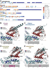
The vertebrate lineages have distinct amino acid sequences for the extended C-terminus of SEPSECS. (A) The extreme C-terminal region of SEPSECS shows poor conservation among vertebrates. Within vertebrates, mammals, birds, reptiles, and amphibians have unique consensus sequences (≥50% conservation). (B–E) Representative structural predictions for the extended C-terminus in non-mammalian, vertebrate clades, using ColabFold (top) and S4PRED (bottom). ColabFold structures (light teal cartoon) are shown aligned to the human SEPSECS (PDBID 7MDL), with the catalytic protomer colored blue and the non-catalytic protomer in red. The S4PRED predictions indicate a helical conformation (highlighted in pink) or a coiled conformation (highlighted in gray), with confidence scores noted above.

Similar articles

References

    1. Hatfield D.L., Schweizer U., Tsuji P., Gladyshev V.. Selenium: Its Molecular Biology and Role in Human Health. 2016; 4th edn.NY: Springer Science & Business Media.
    1. Schwarz K., Foltz C.M.. Factor 3 activity of selenium compounds. J. Biol. Chem. 1958; 233:245–251. - PubMed
    1. Turanov A.A., Kehr S., Marino S.M., Yoo M.-H., Carlson B.A., Hatfield D.L., Gladyshev V.N.. Mammalian thioredoxin reductase 1: roles in redox homoeostasis and characterization of cellular targets. Biochem. J. 2010; 430:285–293. - PMC - PubMed
    1. Hawkes W.C., Alkan Z.. Regulation of Redox Signaling by Selenoproteins. Biol. Trace Elem. Res. 2010; 134:235–251. - PMC - PubMed
    1. Cox A.G., Tsomides A., Kim A.J., Saunders D., Hwang K.L., Evason K.J., Heidel J., Brown K.K., Yuan M., Lien E.C.et al. .. Selenoprotein H is an essential regulator of redox homeostasis that cooperates with p53 in development and tumorigenesis. Proc. Natl. Acad. Sci. 2016; 113:E5562–E5571. - PMC - PubMed

Substances

LinkOut - more resources