

Intrathyroidal Thymoma: A Diagnostic Challenge
- PMID: 39385927
- PMCID: PMC11463876
- DOI: 10.7759/cureus.68987
Intrathyroidal Thymoma: A Diagnostic Challenge
Abstract
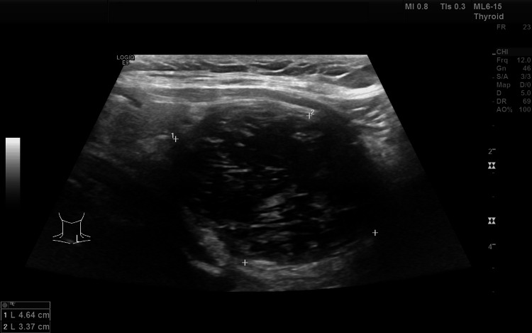
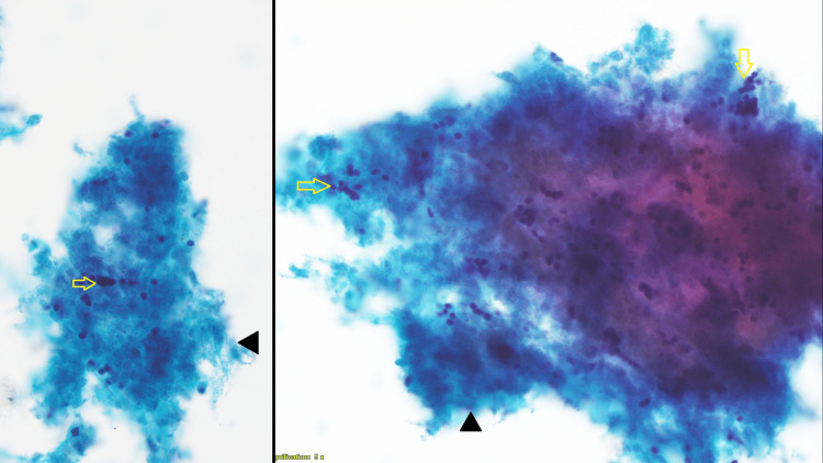
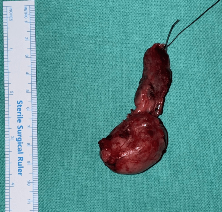
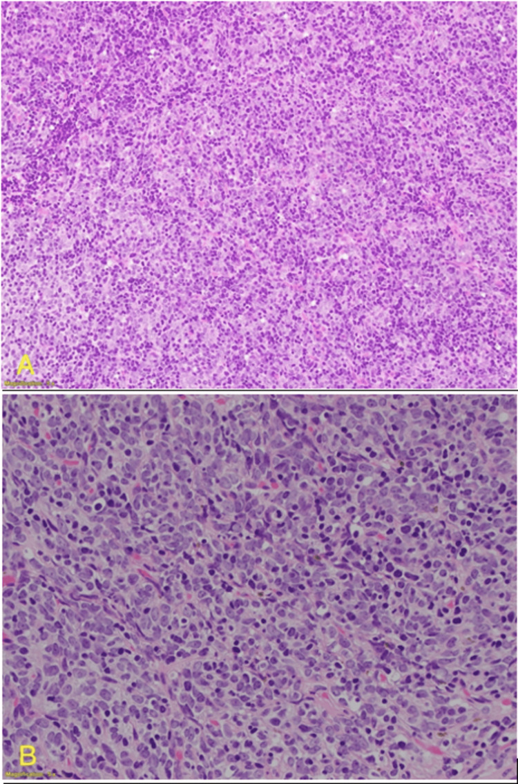
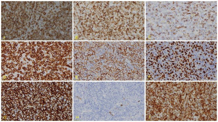
Intrathyroidal thymoma is a rare tumor that can be challenging to diagnose due to its unusual location and resemblance to more common thyroid conditions. We present the case of a 58-year-old woman with an incidentally discovered thyroid nodule during evaluation for an upper respiratory infection. Ultrasonography revealed an exophytic nodule in the left thyroid lobe, categorized as TR 3. Fine-needle aspiration cytology suggested a neoplastic process, leading to a left hemithyroidectomy. Histopathology confirmed a diagnosis of intrathyroidal thymoma, Type B2, with extensive necrosis, and immunohistochemistry validated the findings. This case underscores the diagnostic challenges of intrathyroidal thymoma, emphasizing its consideration in the differential diagnosis of atypical thyroid nodules. Despite the difficulties in preoperative identification, surgical resection and subsequent histopathological examination remain essential for a definitive diagnosis. The patient is currently under surveillance, and there is no evidence of residual thymic tissue or abnormalities in the remaining thyroid tissue.
Keywords: aberrant thymic tissue; ectopic thymoma; fnac; immunohistochemistry; intrathyroidal thymoma; thymoma; thyroid.
Copyright © 2024, Balaji et al.
Conflict of interest statement
Human subjects: Consent was obtained or waived by all participants in this study. Conflicts of interest: In compliance with the ICMJE uniform disclosure form, all authors declare the following: Payment/services info: All authors have declared that no financial support was received from any organization for the submitted work. Financial relationships: All authors have declared that they have no financial relationships at present or within the previous three years with any organizations that might have an interest in the submitted work. Other relationships: All authors have declared that there are no other relationships or activities that could appear to have influenced the submitted work.
Figures
References
-
- Intrathyroidal epithelial thymoma: an entity distinct from squamous cell carcinoma of the thyroid. Miyauchi A, Kuma K, Matsuzuka F, Matsubayashi S, Kobayashi A, Tamai H, Katayama S. World J Surg. 1985;9:128–135. - PubMed
-
- Ectopic primary intrathyroidal thymoma: a clinicopathological and immunohistochemical analysis of 3 cases. Weissferdt A, Moran CA. Hum Pathol. 2016;49:71–76. - PubMed
-
- Tumors of the neck showing thymic or related branchial pouch differentiation: a unifying concept. Chan JK, Rosai J. Hum Pathol. 1991;22:349–367. - PubMed
-
- Ectopic cervical thymus: case report and review of pathogenesis and management. Ahsan F, Allison R, White J. J Laryngol Otol. 2010;124:694–697. - PubMed
-
- Ectopic intrathyroidal thymoma: a case report and review. Cohen JB, Troxell M, Kong CS, McDougall IR. Thyroid. 2003;13:305–308. - PubMed
Publication types
LinkOut - more resources
Full Text Sources