. 2024 Oct 9;9(5):e70007.
doi: 10.1002/lio2.70007. eCollection 2024 Oct.

Immunocytochemistry assessment of vocal fold regeneration after cell-based implant in rabbits

Affiliations

Immunocytochemistry assessment of vocal fold regeneration after cell-based implant in rabbits

Larissa Nicolas et al. Laryngoscope Investig Otolaryngol. .

Abstract

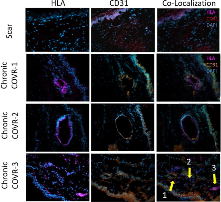
Objective: Cell-based outer vocal fold replacement (COVR) offers a potential treatment for severe vocal fold scarring or cancer reconstruction. Previous work in rabbits using human adipose-derived stem cells (ASC) in fibrin suggested that a hybrid structure emerged within 2 months, containing both implanted and host cells. This project uses immunocytochemistry to better define the phenotypic fate of implanted cells and features of the extracellular environment.

Methods: Immunocytochemistry was performed on sections collected from rabbits 2 months after COVR implantation or scar surgery. Cellular targets included human leukocyte antigen (HLA), CD31, and smooth muscle actin (SMA).

Results: HLA was present in all implanted sections and was used to identify human cells. In adjacent sections, HLA-positive cells were identified expressing CD31. SMA was not identified in the same cells as HLA. These markers were also present in injured vocal folds not receiving COVR. SMA protein content did not differ according to treatment.

Conclusions: Implanted human ASC persist in rabbit vocal folds. Some appear to express CD31, an endothelial marker. Smooth muscle actin, a marker of myofibroblast phenotype, was present in all sections regardless of treatment, and was not identified in hASC. Host cells also infiltrate the structure, producing a hybrid host-graft vocal fold.

Keywords: angiogenesis; cell‐based implant; human adipose derived stem cells; smooth muscle actin; vocal fold regeneration.


Conflict of interest statement

The authors declare no conflicts of interest.

Figures

FIGURE 1
FIGURE 1
Rating of HLA and CD31+ Cells in COVR Implanted Tissue. The chart provides an overview regarding the presence of positively stained cells within paraffin‐embedded tissue. All animals that were analyzed underwent COVR implantation after injury. More than three images were taken from each slide and were then visually inspected for the presence of HLA and CD31‐positive cells. The diagram at the bottom of the chart provides examples of the “Few”, “Some”, and “Many” rating schemes.
FIGURE 2
FIGURE 2
HLA and CD31 Labeling in COVR Implanted Tissue After Two Months. Microscopy from paraffin‐embedded larynges. M and F denote male or female. Pentachrome stained sections at 4X show the full section of the vocal fold (VF), with the black box indicating the specific region of the slide from which images were taken. In the corresponding immunohistochemistry (IHC) images, HLA‐positive cells are indicated with violet, and CD31‐positive cells are marked with green. All images were taken at 40X magnification. The last row in the figure, M‐Day 0, demonstrates more widespread HLA positivity in anthe animal that died immediately postoperatively.
FIGURE 3
FIGURE 3
CD31 and HLA Expression in Chronic COVR vs Scar. All images were captured at 40X magnification. Each image in the Chronic COVR group represents a distinct animal. In the first row (Scar), HLA cells are stained violet, CD31 cells are stained red, and DAPI are stained blue. In the second, third, and fourth rows (Chronic COVR groups), HLA cells are stained violet, whereas CD31 cells are stained orange, and DAPI stained blue. The co‐localization images were obtained by overlaying the respective HLA and CD31 images taken from sequentially cut slides. In the co‐localization image for Chronic COVR‐3, arrows 1 and 2 highlight a potential vascular structure where HLA‐positive and CD31‐positive cells overlap. Arrow 3 indicates a cluster of HLA‐positive cells without co‐localization of the CD31 marker.
FIGURE 4
FIGURE 4
SMA and HLA labeling 2 months after surgery. Each treatment group is represented by a representative labeled section from one animal. Frozen sections were imaged at 40X magnification. All animals demonstrate positive staining for SMA. The presence of HLA is only observed in acute and chronic COVR groups with none visualized in scar. Co‐localization of HLA and SMA within individual cells was not observed.
FIGURE 5
FIGURE 5
SMA Western blot quantification. Each “X” on the graph denotes one animal for which Western Blot was performed.; n = 3 in each of the three groups. Y‐axis values are band intensity calculated via ImageJ analysis and normalized by a loading control band.

Similar articles

References

    1. Hansen JK, Thibeault SL. Current understanding and review of the literature: vocal fold scarring. J Voice. 2006;20(1):110‐120. - PubMed
    1. Kumai Y. Pathophysiology of fibrosis in the vocal fold: current research, future treatment strategies, and obstacles to restoring vocal fold pliability. Int J Mol Sci. 2019;20(10):2551. - PMC - PubMed
    1. Mattei A, Bertrand B, Jouve É, et al. Feasibility of first injection of autologous adipose tissue–derived stromal vascular fraction in human scarred vocal folds. JAMA otolaryngol—Head Neck Surg. 2020;146(4):355. - PMC - PubMed
    1. Ma Y, Long J, Amin MR, et al. Autologous fibroblasts for vocal scars and age‐related atrophy: a randomized clinical trial. Laryngoscope. 2019;130(11):2650‐2658. - PMC - PubMed
    1. Nagubothu SR, Sugars RV, Tudzarovski N, et al. Mesenchymal stromal cells modulate tissue repair responses within the injured vocal fold. Laryngoscope. 2019;130(1):E21‐E29. - PubMed