[Preprint]. 2024 Dec 20:2024.09.24.614809.
doi: 10.1101/2024.09.24.614809.

Caspase-Activated DNase localizes to cancer causing translocation breakpoints during cell differentiation

Affiliations

Caspase-Activated DNase localizes to cancer causing translocation breakpoints during cell differentiation

Dalal Alsowaida et al. bioRxiv. .

Abstract

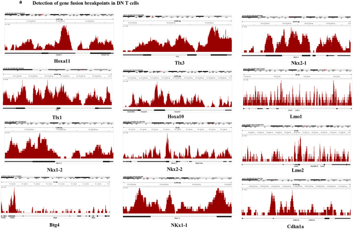
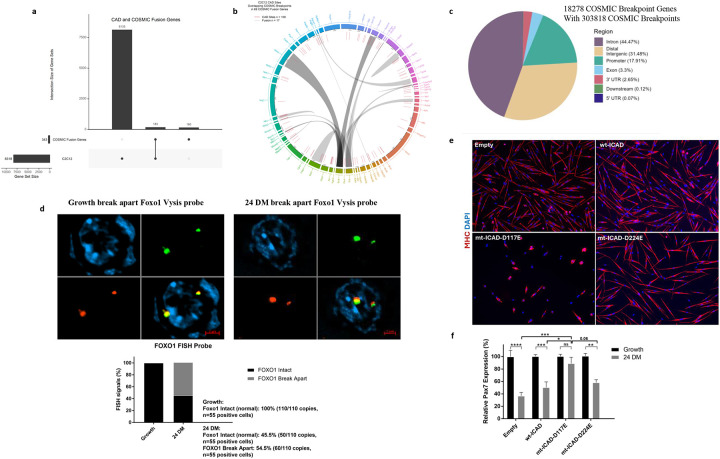
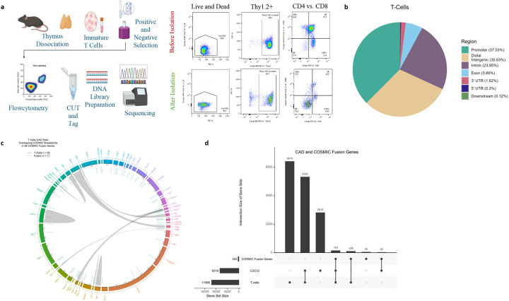
Caspase activated DNase (CAD) induced DNA breaks promote cell differentiation and therapy-induced cancer cell resistance. CAD targeting activity is assumed to be unique to each condition, as differentiation and cancer genesis are divergent cell fates. Here, we made the surprising discovery that a subset of CAD-bound targets in differentiating muscle cells are the same genes involved in the genesis of cancer-causing translocations. In muscle cells, a prominent CAD-bound gene pair is Pax7 and Foxo1a, the mismatched reciprocal loci that give rise to alveolar rhabdomyosarcoma. We show that CAD-targeted breaks in the Pax7 gene are physiologic to reduce Pax7 expression, a prerequisite for muscle cell differentiation. A cohort of these CAD gene targets are also conserved in early differentiating T cells and include genes that spur leukemia/lymphoma translocations. Our results suggest the CAD targeting of translocation prone oncogenic genes is non-pathologic biology and aligns with initiation of cell fate transitions.


Figures

Extended Data Figure 1.
Extended Data Figure 1.. Gene expression analysis of CAD bound genes in differentiating muscle cells.
(A) All peaks for CAD are called by MACS regions extended +/− 500bp from their summits. Followed by mapping them to masked mm10 genome to get nucleotide sequences. The alignments have higher representation of GC-rich sequences in CAD binding genome. (B) KEGG and Gene Ontology (GO) analysis identified within CAD functional genes Top 15 terms are enriched for molecular functions. (C) The volcano plot shows the genes with trending expression changes at 12hr and 24 hr differentiation for which CAD is bound to those gene promoters. (D) Integrative genomics viewer (IGV) snapshots depicting CAD CUT&Tag signal associated peak calls for the Cdkn1a gene from C2C12 skeletal muscle cells.
Extended Data Figure 2.
Extended Data Figure 2.. CAD targeted breakpoint translocation (BPT) genes in differentiating C2C12 muscle cells.
(A) IGV snapshots depicting CAD CUT&Tag signal and associated peak calls for indicated genes in C2C12 skeletal muscle cells. (B) Heatmaps depicting changes in gene expression levels in CAD fusion genes or CAD breakpoint genes during 12-, 24-, 48-, and 72-hours differentiation. Blue color indicates elevated mRNA levels, while red color indicates repressed levels. Color scale represents the log2fold change. Three biological replicates for each timepoint were assessed. Boxplot representing CAD breakpoint genes or CAD fusion genes changing expression during differentiation. (C) Graphical representation of sequence motifs detected in the CAD bound genome using Motif enrichment analysis calculated with STREME; E-values of motifs are indicated). (D) Schematic outline depicting the chromosomal translocations producing the chimeric proteins PAX7/FOXO1 t(1;13). Integrative genome view representing enrichment of CAD at Pax7 and Foxo1 genomic loci. Genome tracks identified by MACS2 peak caller of CAD treated with DMSO or DEVD (growth and 24 hours post-differentiation). (E) Detection of Pax7-Foxo1 fusion transcript in differentiating primary myoblasts. Primers specific to the 5’ region of the Pax7 transcript and the 3’ region of the Foxo1 transcript were used to probe cDNA libraries of differentiating primary myoblasts for the appearance of a fusion transcript. PCR product was observed at ~3000 bp that corresponded to the expected size of a Pax7-Foxo1 transcript. Secondary PCR from the isolated 3000bp band was performed for the 5’ region of the Pax7 transcript and 3’ region of the Foxo1 transcript. (N=2). Quantitative PCR (N=4) of Pax7-Foxo1 gene fusion during 12- and 24 hours post-differentiation. *: p<0.05, ****: p<0.0001 when compared to growth using one-way ANOVA with Dunnett’s multiple comparisons test. (F) FISH analysis detect Pax7-Foxo1 gene fusions in primary myoblast cells during cell growth and 24 hours post differentiation using PAX7-FOXO1 Fusion/Translocation FISH Probe Kit (CT-PAC089, Cytotest). The PAX7-FOXO1 fusion FISH probe set is designed to detect rearrangements between the human PAX7 and FOXO1 genes, located on chromosome band 1p36.13 and 13q14.11, respectively. For Pax7-Foxo1 FISH signals, 100 copies (n=50) of distinct FISH probes (100%) were quantified during growth condition. In differentiated conditions (n=50), 66 copies of distinct FISH probes (66%) and 34 copies of fused (translocated) FISH signals (34%) were quantified. (G) Western blot analysis of His-tagged ICAD constructs were detected with a mouse anti-His primary antibody in transfected mouse primary myoblasts (Tubulin was used to control for loading).
Extended Data Figure 3.
Extended Data Figure 3.. CAD targeted breakpoint translocation (BPT) genes in early differentiating T cell progenitors.
(A) IGV snapshots depicting CAD CUT&Tag signals and associated peak calls for breakpoint translocation (BPT) genes identified in early differentiating T cell progenitors.
Figure 1.
Figure 1.. Caspase Activated DNase (CAD) displays a binding preference for promoters of differentiation expressed genes.
Genomic distribution of CAD signal for CAD binding sites. (A) plotHeatmaps of CUT and Tag show count per million (CPM) normalized signals for reads across 3,000 bp genomic intervals relative to the TSS. Genomic distribution of the binding sites of CAD at Growth, and 24 hours post differentiation from C2C12 muscle cells treated with DMSO or z.DEVD-FMK. Data sets were generated from merged, two independent biological replicates for each condition. Heatmap profiles were generated using deepTools computeMatrix. Peak calling of binding sites was performed using Model-based Analysis of Chip-Seq. (MACS), Sparse Enrichment Analysis for CUT & RUN (SEACR), and GoPeaks. (B) PlotProfile averaged profiles of the compressed matrix of occupancy/signal near TSS produced by computeMatrix. (C) Graphical representation of sequence motifs detected in CAD DNA sequences using Motif enrichment analysis, demonstrates repetitive element enrichment for CAD bound genomic regions (as calculated with STREME; E-values of motifs are indicated). (D) Pie chart illustrating percent genomic distribution of the binding sites of CAD at 24 hours post differentiation from C2C12 muscle cells treated with z.DEVD-FMK, indicates enrichment at promoters, enhancers, distal intergenic regions, exons, introns, two untranslated regions (UTRs). The DiffBind tool was used to obtain the consensus peakset and AnnotatePeak function was used with the ChIPseeker package to determine peaks. (E) Gene Ontology (GO) analysis identified within CAD bound loci for biological processes demonstrates enrichment of genes that regulate gene expression and chromatin modification. RNA-Seq. analysis of CAD promoter genes. (F) Heatmaps depicting changes in gene expression for CAD promoter bound gene targets during 12-, 24-, 48-, and 72-hours differentiation relative to growth conditions (reference point). Blue color indicates elevated mRNA/gene expression, while red color indicates decreased mRNA/gene expression levels (Color scale represents the log2fold change). Three biological replicates for each timepoint were assessed, and DESeq2 results were obtained to get the log2FoldChange and p values. Genes with p. adjusted < 0.05 across all timepoint comparisons were considered significant for expression trend analysis. (G) Box-plot diagram depicts the distributions of gene expression log2Fold change across different differentiation timepoints 12, 24, 48, and 72 hours. The central line in the box marking the median value. (H) The subset of CAD bound genes (promoters) are listed with an increase in gene expression in C2C12 muscle cells at 12- and 24-hour differentiating cells (single gene data points from the box plots in G that are above the median) All genes were filtered first that the adjusted p-value for all timepoints is < 0.05. Then the median was taken for each timepoint 12 and 24 hours to be equal to or greater than 2 log2fold value.
Figure 2.
Figure 2.. CAD is enriched at known cancer breakpoint prone genes and may target these loci to alter expression and propel cell differentiation.
CAD relationship to cancer causing breakpoint translocation genes. (A) Upset plot visualizes the intersection size of gene sets between CAD bound loci from C2C12 muscle cells with breakpoint translocation/fusion genes from COSMIC database. COSMIC fusion genes were mapped to the mm10 orthologs from bioMart and obtained the intersection and union of the CAD and COSMIC genes to plot the Upset plot. CAD bound to ~53.4% of the total BPT genes identified (183 out of 343 genes) (UpSet plot Fig. 2a). (B) Circa plot visualizing cross-comparison between CAD-binding sites and COSMIC fusion genes set. Tracks from inside to the outside correspond to (1) gene synteny between chromosomes, (2) genes For the Circa plot, from outer to inner: Rainbow: represents COSMIC fusion genes for which CAD sites are bound (i.e. CAD-Fusion genes), Red/teal/purple lines represents CAD site locations where COSMIC Breakpoints overlap with the CAD sites (i.e. CAD-Breakpoints) in the fusion genes, lined-up with their respective genes in their gene ranges (and coloured). CAD gene binding in murine muscle cells matched to human BPTs displayed a significant degree of precise syntenic site overlap between CAD and the gene breakpoints (Fig. 2b, 2c; 100 CAD bound sites directly overlap in 69 of the 183 CAD bound genes). The innermost links “Grey” indicate: Gene fusion pairs for CAD-Fusion genes in the circus. Circa plots were made with the circlize package (R package). (C) Pie chart representing genomic distribution of COSMIC breakpoint genes within breakpoints. CAD targets and breaks Pax7 and Foxo1 loci to reduce expression of these genes and advance muscle cell differentiation. (D) FISH analysis to Foxo1 gene integrity in primary myoblast cells during cell growth and 24 hours post differentiation. The FISH probe is designed to detect break apart gene loci involving the FOXO1 (FKH1, FKHR) gene located on chromosome 13q14. For Foxo1 FISH probe, 110 copies (n=55) of intact FISH probes (100%) were quantified during growth conditions. In differentiated muscle cells (n=55), 50 copies of intact FISH probes (45.5%) and 60 copies of break apart FISH signals (54.5%) were quantified. (E) Primary myoblasts expressing wt-hICAD, mt-hICAD-D117E, and mt-hICAD D224E were subject to low serum induction of differentiation for 72 hours, which was monitored with staining for the differentiation specific marker, myosin heavy chain (MHC, red) nuclei were counterstained with DAPI (blue). (F) The same ICAD constructs were used to generate stable transfectants in primary myoblasts, where hICAD-D117E transfected cells (repressed CAD activity) maintained elevated Pax7 gene expression post low serum induction of differentiation. Relative expression of Pax7 at 24 hours following the induction of differentiation was determined and normalized to the Pax7 expression in each individual cell line at growth. n=4 (in triplicate). *: p<0.05, **: p<0.01, ***: p<0.001****: p<0.0001 using two-way ANOVA-Tukey’s multiple comparisons test.
Figure 3.
Figure 3.. CAD binds to genomic sites in murine T cell progenitors that align with a subset of human breakpoint translocation genes.
Schematic representation of the workflow for the CUT and Tag protocol applied to T cells isolated from thymus of wild type mice. Cell suspensions were acquired from mouse thymus then subject to FACS purification (Thy1.2+CD4CD8 ) to isolate early differentiating T cell progenitors (B) Pie chart illustrating the genomic distribution of CAD binding sites in differentiating T cell progenitors, showing the percentage for each genomic location category. Enrichment was noted at core promoters, distal intergenic regions, introns, exons, with a limited distribution at 3’ and 5’ untranslated regions (UTRs). The DiffBind tool was used to obtain the consensus peakset and AnnotatePeak function was used with the ChIPseeker package to determine peaks. (C) Circa plot visualizing the cross-comparison of CAD-binding sites between T cells and the COSMIC fusion gene set. Tracks from inside to the outside correspond to (1) gene synteny between chromosomes, (2) genes For the Circa plot, from outer to inner: Rainbow: represents COSMIC fusion genes for which CAD sites are bound (i.e. CAD-Fusion genes), Red/teal/purple lines represents CAD site locations where COSMIC Breakpoints overlap with the CAD sites (i.e. CAD-Breakpoints) in the fusion genes, lined-up with their respective genes in their gene ranges (and coloured). The innermost links “Grey” indicate: Gene fusion pairs for CAD-Fusion genes in the circus. Circa plots were made with the circulize package (R package). (D) UpSet plot visualizes the intersection size of gene sets between CAD from C2C12 muscle cells and fusion genes from T cell progenitors and the COSMIC fusion gene set.

References

    1. Larsen B.D. & Megeney L.A. Parole terms for a killer: directing caspase 3/CAD induced DNA strand breaks to coordinate changes in gene expression. Cell Cycle 9, 2940–2945 (2010). - PMC - PubMed
    1. Benada J., Alsowaida D., Megeney L.A. & Sørensen C.S. Self-inflicted DNA breaks in differentiation and cancer. Trends in Cell Biology 33, 850–859 (2023). - PubMed
    1. Larsen B.D., Rampalli S., Burns L., Dilworth F.J. & Megeney L.A. Caspase 3/Caspase activated DNase promote cell differentiation by inducing DNA strand breaks. Proc. Nat. Acad. Sci. USA. 107, 4230–4235 (2010). - PMC - PubMed
    1. Maurya D., Rai G., Mandal D. & Mondal B.C. Macrophage differentiation requires DNA damage caused by Caspase-Activated DNase. Cell Reports., 43, 114251 (2024). - PMC - PubMed
    1. Liu X. et al. Caspase-3 promotes genetic instability and carcinogenesis. Mol. Cell. 58, 284–296 (2015). - PMC - PubMed

Publication types