[Preprint]. 2024 Sep 23:2024.09.23.614499.
doi: 10.1101/2024.09.23.614499.

Versatile Cell Penetrating Peptide for Multimodal CRISPR Gene Editing in Primary Stem Cells

Affiliations

Versatile Cell Penetrating Peptide for Multimodal CRISPR Gene Editing in Primary Stem Cells

Josh P Graham et al. bioRxiv. .

Abstract

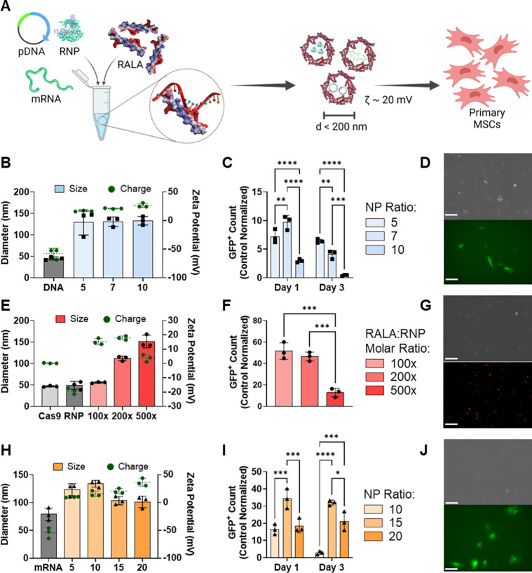
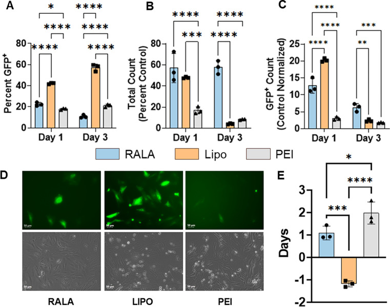
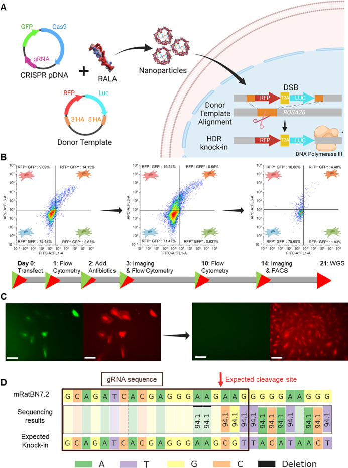
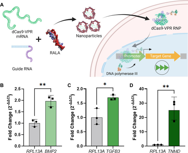
CRISPR gene editing offers unprecedented genomic and transcriptomic control for precise regulation of cell function and phenotype. However, delivering the necessary CRISPR components to therapeutically relevant cell types without cytotoxicity or unexpected side effects remains challenging. Viral vectors risk genomic integration and immunogenicity while non-viral delivery systems are challenging to adapt to different CRISPR cargos, and many are highly cytotoxic. The arginine-alanine-leucine-alanine (RALA) cell penetrating peptide is an amphiphilic peptide that self-assembles into nanoparticles through electrostatic interactions with negatively charged molecules before delivering them across the cell membrane. This system has been used to deliver DNAs, RNAs, and small anionic molecules to primary cells with lower cytotoxicity compared to alternative non-viral approaches. Given the low cytotoxicity, versatility, and competitive transfection rates of RALA, we aimed to establish this peptide as a new CRISPR delivery system in a wide range of molecular formats across different editing modalities. We report that RALA was able to effectively encapsulate and deliver CRISPR in DNA, RNA, and ribonucleic protein (RNP) formats to primary mesenchymal stem cells (MSCs). Comparisons between RALA and commercially available reagents revealed superior cell viability leading to higher numbers of transfected cells and the maintenance of cell proliferative capacity. We then used the RALA peptide for the knock-in and knock-out of reporter genes into the MSC genome as well as for the transcriptional activation of therapeutically relevant genes. In summary, we establish RALA as a powerful tool for safer and effective delivery of CRISPR machinery in multiple cargo formats for a wide range of gene editing strategies.

Keywords: CRISPR gene editing; Cell-penetrating peptide; mesenchymal stem cells; non-viral CRISPR delivery; regenerative medicine.


Conflict of interest statement

Competing Interests: The authors have no competing interests to share.

Figures

Figure 1.
Figure 1.. RALA encapsulates and delivers CRISPR machinery in pDNA, mRNA and RNP molecular formats into primary MSCs.
A) Schematic of RALA-CRISPR nanoparticle formation. Individual CRISPR cargos (pDNA, mRNA, or RNP) are mixed at specific ratios with the RALA peptide resulting in positively charged nanoparticles with diameters less than 200 nm. B) RALA-pDNA nanoparticle size (left y axis) and zeta potential (right y axis) at different N:P and molar ratios. C) Transfection yield (percent transfected relative to control count) after transfection with N:P ratios of 5, 7 and 10 on days 1 and 3 after transfection, and D) fluorescent images of MSCs 24 hours after transfection with RALA-pDNA using N:P ratio of 5. Scale bar = 100 μm. E) RALA-RNP nanoparticle size (left y axis) and zeta potential (right y axis) at 100x, 200x, 500x RALA: RNP molar ratios, F) Transfection yield (percent transfected relative to control count) after transfection with each ratio 8 hours after transfection, and G) fluorescent images of 100x RALA:RNP transfection with fluorescently labelled gRNA using a Cy5 filter. Scale bar = 100 μm. H) RALA-mRNA nanoparticle size (left y axis) and zeta potential (right y axis) at N:P ratios of 5, 10, 15 and 20, I) Transfection yield (percent transfected relative to control count) after transfection with each ratio on days 1 and 3, and J) fluorescent images of N:P = 15 transfection one day after transfection with FITC filter. Scale bar = 100 μm. * denotes significance p<0.05 (n = 3), **p<0.01 (n = 3), ***p<0.001 (n = 3), ****p<0.0001 (n = 3).
Figure 2.
Figure 2.. RALA outperforms commercial vectors for the delivery of pDNA encoding for CRISPR components.
The transfection of pDNA encoding for Cas9-T2A-GFP using RALA, Lipofectamine 3000, or PEI was characterized via flow cytometry. A) Transfection rate defined as the percentage of GFP+ cells in the transfected population. B) Cell viability defined as the total cell counts as a percentage of the non-transfected control count. C) Transfection yield defined as the percentage of GFP+ cells compared to the number of non-transfected control cells. The transfection was further characterized through D) representative fluorescent images of each transfection and E) the calculation of the cell doubling time from days 1 to 3. * denotes significance p<0.05 (n = 3), **p<0.01 (n = 3), ***p<0.001 (n = 3), ****p<0.0001 (n = 3).
Figure 3.
Figure 3.. CRISPR knock-in through RALA-mediated pDNA co-delivery.
A) Schematic of RFP-LUC knock-in strategy through pDNA co-delivery. B) Flow cytometry profiling of RFP knock-in cells on days 1, 3, and 10 respectively showing gating representing RFP+GFP, RFP+GFP+, RFPGFP+, and RFPGFP cells moving clockwise from the top right. Percentages of the total population are listed. C) Representative fluorescent images of RFP knock-in cells taken on days 3 and 14 after transfection. Left image is FITC channel for GFP signal, right image is Cy5 channel for miRFP670nano3 imaging. Scale bar = 100 μm. D) WGS results at the gene knock-in location. The Rattus norvegicus genome assembly (top, mRatBN7.2) is compared to the expected gene knock-in sequence. The gRNA sequence used for knock-in is shown with a black box and the expected cleavage site is marked with a red arrow.
Figure 4.
Figure 4.. RFP knock-out using RALA-RNP nanoparticles.
A) Schematic of gene knock-out strategy. Cas9 and gRNA targeting the RFP sequence were incubated for 15 minutes to form RNP complexes and then mixed with the RALA peptide to form nanoparticles. These nanoparticles are transfected and the RNPs are transported to the nucleus where they target the RFP gene leading to a loss of fluorescent signal. B) The RFP signal of each population was analyzed one week after transfection using flow cytometry to determine the rate of knock-out. C) The editing efficiency was defined as the percentage of RFP cells as quantified from flow cytometry results. D) Fluorescent images taken after passaging knock-out cells taken with Cy5 filter. Scale bar 100 μm. E) Average radiance (p/s/cm2/sr) images to visualize the bioluminescence of knock-out cells after passaging and F) quantification of average radiance. * Denotes significance p<0.05 (n = 3), **p<0.01 (n = 3), ***p<0.001 (n = 3), ****p<0.0001 (n = 3).
Figure 5.
Figure 5.. RALA mRNA delivery outperforms commercial transfection reagents Lipofectamine MessengerMAX and PEI.
A) Transfection rate defined as the percentage of GFP+ cells in the transfected population at days 1 and 3 post-transfection. B) Cell viability defined as the total cell counts as a percentage of the non-transfected control count. C) Transfection yield defined as the percentage of GFP+ cells compared to the number of non-transfected control cells. The transfection was further characterized through D) representative images of each transfection and E) the cell doubling time from days 1 to 3. * denotes significance p<0.05 (n = 3), **p<0.01 (n = 3), ***p<0.001 (n = 3), ****p<0.0001 (n = 3).
Figure 6.
Figure 6.. Activation of therapeutic gene expression using dCas9-VPR mRNA.
A) Schematic of CRISPR-mediated activation of gene expression (CRISPRa) strategy followed in this study. dCas9-VPR mRNA is co-delivered with gRNA and immediately forms an RNP upon cell delivery and mRNA translation. This RNP translocates to the nucleus where it binds upstream of the target gene TSS and recruits RNA polymerase III to the promoter region to upregulate transcription. Fold changes resultant of RALA gene activation for B) BMP2, C) TGFB3, and D) TNMD. All genes are compared to a control transfected with no gRNA and the RPL13A housekeeping gene. * denotes significance p<0.05 (n = 3), **p<0.01 (n = 3), ***p<0.001 (n = 3), ****p<0.0001 (n = 3).

References

    1. Graham J., Werba L., Federico I., and Gonzalez-Fernandez T., “CRISPR Strategies for Stem Cell Engineering: A New Frontier in Musculoskeletal Regeneration,” Eur. Cell. Mater., vol. 46, pp. 91–118, Nov. 2023, doi: 10.22203/eCM.v046a05. - DOI
    1. Jinek M., Chylinski K., Fonfara I., Hauer M., Doudna J. A., and Charpentier E., “A Programmable Dual-RNA–Guided DNA Endonuclease in Adaptive Bacterial Immunity,” Science, vol. 337, no. 6096, pp. 816–821, Aug. 2012, doi: 10.1126/science.1225829. - DOI - PMC - PubMed
    1. Qi L. S. et al. , “Repurposing CRISPR as an RNA-guided platform for sequence-specific control of gene expression,” Cell, vol. 184, no. 3, p. 844, Feb. 2021, doi: 10.1016/j.cell.2021.01.019. - DOI - PubMed
    1. Ran F. A., Hsu P. D., Wright J., Agarwala V., Scott D. A., and Zhang F., “Genome engineering using the CRISPR-Cas9 system,” Nat. Protoc., vol. 8, no. 11, pp. 2281–2308, Nov. 2013, doi: 10.1038/nprot.2013.143. - DOI - PMC - PubMed
    1. Ikmi A., McKinney S. A., Delventhal K. M., and Gibson M. C., “TALEN and CRISPR/Cas9-mediated genome editing in the early-branching metazoan Nematostella vectensis,” Nat. Commun., vol. 5, no. 1, p. 5486, Nov. 2014, doi: 10.1038/ncomms6486. - DOI - PubMed

Publication types

LinkOut - more resources