[Preprint]. 2024 Sep 27:2024.09.25.614546.
doi: 10.1101/2024.09.25.614546.

Expression-based selection identifies a microglia-tropic AAV capsid for direct and CSF routes of administration in mice

Affiliations

Expression-based selection identifies a microglia-tropic AAV capsid for direct and CSF routes of administration in mice

Miguel C Santoscoy et al. bioRxiv. .

Abstract

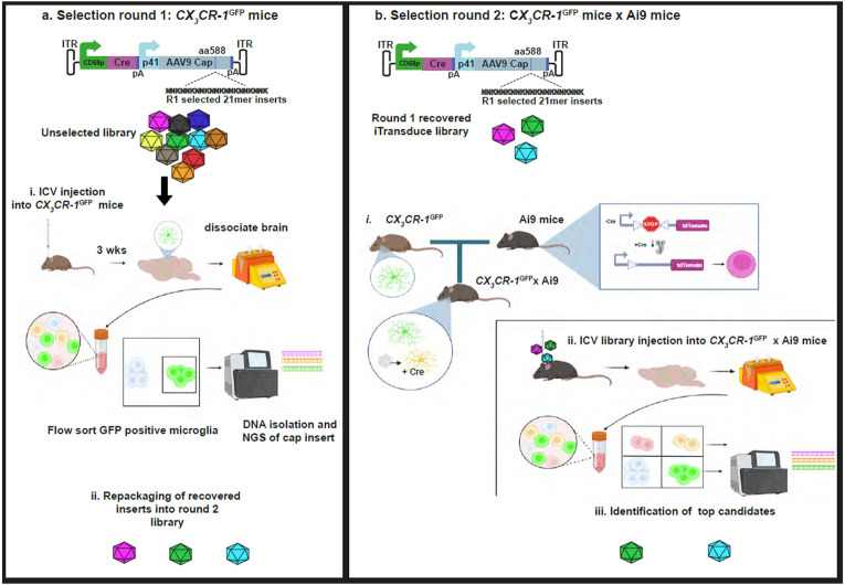
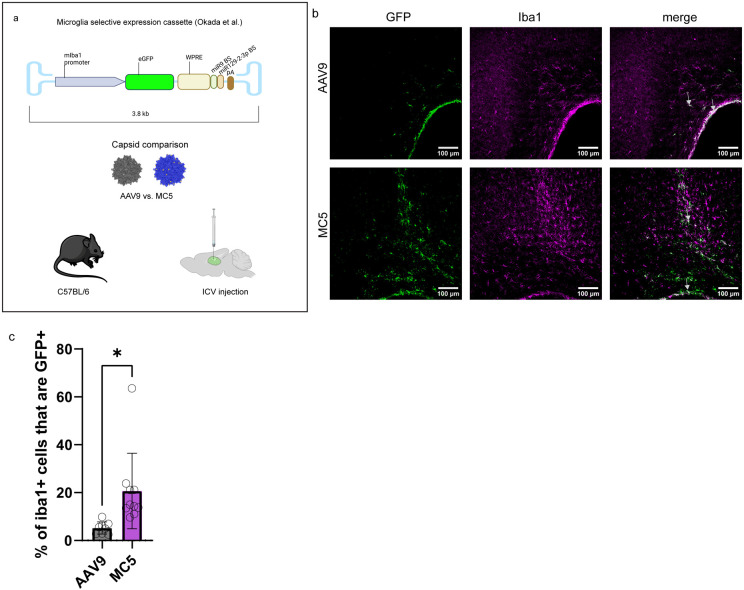
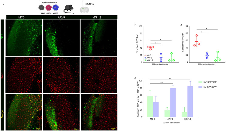
Microglia are critical innate immune cells of the brain. In vivo targeting of microglia using gene-delivery systems is crucial for studying brain physiology and developing gene therapies for neurodegenerative diseases and other brain disorders such as NeuroAIDS. Historically, microglia have been extremely resistant to transduction by viral vectors, including adeno-associated virus (AAV) vectors. Recently, there has been some progress demonstrating the feasibility and potential of using AAV to transduce microglia after direct intraparenchymal vector injection. Data suggests that combining specific AAV capsids with microglia-specific gene expression cassettes to reduce neuron off-targeting will be key. However, no groups have developed AAV capsids for microglia transduction after intracerebroventricular (ICV) injection. The ICV route of administration has advantages such as increased brain biodistribution while avoiding issues related to systemic injection. Here, we performed an in vivo selection using an AAV peptide display library that enables recovery of capsids that mediate transgene expression in microglia. Using this approach, we identified a capsid, MC5, which mediated enhanced transduction of microglia after ICV injection compared to AAV9. Furthermore, MC5 enhanced both the efficiency (85%) and specificity (93%) of transduction compared to a recently described evolved AAV9 capsid for microglia targeting after direct injection into the brain parenchyma. Exploration of the use of MC5 in a mouse models of Alzheimer's disease revealed transduced microglia surrounding and within plaques. Overall, our results demonstrate that the MC5 capsid is a useful gene transfer tool to target microglia in vivo by direct and ICV routes of administration.


Conflict of interest statement

Competing interests: C.A.M. has a financial interest in Sphere Gene Therapeutics, Inc., Chameleon Biosciences, Inc., and Skylark Bio, Inc., companies developing gene therapy platforms. C.A.M.’s interests were reviewed and are managed by MGH and Mass General Brigham in accordance with their conflict-of-interest policies. C.A.M., M.C.S., K.S.H., and P.E. have filed a patent application with claims involving the MC5 capsid.

Figures

Figure 1.
Figure 1.. Overview of microglia-transducing AAV capsid selection method.
a. Round 1 selection. The iTransduce library selection cassette contains a CD68 promoter driving Cre and an AAV p41 promoter driving AAV9 capsid with randomized 21-mer inserts (7-mer peptides) after amino acid 588. This ITR flanked cassette is packaged into the peptide display library. i. CX3CR1GFP mice which express GFP in microglia are injected ICV with the unselected library and 3 weeks later brain is dissociated and GFP+ microglia are flow sorted. The capsid gene region flanking the peptide inserts is PCR amplified, analyzed by NGS. ii. The amplified inserts are then repackaging into a library from round 2. b. Round 2 selection. In the second round we use the CD68-driven Cre to select capsids that transduce microglia. i. CX3CR1GFP mice are crossed with Ai9 mice. Microglia are GFP+ and capsids that express Cre, induce tdTomato expression (microglia express both GFP and tdTomato). ii. Mice are injected ICV with the condensed library from round 1 and brains dissociated 3 weeks later. Transduced microglia (double positive) and GFP+ microglia are flow sorted and NGS performed on both populations. iii. Candidate peptides are chosen from these data.
Figure 2.
Figure 2.. The MC5 capsid’s 7-mer peptide is a putative syndecan-4 motif.
a. Alignment of the MC5 amino acids (aa) with murine and human syndecan-4. Conserved aa’s are shown in magenta, identity between murine and human only in blue, and non-conserved residues in black. b. Schematic of murine syndecan-4 depicting key domains as well as the IPENAQP motif with high identity to the MC5 peptide.
Figure 3.
Figure 3.. The MC5 capsid is more efficient than the parental AAV9 capsid at transduction of microglia after ICV injection in adult mice.
a. Schematic of the experiment. The microglia selective transgene expression cassette from Okada et al. was utilized and packaged into AAV9 or MC5. Adult C57BL/6 mice (n=4-5 per group) were injected ICV with either vector. b. Confocal imaging surrounding the lateral ventricle to detect vector transduction of cells (GFP, green) and microglia (Iba1, magenta). Arrows point to representative transduced microglia in brain parenchyma and lining the ventricles. c. Transduction efficiency of microglia for each capsid. Individual brain sections containing the lateral ventricle (two per mouse) are shown as individual data points. Error bars represent standard deviation of the mean. *p=0.015.
Figure 4.
Figure 4.. Transduction of microglia by MC5, AAV9, and MG1.2 in the hippocampus after direct injection of the highest dose tested (2.1x10⁹ vg).
a. Immunofluorescence detection of AAV capsid transduction (GFP) and microglia (Iba1) in mice injected into the hippocampus (n=3 mice/group). GFP is shown in green and Iba1 in red. Colocalization is visualized as yellow in the merge image. Scale bar= 50 μm. b. Transduction efficiency of microglia in the hippocampus by each capsid. MC5 vs. AAV9 *,p=0.0216; MC5 vs. MG1.2 *,p= 0.0192. c. Transduction specificity for microglia of each capsid. MC5 vs. AAV9, *, p=0.0248; MC5 vs. MG1.2, *, p= 0.0137. d. Transduction specificity of microglia (Iba1+) and other cells (Iba1−) cells for each capsid in the hippocampus. MC5 vs. AAV9,**, p=0.0067; MC5 vs. MG1.2, **, p=0.0026.
Figure 5.
Figure 5.. MC5 mediates enhanced transduction efficiency and specificity towards microglia after intracranial injection in hippocampus (1.0x10⁹ vg).
a. Immunofluorescence detection of AAV capsid transduction (GFP) and microglia (Iba1) in mice injected into the hippocampus (n=3 mice/group). GFP is shown in green and Iba1 in red. Colocalization is visualized as yellow in the merge image. Scale bar= 50 μm. b. High magnification of MC5-transduced microglia. c. Transduction efficiency of microglia in the hippocampus by each capsid. ***,p=0.001. d. Transduction specificity for microglia of each capsid. *,p=0.0363; ***, p=0.001. e. Transduction specificity of microglia (Iba1+) and other cells (Iba1−) cells for each capsid in the hippocampus. *,p=0.012; ***, p=0.001.
Figure 6.
Figure 6.. Transduction of microglia by MC5 in the hippocampus after direct injection at three tested doses.
a. Immunofluorescence detection of AAV capsid transduction (GFP) and microglia (Iba1) in mice injected into the hippocampus (n=3 mice/group). GFP is shown in green and Iba1 in red. Colocalization is visualized as yellow in the merge image. Scale bar= 50 μm. b. Transduction efficiency of microglia in the hippocampus by MC5. ***, p=0.001. c. Transduction specificity for microglia of MC5. *, p= 0.0142. d. Transduction specificity of microglia (Iba1+) and other cells (Iba1−) cells for MC5 in the hippocampus. **, p=0.0028.
Figure 7.
Figure 7.. MC5 transduces microglia in APP/PS1 mice with amyloid β plaques.
At 7 days post-injection, GFP-positive plaque-associated microglia were observed throughout cortex in 8-month-old APP/PS1 mice. Image is an 8-micron thick z-projection image. Scale bar= 20 μm.

References

    1. Okada Y, Hosoi N, Matsuzaki Y, Fukai Y, Hiraga A, Nakai J, Nitta K, Shinohara Y, Konno A, Hirai H. Development of microglia-targeting adeno-associated viral vectors as tools to study microglial behavior in vivo. Commun Biol. 2022;5(1):1224. - PMC - PubMed
    1. Lin R, Zhou Y, Yan T, Wang R, Li H, Wu Z, Zhang X, Zhou X, Zhao F, Zhang L, et al. Directed evolution of adeno-associated virus for efficient gene delivery to microglia. Nat Methods. 2022;19(8):976–85. - PubMed
    1. Hanlon KS, Meltzer JC, Buzhdygan T, Cheng MJ, Sena-Esteves M, Bennett RE, Sullivan TP, Razmpour R, Gong Y, Ng C, et al. Selection of an Efficient AAV Vector for Robust CNS Transgene Expression. Mol Ther Methods Clin Dev. 2019;15:320–32. - PMC - PubMed
    1. Lek A, Wong B, Keeler A, Blackwood M, Ma K, Huang S, Sylvia K, Batista AR, Artinian R, Kokoski D, et al. Unexpected Death of a Duchenne Muscular Dystrophy Patient in an N-of-1 Trial of rAAV9-delivered CRISPR-transactivator. medRxiv. 2023:2023.05.16.23289881.
    1. Wilson JM, Flotte TR. Moving Forward After Two Deaths in a Gene Therapy Trial of Myotubular Myopathy. Hum Gene Ther. 2020;31(13-14):695–6. - PubMed

Publication types

LinkOut - more resources