[Preprint]. 2025 Mar 7:2024.09.26.615255.
doi: 10.1101/2024.09.26.615255.

Functional redundancy between penicillin-binding proteins during asymmetric cell division in Clostridioides difficile

Affiliations

Functional redundancy between penicillin-binding proteins during asymmetric cell division in Clostridioides difficile

Shailab Shrestha et al. bioRxiv. .

Abstract

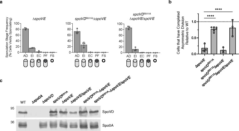
Peptidoglycan synthesis is an essential driver of bacterial growth and division. The final steps of this crucial process involve the activity of the SEDS family glycosyltransferases that polymerize glycan strands and the class B penicillin-binding protein (bPBP) transpeptidases that cross-link them. While many bacteria encode multiple bPBPs to perform specialized roles during specific cellular processes, some bPBPs can play redundant roles that are important for resistance against certain cell wall stresses. Our understanding of these compensatory mechanisms, however, remains incomplete. Endospore-forming bacteria typically encode multiple bPBPs that drive morphological changes required for sporulation. The sporulation-specific bPBP, SpoVD, is important for synthesizing the asymmetric division septum and spore cortex peptidoglycan during sporulation in the pathogen Clostridioides difficile. Although SpoVD catalytic activity is essential for cortex synthesis, we show that it is unexpectedly dispensable for SpoVD to mediate asymmetric division. The dispensability of SpoVD's catalytic activity requires the presence of its SEDS partner, SpoVE, and is facilitated by another sporulation-induced bPBP, PBP3. Our data further suggest that PBP3 interacts with components of the asymmetric division machinery, including SpoVD. These findings suggest a possible mechanism by which bPBPs can be functionally redundant in diverse bacteria and facilitate antibiotic resistance.


Figures

Figure 1 |
Figure 1 |. SpoVD catalytic activity is partially dispensable for its function during asymmetric division.
a Schematic of a SEDS-bPBP peptidoglycan synthase complex. The SEDS glycosyltransferase polymerizes nascent glycan strands from lipid-linked PG precursors in the cytoplasmic membrane, while the class B PBP (bPBP) crosslinks the stem peptides between the growing strands. b Cytological profile of individual cells representing each of the five morphological stages of sporulation as indicated. Representative phase-contrast and fluorescence micrographs are WT cells sampled from sporulation-inducing 70:30 plates after 18 hours of growth. The nucleoid was stained using Hoechst, and the cell membrane was stained using FM4–64. Cells undergoing asymmetric division (AD) have a flat polar septum; cells undergoing engulfment (EI) have a curved polar septum; cells that have completed engulfment (EC) are indicated by bright-membrane staining around a fully engulfed forespore; Phase-visible forespores (PF) indicate forespores completing maturation visible as phase-dark or phase-bright forespores (yellow arrow) associated with the mother cell; mature free spores (FS) are observable as independent phase-bright particles. c, d Quantification of the cytological profiling of cells sampled from sporulation-inducing plates after 20–22 hours of growth. White circles indicate data from each replicate, bars indicate the average means, and error bars indicate standard deviation. >1,000 total cells and >100 visibly sporulating cells per sample from a minimum of three biological replicates. c shows the distribution of visibly sporulating cells among the indicated stages of sporulation. d shows the proportion of cells that complete and progress beyond asymmetric division, i.e., all visibly sporulating cells, as a percentage of the total cells profiled. Note that the data is normalized to WT (dotted line). **** p < 0.0001. Statistical significance was determined using a one-way ANOVA and Tukey’s test. e Western blot analyses of SpoVD levels in the indicated strains 14 hours after growth on sporulation-inducing plates. The anti-Spo0A antibody was used as a proxy for measuring sporulation induction.
Figure 2 |
Figure 2 |. The catalytically inactive SpoVD requires its SEDS partner, SpoVE, to facilitate asymmetric division.
a, b Quantification of the cytological profiling of cells sampled from sporulation-inducing plates after 20–22 hours of growth. White circles indicate data from each replicate, bars indicate average means, and error bars indicate standard deviation. >1,000 total cells and >100 visibly sporulating cells per sample from a minimum of three biological replicates. a shows the distribution of visibly sporulating cells among the indicated stages of sporulation. See Figure 1 for the distribution of WT cells. b shows the proportion of cells that complete and progress beyond asymmetric division, i.e., all visibly sporulating cells, as a percentage of the total cells profiled. Note that the data is normalized to WT (dotted line) and that the spoVDS311A data derives from Figure 1. *** p < 0.001, **** p < 0.0001. Statistical significance was determined using a one-way ANOVA and Tukey’s test. c Western blot analyses of SpoVD levels in the indicated strains 14 hours after growth on sporulation-inducing plates. The anti-Spo0A antibody was used as a proxy for measuring sporulation induction.
Figure 3 |
Figure 3 |. PBP3 is a sporulation-specific bPBP that is involved in spore formation.
a Protein schematic comparing SpoVD and PBP3. Functional domains and catalytic sites were predicted using HMMER. TM, transmembrane domain; PASTA, PBP, and serine/threonine kinase-associated domain. The catalytic serine residues are shown in green. b Western blot showing the levels of the SpoVD, PBP3, and Spo0A in cells sampled from sporulation-inducing plates after ~14 hours of growth. SpoVD and PBP3 are not detected in the Δspo0A strain, which cannot initiate sporulation. c Efficiency of heat-resistant spore formation (sporulation efficiency) of the pbp3 mutant and complemented strains relative to WT. Means with standard deviation are indicated. Cells were collected from sporulation-inducing 70:30 plates ~20–22 hours after inoculation. Data from a minimum of four biological replicates. * p < 0.05, *** p < 0.001. Statistical significance was determined using a one-way ANOVA and Tukey’s test. d Representative phase-contrast micrographs of WT, pbp3 mutant, and complemented cells collected from sporulation-inducing 70:30 plates after ~20 hours of growth. Examples of phase-bright spores are indicated by yellow arrows. The blue arrow highlights an elongated forespore in the Δpbp3 mutant. Scale bar, 5 μm.
Figure 4 |
Figure 4 |. PBP3 partially compensates for loss of SpoVD catalytic activity during asymmetric division.
a, b Quantification of the cytological profiling of cells sampled from sporulation-inducing plates after 20–22 hours of growth. White circles indicate data from each replicate, bars indicate average means, and error bars indicate standard deviation. >1,000 total cells and >100 visibly sporulating cells per sample. Data from a minimum of three independent experiments. **** p < 0.0001. Statistical significance was determined using a one-way ANOVA and Tukey’s test. a shows the distribution of visibly sporulating cells among the indicated stages of sporulation. See Figure 1b for the distribution of WT cells. b shows the proportion of cells that complete and progress beyond asymmetric division, i.e., all visibly sporulating cells, as a percentage of the total cells profiled. Note that the data is normalized to WT (dotted line) and that the spoVDS311A data derives from Figure 1. c Western blot showing the levels of the SpoVD, PBP3, and Spo0A in cells sampled from sporulation-inducing plates after ~14 hours of growth. SpoVD and PBP3 are not detected in the Δspo0A strain, which cannot initiate sporulation.
Fig. 5 |
Fig. 5 |. PBP3 interacts with multiple components of the polar divisome.
a Bacterial two-hybrid analysis of interactions between PBP3 and other PG synthases or components of the polar divisome. The β-galactosidase activity was normalized to the negative control. N-terminal T18 or T24 fusion to PBP3 was paired with reciprocal N-terminal fusions to the indicated proteins. Data from three technical replicates. b The schematic shows interactions detected in the bacterial two-hybrid analyses. Components of the predicted polar divisome are indicated. PBP1 may also be a part of the polar divisome based on the co-immunoprecipitation analyses using SpoVD-FLAG3 as bait. c, d Co-immunoprecipitations performed on cells sampled from sporulation-inducing plates after 12 hours of growth. c PBP3-FLAG3 was used as bait in the Δpbp3/pbp3-FLAG3 strain background; d SpoVD-FLAG3 was used as bait in the ΔspoVD/spoVD-FLAG3 strain background. Δpbp3/pbp3 and ΔspoVD/spoVD strains were used as negative controls (No Tag). The presence of SpoVD, PBP3, and PBP1 in the pull-downs was probed using antibodies against the indicated proteins and western blotting. The FLAG-tagged proteins were detected using an anti-FLAG antibody. SpoIID was used as a control protein because it is also a PG-associated transmembrane protein localized to the forespore membrane, but it is not predicted to be a part of the polar divisome.
Fig. 6 |
Fig. 6 |. Prevalence of bPBP and SEDS enzymes in Firmicutes.
Heatmaps showing the distribution of class B penicillin-binding protein (bPBP) and SEDS protein numbers encoded in the genomes of sporulating (n = 328) and non-sporulating (n = 166) Firmicutes organisms. Sporulation ability was inferred by the presence of broadly conserved sporulation-specific genes spo0A and spoIIE in the genome. The dataset comprises 494 diverse Firmicutes organisms, as reported in Shrestha et al., 2023.

References

    1. Alabdali Y. A. J., Oatley P., Kirk J. A., & Fagan R. P. (2021). A cortex-specific penicillin-binding protein contributes to heat resistance in Clostridioides difficile spores. Anaerobe, 70, 102379. 10.1016/j.anaerobe.2021.102379 - DOI - PMC - PubMed
    1. Cameron T. A., & Margolin W. (2024). Insights into the assembly and regulation of the bacterial divisome. Nature Reviews Microbiology, 22(1), 33–45. 10.1038/s41579-023-00942-x - DOI - PMC - PubMed
    1. Castanheira S., Cestero J. J., Rico-Pérez G., García P., Cava F., Ayala J. A., Pucciarelli M. G., & García-del Portillo F. (2017). A Specialized Peptidoglycan Synthase Promotes Salmonella Cell Division inside Host Cells. mBio, 8(6), e01685–17. 10.1128/mBio.01685-17 - DOI - PMC - PubMed
    1. Castanheira S., López-Escarpa D., Pucciarelli M. G., Cestero J. J., Baquero F., & García-del Portillo F. (2020). An alternative penicillin-binding protein involved in Salmonella relapses following ceftriaxone therapy. eBioMedicine, 55, 102771. 10.1016/j.ebiom.2020.102771 - DOI - PMC - PubMed
    1. Dahlstrom K. M., Giglio K. M., Collins A. J., Sondermann H., & O’Toole G. A. (2015). Contribution of Physical Interactions to Signaling Specificity between a Diguanylate Cyclase and Its Effector. mBio, 6(6), 10.1128/mbio.01978-15. https://doi.org/10.1128/mbio.01978-15 - DOI - DOI - PMC - PubMed

Publication types