Comparative Study
. 2024 Nov 15;227(22):jeb247545.
doi: 10.1242/jeb.247545. Epub 2024 Nov 27.

Spatial arrangement of the whiskers of harbor seals (Phoca vitulina) compared with whisker arrangements of house mice (Mus musculus) and brown rats (Rattus norvegicus)

Affiliations
Comparative Study

Spatial arrangement of the whiskers of harbor seals (Phoca vitulina) compared with whisker arrangements of house mice (Mus musculus) and brown rats (Rattus norvegicus)

Matthew M Graff et al. J Exp Biol. .

Abstract

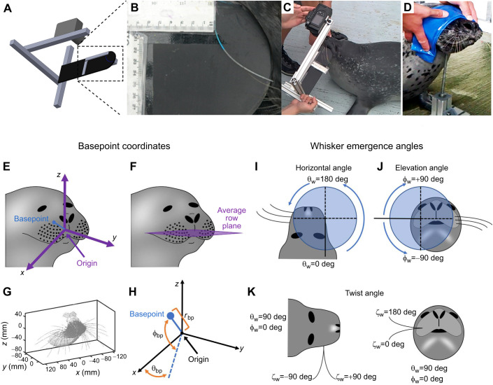
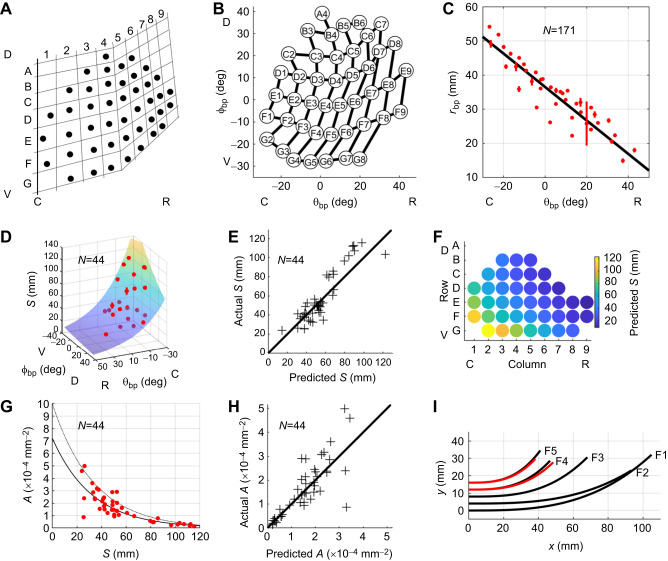
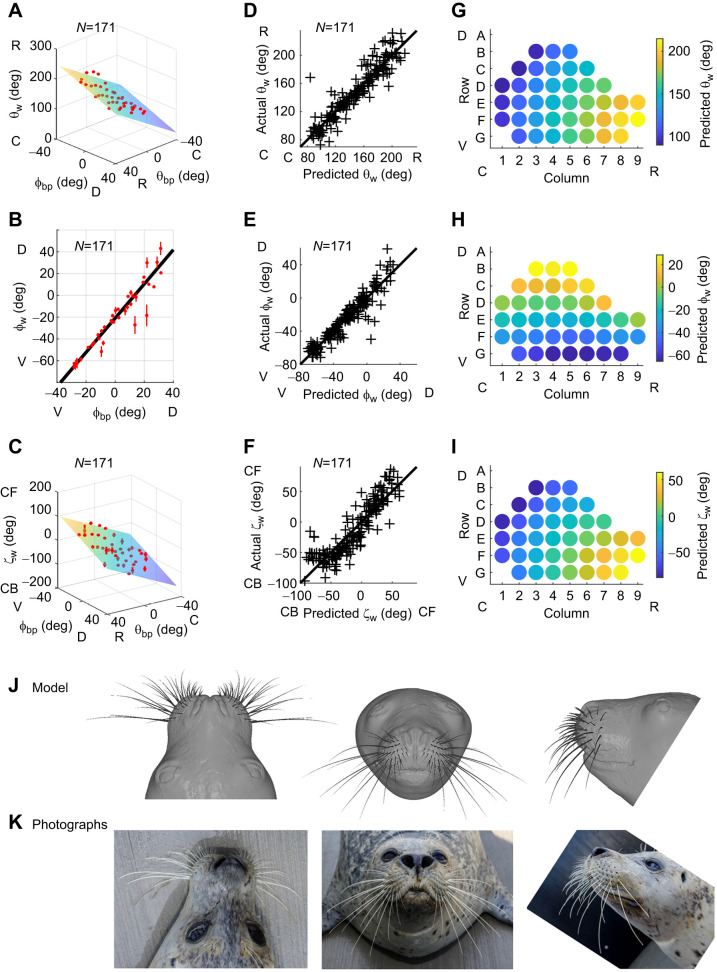
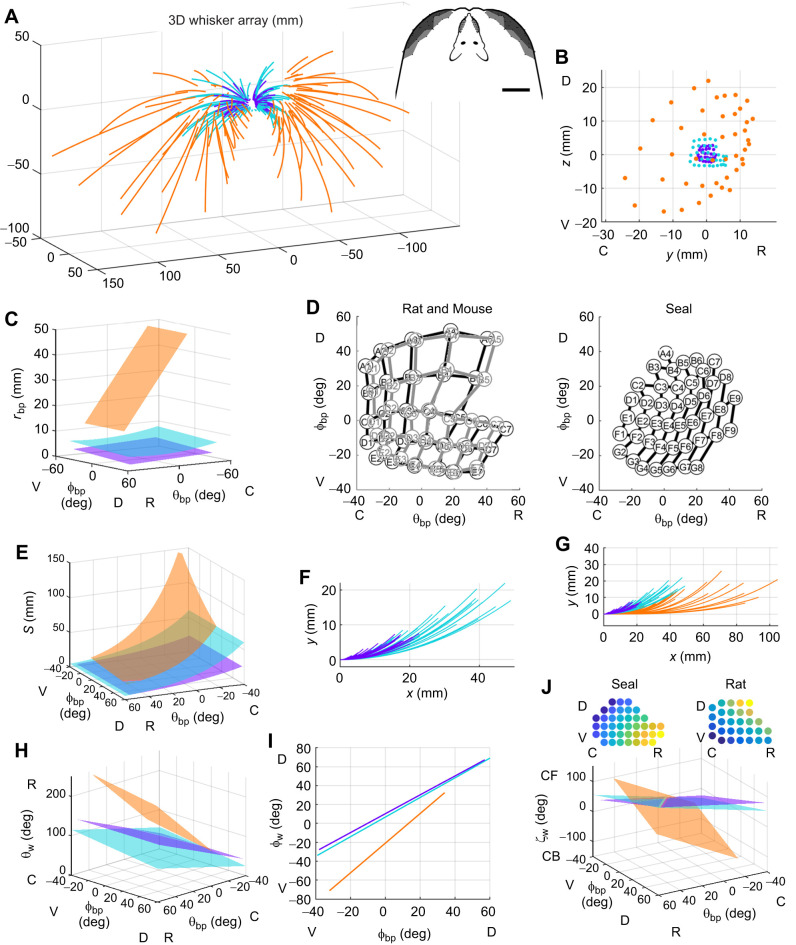
Whiskers (vibrissae) are important tactile sensors for most mammals. We introduce a novel approach to quantitatively compare 3D geometry of whisker arrays across species with different whisker numbers and arrangements, focusing on harbor seals (Phoca vitulina), house mice (Mus musculus) and Norway rats (Rattus norvegicus). Whiskers of all three species decrease in arclength and increase in curvature from caudal to rostral. They emerge from the face with elevation angles that vary linearly with dorsoventral position, and with curvature orientations that vary diagonally as linear combinations of dorsoventral and rostrocaudal positions. In seals, this diagonal varies linearly with horizontal emergence angles, and is orthogonal to the diagonal for rats and mice. This work provides the first evidence for common elements of whisker arrangements across species in different mammalian orders. Placing the whisker array model on a CAD model of a seal head enables future mechanical studies of whisker-based sensing, including wake tracking.

Keywords: Comparative anatomy; Flow sensing; Multimodal; Somatosensory; Touch; Trigeminal; Whisker.


Conflict of interest statement

Competing interests The authors declare no competing or financial interests.

Figures

Fig. 1.
Fig. 1.
Imaging of the head and vibrissal array of a harbor seal in 3D and 2D, and subsequent selection of coordinate systems for whisker basepoints and emergence angles. (A) A schematic diagram of the custom-built camera frame system used to obtain 2D photographs of the whiskers. The orientation of the background was manually adjusted so that the whisker of interest lay flat on the background. (B) An expanded view of the camera frame system shows an example 2D photograph of a whisker identified with non-toxic watercolor paint. (C) A photograph showing how the custom-built camera frame system was used in practice. (D) A blindfolded seal is shown perching its head on a chinrest to permit 3D laser scanning. (E) Schematic diagram of the standard position and orientation of the vibrissal array. The origin (0,0,0) is the mean position of the basepoints of the left and right arrays. The right basepoints surround the positive x-axis. The blue line and dot indicate one example of a basepoint location, for comparison with H. (F) The horizontal (xy) plane is defined by the average row plane of the basepoints. (G) An example point cloud of a seal's face in standard position and orientation. (H) Each basepoint location is described in spherical coordinates (θbp, φbp, rbp). The angle θbp ranges from −90 deg (caudal) to +90 deg (rostral), where θbp=0 deg lies in the xz plane. The angle φbp ranges from −90 deg (ventral) to +90 deg (dorsal), where φbp=0 deg lies in the xy plane. The basepoint coordinates of one example whisker (blue dot) are shown. (I) The variable θw describes the rostrocaudal angle at which the proximal, approximately linear portion of the whisker emerges from the mystacial pad. Values range from 0 deg to 360 deg. (J) The variable φw describes the dorsoventral angle at which the proximal, approximately linear portion of the whisker emerges from the mystacial pad. Values range from −90 deg to 90 deg. (K) The variable ζw describes the whisker's twist about its own axis. Values range between ±180 deg. Photos in B–D have been brightened and contrast enhanced.
Fig. 2.
Fig. 2.
Locations of whisker basepoints and their relationship to 2D whisker geometry. (A) Qualitative description of the shape of the harbor seal whisker array. Figure recreated from Dehnhardt and Kaminski (1995). (B) Plotting φbp against θbp quantifies array geometry in a manner that closely resembles the qualitative description in A. (C) Radial basepoint coordinate (rbp) plotted as a function of θbp for N=171 basepoints. Black line represents Eqn 1. Mean values of rbp when grouped by row and column identity are shown as red dots; red vertical bars indicate standard errors. (D) Arclength (S) plotted as a function of θbp and φbp. The surface represents Eqn 2. Mean values of S when grouped by row and column identity are shown as red dots; red vertical bars indicate standard errors. (E) Experimentally measured values for arclength (S) compared with values predicted from Eqn 2. Black diagonal line indicates equality. D and E contain data from N=44 whiskers. (F) A colormap illustrates variations in arclength predicted by Eqn 2 across the array. (G) Cubic curvature coefficient (A) can be described as an exponentially decreasing function of arclength. Solid black line represents Eqn 4 and dotted gray line represents Eqn 5. Red dots are experimentally measured values of A. (H) Experimentally measured values for curvature coefficient (A) compared with values predicted from Eqn 4. Black diagonal line indicates equality. G and H contain data from N=44 whiskers. (I) Black traces show shapes of the F-row whiskers from a single seal. Whiskers F4 and F5 from two other seals are shown in red to illustrate shape variability. C, caudal; R, rostral; V, ventral; D, dorsal.
Fig. 3.
Fig. 3.
Quantifying the relationship between whisker angles of emergence and basepoint coordinates allows construction of a CAD model of the whisker array. (A–C) The horizontal angle of emergence (θw) fitted to Eqn 6, the elevation angle of emergence (φw) fitted to Eqn 7 and the twist angle of emergence (ζw) fitted to Eqn 8. For all plots, means±s.e.m. when grouped by whisker identity are shown in red. (D–F) Actual values of θw, φbp and ζw compared with values predicted from Eqns 6, 7 and 8. All plots reveal relatively uniform dispersion of actual versus predicted values about y=x, indicating that correct models were selected. A–F show data for N=171 whiskers. (G–I) The plots show how values for θw, φbp and ζw will vary across the array, as predicted by Eqns 6, 7 and 8. (J) A 3D model of the average seal whisker array is shown superimposed on a CAD model of the seal's head. The head model was created using 3D laser scans of seal 2, with the exception of the eyes and portions of the top of the head (see Materials and Methods). (K) Photographs of seal 2 permit visual comparison between the real animal and CAD models for both head and whisker array. C, caudal; R, rostral; D, dorsal; V, ventral; CF, concave forwards; CB, concave backwards.
Fig. 4.
Fig. 4.
The harbor seal whisker array compared with arrays of rats and mice. In all panels, data for harbor seals are in orange, data for rats (from Belli et al., 2018) are in teal, and data from mice (from Bresee et al., 2023) are in purple. (A) Full 3D geometric models of seal, rat and mouse whisker arrays shown in standard position and orientation. Inset: top-down views comparing rat and seal arrays. Scale bar: 20 mm. (B) Whisker basepoints plotted in the sagittal (yz) plane. (C) Plotting rbp as a function of basepoint position highlights size differences between species. (D) To normalize for head size, mean whisker basepoint locations for all three species are plotted in θbp and φbp. In these angular coordinates, seal whisker basepoints (right; same data as in Fig. 2B) are spaced more densely than mouse and rat whisker basepoints (left, overlayed). (E) Whisker arclength (S) plotted as a function of basepoint position (θbp and φbp). (F) Seal whiskers are fitted with cubic equations, while mouse and rat whiskers are fitted with quadratic equations. (G) The large overlap in mouse and rat whisker shapes, revealed in an expanded view of F. Proximal portions of all whiskers are aligned with the x-axis. (H–J) Rostral–caudal emergence angle (θw), elevation emergence angle (φw) and twist emergence angle (ζw) plotted as functions of basepoint position (θbp and φbp). The insets in J show heatmaps for the values of ζw for seal and rat. For both animals, the gradient of twist angle varies linearly with θbp and φbp, but the gradient of the seal twist angle is orthogonal to that of the rat. C, caudal; R, rostral; D, dorsal; V, ventral; CF, concave forwards; CB, concave backwards.

Update of

References

    1. Ahl, A. S. (1986). The role of vibrissae in behavior - a status review. Vet. Res. Commun. 10, 245-268. 10.1007/bf02213989 - DOI - PubMed
    1. Belli, H. M. (2017). Sensing with whiskers: from geometry and mechanics to the statistics of the array. PhD thesis, Northwestern University. ProQuest/UMI, 253 pages (Publication No. 10681095). 10.21985/N2WF4K - DOI
    1. Belli, H. M., Bresee, C. S., Graff, M. M. and Hartmann, M. J. Z. (2018). Quantifying the three-dimensional facial morphology of the laboratory rat with a focus on the vibrissae. PLoS One 13, e0194981. 10.1371/journal.pone.0194981 - DOI - PMC - PubMed
    1. Bookstein, F. L. (2018). A Course in Morphometrics for Biologists. Cambridge University Press.
    1. Brecht, M., Preilowski, B. and Merzenich, M. M. (1997). Functional architecture of the mystacial vibrissae. Behav. Brain Res. 84, 81-97. 10.1016/s0166-4328(97)83328-1 - DOI - PubMed

Publication types

LinkOut - more resources