. 2024;44(12):562-576.
doi: 10.1080/10985549.2024.2408016. Epub 2024 Oct 10.

The DNA Damage Repair Function of Fission Yeast CK1 Involves Targeting Arp8, a Subunit of the INO80 Chromatin Remodeling Complex

Affiliations

The DNA Damage Repair Function of Fission Yeast CK1 Involves Targeting Arp8, a Subunit of the INO80 Chromatin Remodeling Complex

Sierra N Cullati et al. Mol Cell Biol. 2024.

Abstract

The CK1 family are conserved serine/threonine kinases with numerous substrates and cellular functions. The fission yeast CK1 orthologues Hhp1 and Hhp2 were first characterized as regulators of DNA repair, but the mechanism(s) by which CK1 activity promotes DNA repair had not been investigated. Here, we found that deleting Hhp1 and Hhp2 or inhibiting CK1 catalytic activities in yeast or in human cells increased double-strand breaks (DSBs). The primary pathways to repair DSBs, homologous recombination and nonhomologous end joining, were both less efficient in cells lacking Hhp1 and Hhp2 activity. To understand how Hhp1 and Hhp2 promote DNA damage repair, we identified new substrates of these enzymes using quantitative phosphoproteomics. We confirmed that Arp8, a component of the INO80 chromatin remodeling complex, is a bona fide substrate of Hhp1 and Hhp2 important for DNA repair. Our data suggest that Hhp1 and Hhp2 facilitate DNA repair by phosphorylating multiple substrates, including Arp8.

Keywords: Arp8; CK1, casein kinase 1; DNA repair; INO80; Schizosaccharomyces pombe; homologous recombination; nonhomologous end joining; phosphoproteomics.


Conflict of interest statement

No potential conflict of interest was reported by the author(s).

Figures

Figure 1.
Figure 1.
Deletion of hhp1 alone or in combination with hhp2 activates rad3-dependent DNA damage checkpoints. (A and C) Live-cell images of the indicated strains. Scale bars, 5 µm. (B and D) Rad52-GFP (B) and Cdc24-mNG (D) foci in > 600 cells for each strain were counted over two to four biological replicates. Error bars indicate ±95%CI. ****, P < 0.001 by chi-square.
Figure 2.
Figure 2.
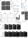
Loss of Hhp1 and Hhp2 activity triggers the DNA damage checkpoint. (A) DIC live-cell images of the indicated strains at 5 h after treatment with DMSO or 25 μM 1NM-PP1. Scale bar: 5 μm. (B) Length at septation was quantified for cells imaged as in A, n ≥ 100 cells. Bars represent means. ****, P < 0.0001 by one-way ANOVA; ns, not significant. (C) Length of cells without septum that are longer than the median length of the cells at septation from B. n ≥ 500 cells without septum were quantified and longer cells than the median are represented. Black solid bars represent means and magenta broken lines represent the median of septated cells from each treatment. *P < 0.05 by F-test; ****P < 0.0001 by Welch’s t test. Representative images are shown below. (D) Fluorescence microscopy images of rad52-GFP and hhp1-as hhp2-as rad52-GFP cells 5 h after treatment with DMSO or 25 μM 1NM-PP1. Rad52-GFP images are max projections. Scale bar: 5 μm. Rad52-GFP foci in > 1000 cells for each strain were counted over two biological replicates and are represented in the right graph. Error bars indicate ±95%CI. ****P < 0.001 by chi-square. (E) Serial 10-fold dilutions of the indicated strains were spotted on YE with or without 1NM-PP1 and hydroxyurea (HU), camptothecin (CPT), or methyl methanesulfonate (MMS), and incubated at 32 °C. cdc2-as was used as a positive control for inhibitor effectiveness; inhibition of Cdc2 is lethal.
Figure 3.
Figure 3.
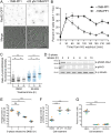
CK1 kinase activity promotes DSB repair in both yeast and human cells. (A and B) hhp1-as hhp2-as rad52-GFP cells were synchronized in 12 mM HU for 3 h then released into YE with or without 25 μM 1NM-PP1. Samples were fixed and imaged at the indicated timepoints. (A) Fluorescence microscopy and DIC images of the indicated strains after 240 min. Rad52-GFP images are max projections. DIC images are single medial slices. Scale bar: 10 μm. (B) Rad52-GFP foci were counted at the indicated timepoints using sum projections of cells imaged as in A. Data are means ± SEM from three replicates of n ≥ 150 each. (C) Asynchronous HeLa cells were treated with DMSO or 0.5 µM SR-3029 (CK1δ and CK1ε inhibitor) for the indicated times. Cells were fixed and stained for γH2AX as a marker of DSBs and DRAQ5 fluorescent probe for total DNA content. (D–G) HeLa cells were synchronized in S-phase using a thymidine-aphidicolin block. (D) Western blot of whole cell lysates with the indicated antibodies after release from S-phase arrest into DMSO. (E) Average number of γH2AX foci per cell after release from an S-phase arrest into DMSO. (F) Average number of γH2AX foci per cell 10 h after release from an S-phase arrest into DMSO, 0.5 µM SR-1227, or 0.5 µM SR-3029. (G) Mitotic index 10 h after release from an S-phase arrest into DMSO, 0.5 µM SR-1227, or 0.5 µM SR-3029.
Figure 4.
Figure 4.
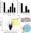
Hhp1 and Hhp2 phosphorylate multiple substrates to promote DSB repair through HR and NHEJ. (A) Analysis of spontaneous HR function using the RDUX200(+) reporter. Data presented as recombination frequency (ratio of colonies grown on selective media to rich media), normalized to wild-type. (B) Analysis of NHEJ function using a plasmid recircularization assay. Data presented as recircularization efficiency (ratio of colonies grown from cells transformed with linearized DNA to circular DNA on selective media), normalized to wild-type. *P < 0.05; **P < 0.01; ***P < 0.001; ****P < 0.0001 by one-way ANOVA. (C) Volcano plot comparing statistically significant differences in phosphorylation sites between hhp1-as hhp1-as relative to wild-type quantified by mass spectrometry-based phosphoproteomics. Dashed lines indicate a Benjamini–Hochberg-corrected q value < 0.01 and a fold change beyond ± 1.5. (D) Linear substrate motif analysis of phosphorylation sites with q < 0.01. (E) Overlap between candidate substrates identified by phosphoproteomics (yellow) and HU-sensitive alleles annotated in PomBase (blue). See also Tables S1–S3 (Supplementary material).
Figure 5.
Figure 5.
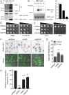
Arp8, a subunit of the INO80 complex, is a substrate of Hhp1 and Hhp2. (A) Arp8-3 × FLAG was immunoprecipitated from S. pombe then incubated with or without recombinant CK1δ at 30 °C for 1 h. Phosphorylated proteins were detected by autoradiography (32P) and protein levels by Western blot with the indicated antibodies. (B and C) Recombinant Arp8 mutants were incubated with recombinant Hhp2ΔC at 30 °C for 1 h. Phosphorylated proteins were detected by autoradiography (32P) and protein levels by Coomassie. (C) Bars represent means ± SD from four independent replicates. 32P signal was normalized to protein level for each lane. (D) Serial 10-fold dilutions of the indicated strains were spotted on EMM ± HU and incubated at 32 °C. (E) Serial 10-fold dilutions of the indicated strains were spotted on YE and incubated at the indicated temperatures. (F and G) arp8Δ and arp8-4A exhibit increased basal levels of Rad52-GFP foci compared to wild-type. (F) Live-cell images of the indicated strains. Merge represents DIC and GFP channels. Rad52-GFP images are sum projections. DIC images are single medial slices. Scale bar, 5 µm. (G) Rad52-GFP foci in > 1300 cells for each strain were counted over three biological replicates. Error bars indicate ±95%CI. ****P < 0.001; **P < 0.01 by chi-square. (H) Spontaneous HR recombination frequency of the indicated strains, normalized to wild-type. ***P < 0.001; ****P < 0.0001 by one-way ANOVA.

References

    1. Bierne H, Michel B.. When replication forks stop. Mol Microbiol. 1994;13:17–23. doi:10.1111/j.1365-2958.1994.tb00398.x. - DOI - PubMed
    1. Sabatinos SA, Green MD, Forsburg SL.. Continued DNA synthesis in replication checkpoint mutants leads to fork collapse. Mol Cell Biol. 2012;32:4986–4997. doi:10.1128/MCB.01060-12. - DOI - PMC - PubMed
    1. Meister P, Taddei A, Vernis L, Poidevin M, Gasser SM, Baldacci G.. Temporal separation of replication and recombination requires the intra-S checkpoint. J Cell Biol. 2005;168:537–544. doi:10.1083/jcb.200410006. - DOI - PMC - PubMed
    1. Petermann E, Orta ML, Issaeva N, Schultz N, Helleday T.. Hydroxyurea-stalled replication forks become progressively inactivated and require two different RAD51-mediated pathways for restart and repair. Mol Cell. 2010;37:492–502. doi:10.1016/j.molcel.2010.01.021. - DOI - PMC - PubMed
    1. Zeman MK, Cimprich KA.. Causes and consequences of replication stress. Nat Cell Biol. 2014;16:2–9. doi:10.1038/ncb2897. - DOI - PMC - PubMed

Publication types

MeSH terms

Substances

LinkOut - more resources