. 2024 Nov 13;15(11):e0175624.
doi: 10.1128/mbio.01756-24. Epub 2024 Oct 10.

Lipophilic bisphosphonates reduced cyst burden and ameliorated hyperactivity of mice chronically infected with Toxoplasma gondii

Affiliations

Lipophilic bisphosphonates reduced cyst burden and ameliorated hyperactivity of mice chronically infected with Toxoplasma gondii

Melissa A Sleda et al. mBio. .

Abstract

The current treatments for toxoplasmosis are only active against fast-growing tachyzoites, present in acute infections, with little effect on slow-growing bradyzoites within tissue cysts, present in latent chronic infections. The mitochondrion of Toxoplasma gondii is essential for its survival, and one of the major anti-parasitic drugs, atovaquone, inhibits the mitochondrial electron transport chain at the coenzyme Q:cytochrome c oxidoreductase site. Coenzyme Q (also known as ubiquinone [UQ]) consists of a quinone head and a lipophilic, isoprenoid tail that anchors UQ to membranes. The synthesis of the isoprenoid unit is essential for cell growth and is inhibited by lipophilic bisphosphonates, which inhibit the parasite growth. In this work, we investigated the effect of lipophilic bisphosphonates on the chronic stages of T. gondii. We discovered that three lipophilic bisphosphonates (BPH-1218, BPH-1236, and BPH-1238), effective for the acute infection, were also effective in controlling the development of chronic stages. We showed effectiveness by testing them against in vitro cysts and in vivo derived tissue cysts and, most importantly, these compounds reduced the cyst burden in the brains of chronically infected mice. We monitored the activity of infected mice non-invasively and continuously with a novel device termed the CageDot. A decrease in activity accompanied the acute phase, but mice recovered to normal activity and showed signs of hyperactivity when the chronic infection was established. Moreover, treatment with atovaquone or BPH-1218 ameliorated the hyperactivity observed during the chronic infection.IMPORTANCETreatment for toxoplasmosis is challenged by a lack of effective drugs to eradicate the chronic stages. Most of the drugs currently used are poorly distributed to the central nervous system, and they trigger allergic reactions in a large number of patients. There is a compelling need for safe and effective treatments for toxoplasmosis. Bisphosphonates (BPs) are analogs of inorganic pyrophosphate and are used for the treatment of bone disorders. BPs target the isoprenoid pathway and are effective against several experimental parasitic infections. Some lipophilic BPs can specifically inhibit the mitochondrial activity of Toxoplasma gondii by interfering with the mechanism by which ubiquinone is inserted into the inner mitochondrial membrane. In this work, we present the effect of three lipophilic BPs against T. gondii chronic stages. We also present a new strategy for the monitoring of animal activity during disease and treatment that is non-invasive and continuous.

Keywords: Toxoplasma gondii; bisphosphonate; bradyzoite; hyperactivity.


Conflict of interest statement

The authors declare no conflict of interest.

Figures

Fig 1
Fig 1
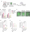
Lipophilic bisphosphonates inhibited the viability of in vitro differentiated bradyzoites. (A) Schematic representation of the in vitro differentiation of type II Me49 bradyzoites. Human foreskin fibroblast (HFF) cells were incubated with 3 µM compound 1 for 2 hours before infection with 2 × 10⁵ tachyzoites of Me49 followed by incubation at ambient CO2 for 3 days. After the tachyzoites differentiated into bradyzoites, compounds were added at the indicated concentrations and incubated for an additional 4 days at ambient CO2. At the end of the incubation, cells were fixed, permeabilized, and the cysts stained with DBA and anti-SAG1. (B) Representative images from control and drug-treated cysts with drugs at 5× EC50. (C) Quantification of vacuole sizes from cultures exposed to 3 x EC50 (top) or 5 x EC50 (bottom) concentrations. (D) Schematic representation of the in vitro bradyzoite viability assay. Me49 tachyzoites were differentiated into bradyzoites and drug treated as shown in panel A. After drug treatment, the cysts were collected as described in Materials and Methods. HFF monolayers were infected with 10,000 bradyzoites and incubated at 5% CO2 for 14–16 days. The plaque image shown is duplicated from Fig. 1E (dimethyl sulfoxide [DMSO] control) and was used as a representative image of plaques. (E) Representative images of plaques formed in the presence of 3× (left panel) and 5× (right panel) the indicated EC50s (shown in parentheses). (F) Quantification of the number of plaques from the 3× (top graph) and 5× (bottom graph) EC50 incubations. Data for panels C and F are from three independent biological replicates. Two-way analysis of variance statistical analysis was performed to compare the multiple drug-treated groups to the control (DMSO).
Fig 2
Fig 2
Lipophilic bisphosphonates inhibited growth of ex vivo derived bradyzoites and reduced tissue cyst size. (A) Schematic representation of the ex vivo bradyzoite collection. Brains of chronically infected mice were collected, homogenized, and treated with acid/pepsin to release bradyzoites. The liberated bradyzoites were plated on human foreskin fibroblast (HFF) monolayers and treated with drugs for 14 days (growth) or for 4 hours (viability). The plaque image shown is duplicated from Fig. 2B (DMSO control) and was used as the representative image of plaques. (B) Representative images of plaques from ex vivo growth and viability from Me49-RFP. (C) Quantification of the number of plaques from the ex vivo growth assay of Me49-RFP at 3× the EC50. (D) Quantification of the number of plaques from the ex vivo viability assay of Me49-RFP at 3× the EC50. Data from panels C and D were from three independent biological replicates. Two-way analysis of variance statistical analysis was performed to compare the multiple drug-treated groups to the control (DMSO). Values in panels C and D are means ± SEM.
Fig 3
Fig 3
Lipophilic bisphosphonates reduced the cyst burden of chronically infected mice. (A) Schematic representation of the infection for treatment of chronically infected mice. CBA/j mice were infected i.p. with 500 Me49-RFP tachyzoites. After 28 days post-infection, two mice were checked for cysts and the remaining mice treated daily for 16 days. After 2 weeks, the brains were collected, and cysts were quantified. (B) Representative images of cysts isolated from control and drug-treated mice. (C) Quantification of cyst size. (D) Quantification of the number of cysts. (E) Representative images of plaques from ex vivo bradyzoites from the control and drug-treated mice. Data from panels C and D are from three independent biological replicates. Two-way analysis of variance statistical analysis was performed to compare the multiple drug-treated groups to the control.
Fig 4
Fig 4
Continuous monitoring of mice activity revealed hyperactivity during chronic infection. (A) Image of the CageDot device attached to the bottom of the mouse cage. (B) Continuous tracing for the entire experiment 1 (56 days) showing the hourly average for each cage. The traces represent the day and night cycles for each cage for all days of the experiment. Day and night data were used for the analysis presented in panel C, and night data were used for all the other analyses. Each cage contained four mice, and the data are the average for all of the mice in the cage. (C) Quantification of energy averages between daytime vs night time (4-hour period for each) for the uninfected cage showing the changes between night and day. (D) Continuous tracings of uninfected and Pru-GFP-infected Balb/c mice for a 54-day experiment (GFP, green fluorescent protein). The acute data represent the activity computed at days 10–20, and the chronic stage was from data collected at days 30–40. Some preliminary monitoring results were previously communicated as an example for the use of the CageDot (31) . (E) Quantification of the acute and chronic stage of disease for uninfected and infected mice. Average of three biological replicates. Values in panels C and E are means ± SEM. Student’s t-test statistical analysis for data in panels C and E.
Fig 5
Fig 5
The hyperactivity of chronically infected mice was reduced upon treatment with BPH-1218. (A) Model for chronic infection. Balb/c mice were infected i.p. with 1,500 Pru-GFP tachyzoites. After 28 days, two mice brains were checked for tissue cysts and the rest were treated daily for 16 days. Two weeks after the end of treatment, the brains were collected and cysts enumerated. (B) Nightly average energy from the last day of treatment and 2 weeks post-treatment. (C) Representative images of Pru-GFP cysts from the control and drug-treated mice. (D) Quantification of the cysts from the control and drug-treated mice. (E) Quantification of the cyst size from the control and drug-treated mice. (G) RT-PCR quantification for parasite tubulin and normalized to parasite number. Data from panels B, D, E, and F are averages from three independent biological replicates. Except for panel B, the uninfected cage at 2 wpt had only two replicates due to a recording error with the CageDot device. Two-way analysis of variance statistical analysis was performed where statistics are shown.

References

    1. Hill D, Dubey JP. 2002. Toxoplasma gondii: transmission, diagnosis and prevention. Clin Microbiol Infect 8:634–640. doi:10.1046/j.1469-0691.2002.00485.x - DOI - PubMed
    1. Parise ME, Hotez PJ, Slutsker L. 2014. Neglected parasitic infections in the United States: needs and opportunities. Am J Trop Med Hyg 90:783–785. doi:10.4269/ajtmh.13-0727 - DOI - PMC - PubMed
    1. Weiss LM, Kim K. 2000. The development and biology of bradyzoites of Toxoplasma gondii . Front Biosci 5:D391–D405. doi:10.2741/weiss - DOI - PMC - PubMed
    1. Cabral CM, Tuladhar S, Dietrich HK, Nguyen E, MacDonald WR, Trivedi T, Devineni A, Koshy AA. 2016. Neurons are the primary target cell for the brain-tropic intracellular parasite Toxoplasma gondii. PLoS Pathog 12:e1005447. doi:10.1371/journal.ppat.1005447 - DOI - PMC - PubMed
    1. Martinez VO, de Mendonça Lima FW, de Carvalho CF, Menezes-Filho JA. 2018. Toxoplasma gondii infection and behavioral outcomes in humans: a systematic review. Parasitol Res 117:3059–3065. doi:10.1007/s00436-018-6040-2 - DOI - PubMed

MeSH terms

LinkOut - more resources