. 2024 Oct 10;81(1):430.
doi: 10.1007/s00018-024-05468-w.

Myosin-5a facilitates stress granule formation by interacting with G3BP1

Affiliations

Myosin-5a facilitates stress granule formation by interacting with G3BP1

Rui Zhou et al. Cell Mol Life Sci. .

Abstract

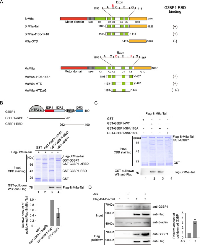
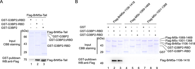
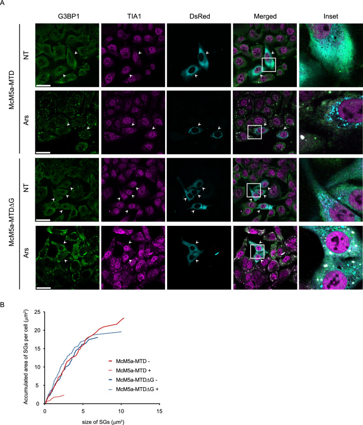
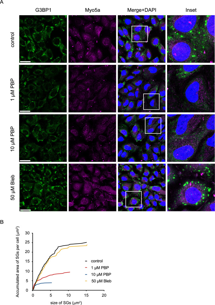
Stress granules (SGs) are non-membranous organelles composed of mRNA and proteins that assemble in the cytosol when the cell is under stress. Although the composition of mammalian SGs is both cell-type and stress-dependent, they consistently contain core components, such as Ras GTPase activating protein SH3 domain binding protein 1 (G3BP1). Upon stress, living cells rapidly assemble micrometric SGs, sometimes within a few minutes, suggesting that SG components may be actively transported by the microtubule and/or actin cytoskeleton. Indeed, SG assembly has been shown to depend on the microtubule cytoskeleton and the associated motor proteins. However, the role of the actin cytoskeleton and associated myosin motor proteins remains controversial. Here, we identified G3BP1 as a novel binding protein of unconventional myosin-5a (Myo5a). G3BP1 uses its C-terminal RNA-binding domain to interact with the middle portion of Myo5a tail domain (Myo5a-MTD). Suppressing Myo5a function in mammalian cells, either by overexpressing Myo5a-MTD, eliminating Myo5a gene expression, or treatment with myosin-5 inhibitor, inhibits the arsenite-induced formation of both small and large SGs. This is different from the effect of microtubule disruption, which abolishes the formation of large SGs but enhances the formation of small SGs under stress conditions. We therefore propose that, under stress conditions, Myo5a facilitates the formation of SGs at an earlier stage than the microtubule-dependent process.

Keywords: Actin; Arsenite; Intrinsically disordered region; Microtubule; Motor protein.


Conflict of interest statement

The authors have no relevant financial or non-financial interests to disclose.

Figures

Fig. 1
Fig. 1
Identification of the interaction between Myo5a and G3BP1 (A) Diagrams of the two isoforms of mouse Myo5a and their truncated proteins used in this study. BrM5a, brain-type Myo5a; McM5a, melanocyte-type Myo5a. Part of Myo5a tail domain is encoded by alternatively spliced exons (A, B, C, E and G for BrM5a; A, C, D, E, F, G for McM5a). Right column summarize the interaction between Myo5a constructs and G3BP1-RBD based on the GST pulldown assay results. + , strong interaction; ± , weak interaction; -, no interaction. (B) GST pulldown of GST-tagged G3BP1 (full-length and truncated constructs) with Flag-tagged BrM5a-Tail. Upper panel shows the diagrams of mouse G3BP1. NTF2-L, nuclear transport factor 2-Like domain; IDR, intrinsically disordered regions; RRM, RNA recognition motif. Lower panel shows the relative amounts of the pulled down Flag-BrM5a-Tail (average and std. of three independent assays). (C) GST pulldown assay was performed using GST-G3BP1-WT, -S84/166A and -S84/166E with Flag-BrM5a-Tail. For all GST pulldown assay, the inputs were analyzed with SDS-PAGE and visualized by Coomassie brilliant blue (CBB) staining; the GSH-Sepharose-bound proteins were eluted by GSH and analyzed by Western blot using anti-FLAG antibody. (D) Arsenite treatment of U2OS cells enhanced the interaction between Flag-BrM5a-Tail and endogenous G3BP1. U2OS cells were transfected with Flag-BrM5a-Tail, and 2 days later treated with 500 μM sodium arsenite (Ars) for 1 h before lysis. Cell lysates were incubated with anti-FLAG M2 affinity agarose and the bound proteins were eluted by FLAG peptide. The inputs and the eluted proteins were detected by Western blot using the indicated antibody. Note: Except for (B), in which insect Sf9-expressed G3BP1 proteins were used, all other GST pulldown assays in this study were performed using bacteria-expressed G3BP1 proteins
Fig. 2
Fig. 2
Characterization of the interaction between G3BP1 and the middle portion of Myo5a tail (A) GST pulldown af GST-tagged G3BP1-RBD with Flag-tagged McM5a-Tail (McM5a-MTDΔG, McM5a-MTD, and McM5a-1106–1467). The inputs were analyzed with SDS-PAGE and visualized by Coomassie brilliant blue (CBB) staining; the GSH-Sepharose-bound proteins were eluted by GSH and analyzed by Western blot using anti-FLAG antibody. (B) The dissociation constant (Kd) of G3BP1-RBD for McM5a-MTD measured by MST assay. The normalized fluorescence is plotted against the concentration of the ligands. G3BP1-RBD bound to McM5a-MTD with a Kd of 1.43 ± 0.24 μM. G3BP1-RBD was unable to interact with McM5a-MTDΔG
Fig. 3
Fig. 3
Both G3BP1-RBD and G3BP2-RBD interact with the intermediate tail of Myo5a, but not those of Myo5b and Myo5c. (A) G3BP2 interacts with BrM5a-Tail via the RBD. The GST pulldown assays were performed using GST-tagged G3BP2 variants with Flag-tagged BrM5a-Tail. (B) Interactions between the RBD of G3BP1/2 and the intermediate tail of three myosin-5 isoforms. GST pulldown assays were performed using 2.5 μM GST-tagged G3BP1-RBD or G3BP2-RBD with 2.5 μM Flag-tagged intermediate tail of three myosin-5 isoforms. For all GST pulldown assay, the inputs were analyzed with SDS-PAGE and visualized by CBB staining; the GSH-Sepharose-bound proteins were eluted by GSH and analyzed by Western blot using anti-FLAG antibody
Fig. 4
Fig. 4
Overexpressing McM5a-MTD in U2OS cells inhibits the arsenite-induced SGs. (A) U2OS cells were transfected with DsRed-McM5a-MTD or DsRed-McM5a-MTDΔG, and 2 days later non-treated (NT) or treated with 500 μM sodium arsenite (Ars) for 1 h prior to fixation. The fixed cells were stained for G3BP1 (Green) and TIA1 (Magenta) as SG markers, SG formation was analyzed by confocal microscopy. The right lane is an enlargement of the image in the white box. Scale bars, 40 μm. (B) Cumulative area of SGs per cell of transfected or non-transfected cells under arsenite-induced conditions. The area of individual SG in each cell (12 cells totally for each condition) was quantified by ImageJ and a cumulative area of SGs per cell was plotted. The SG size on the X-axis represents the SGs from the 12 analyzed cells considered individually
Fig. 5
Fig. 5
The myosin-5 inhibitor PBP inhibits the arsenite-induced SG formation in U2OS cells. (A) U2OS cells were left untreated (control) or treated with myosin inhibitors (the myosin-5 inhibitor PBP or the myosin-2 inhibitor blebbistatin), and then induced with 500 μM sodium arsenite for 1 h prior to fixation. The fixed cells were stained for G3BP1 (Green) and Myo5a (Magenta). Right lane shows the enlargement of the image in the white box. Scale bar, 40 μm. (B) Cumulative area of SGs per cell under arsenite-induced conditions. The area of individual SG in each cell (at least 25 cells for each condition) was quantified by ImageJ and a cumulative area of SGs per cell was plotted. The SG size on the X-axis represents the SGs from all the analyzed cells considered individually
Fig. 6
Fig. 6
Knockout of Myo5a gene in U2OS cells inhibits the arsenite-induced SG assembly. (A) The wild-type and two Myo5a−/− U2OS cell lines were non-treated (NT) or treated with 500 μM sodium arsenite (Ars) for 1 h. Cells were fixed and processed with immunostaining for G3BP1 (Green) and TIA (Magenta). Scale bar, 40 μm. (B) Cumulative area of SGs per cell (at least 25 cells from both wild-type or two Myo5a−/− U2OS cell lines) under arsenite-induced conditions. The SG size on the X-axis represents the SGs from all the analyzed cells considered individually. (C) Detections of Myo5a and G3BP1 in the cell lysates of the wild-type and two Myo5a−/− U2OS cell lines by Western blot
Fig. 7
Fig. 7
Effects of Nocodazole on SG formation in wild-type and Myo5a−/− U2OS cells. (A) The distribution of SGs in wild-type U2OS cells and two Myo5a−/− U2OS cell lines. U2OS cells were pretreated with DMSO or nocodazole, then induced with 500 μM sodium arsenite for 1 h. Cells were fixed and stained for G3BP1 (Green) and Tubulin (Magenta). Right lane shows the enlargement of the image in the white boxes. Scale bar, 40 μm. (B) Cumulative area of SGs per cell in the wild-type and the two Myo5a−/− U2OS cell lines with or without nocodazole treatment. The SG size on the X-axis represents the SGs from all the analyzed cells considered individually. (C) Proposed model for SGs assembly. G3BP1 is in the compact state under normal conditions. Upon stress, polysomes disassemble and mRNAs are released in an unfolded protein-free state, RNA-bound G3BP1 adopts an expanded conformation and aggregates with mRNA and other SG factors to form mini SGs (~ 100 nm), which then fuse with each other to form small SGs in micron scale, and eventually to form large SGs. The formation of small SGs is facilitated by the transport by Myo5a along actin filaments and the formation of large SGs depends on the transport of small SGs along microtubules

References

    1. Protter DSW, Parker R (2016) Principles and properties of stress granules. Trends Cell Biol 26(9):668–679 - PMC - PubMed
    1. Lyon AS, Peeples WB, Rosen MK (2021) A framework for understanding the functions of biomolecular condensates across scales. Nat Rev Mol Cell Biol 22(3):215–235 - PMC - PubMed
    1. Hofmann S, Kedersha N, Anderson P, Ivanov P (2021) Molecular mechanisms of stress granule assembly and disassembly. Biochim Biophys Acta Mol Cell Res 1868(1):118876 - PMC - PubMed
    1. Kedersha N, Cho MR, Li W, Yacono PW, Chen S, Gilks N et al (2000) Dynamic shuttling of TIA-1 accompanies the recruitment of mRNA to mammalian stress granules. The J Cell Biol 151(6):1257–1268 - PMC - PubMed
    1. Kedersha NL, Gupta M, Li W, Miller I, Anderson P (1999) RNA-binding proteins TIA-1 and TIAR link the phosphorylation of eIF-2 alpha to the assembly of mammalian stress granules. The J Cell Biol 147(7):1431–1442 - PMC - PubMed

MeSH terms

LinkOut - more resources