. 2024 Oct 10;142(11):1028-1036.
doi: 10.1001/jamaophthalmol.2024.3828. Online ahead of print.

Sterile Intraocular Inflammation Associated With Faricimab

Affiliations

Sterile Intraocular Inflammation Associated With Faricimab

Mariano Cozzi et al. JAMA Ophthalmol. .

Erratum in

  • Error in Text and Table.
    [No authors listed] [No authors listed] JAMA Ophthalmol. 2025 Jan 1;143(1):85. doi: 10.1001/jamaophthalmol.2024.5503. JAMA Ophthalmol. 2025. PMID: 39636618 Free PMC article. No abstract available.

Abstract

Importance: Randomized clinical trials are conducted to establish both drug safety and efficacy. However, evidence of adverse events associated with these drugs in the clinical practice setting can be of value at generating hypotheses regarding less common safety issues, even if causality cannot be determined.

Objective: To present and analyze cases of intraocular inflammation associated with faricimab therapy in patients referred to a single European institution.

Design, setting, and participants: This was a review starting in April of 2024 of an observational case series. Patients were from a single academic-based tertiary referral center in Switzerland. Included in the analysis were patients referred for intraocular inflammation soon after receiving a faricimab intravitreal injection between June 1, 2022, and March 5, 2024.

Exposure: Faricimab, 6 mg (0.05 mL of a 120-mg/mL solution), administrated for neovascular age-related macular degeneration or diabetic macular edema.

Main outcomes and measures: The systemic and ocular histories and imaging data available were reviewed. The following were evaluated: visual acuity measured with habitual correction using the Early Treatment of Diabetic Retinopathy Study charts before and after the event; intraocular pressure; patient symptoms; anterior, intermediate, or posterior location of the intraocular inflammation; and the presence of retinal vasculitis. Multimodal imaging including color fundus photographs, fluorescein angiograms, indocyanine green angiograms, and optical coherence tomography were reviewed.

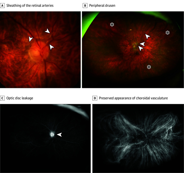
Results: A total of 12 eyes from 7 patients (mean [SD] age, 73.3 [16.7] years; 4 female [57.1%]) over 22 months were identified as having noninfectious intraocular inflammation after intravitreal faricimab injections. Among these cases, in 2 eyes, retinal vasculitis was present together with anterior and posterior inflammation. One of the 2 eyes had an occlusive form of vasculitis of the arteries and veins, leading to subsequent macular capillary nonperfusion and clinically relevant irreversible vision deterioration from 20/80 to 20/2000. The remaining eyes were characterized by moderate anterior segment inflammation without substantial vision changes. The intraocular inflammation event occurred after a median (IQR) of 3.5 (2.0-4.3) faricimab injections. The median (IQR) interval between the last faricimab injection and the diagnosis of inflammation was 28 (24-38) days. Increased intraocular pressure of 30 mm Hg or higher was found in 3 eyes.

Conclusions and relevance: This case series highlights the occurrence of rare, but potentially severe, intraocular inflammation associated with faricimab therapy. Although these findings do not prove causality and can only generate hypotheses for future investigations, these results suggest the importance of continuous surveillance and monitoring for patients undergoing faricimab therapy to promptly identify and manage potential adverse events.


Conflict of interest statement

Conflict of Interest Disclosures: Dr Cozzi reported receiving personal fees from Bayer, Nidek, Novartis, Zeiss, and Heidelberg Engineering outside the submitted work. Dr Muth reported receiving consultant fees from Roche, Custom Surgical, Annexon Biosciences, Bayer, and Canon outside the submitted work. Dr Zweifel reported receiving consultant and advisor support from Alcon, Allergan, Apellis, Bayer, Endogena, Novartis, Roche, and Zeiss and grant support from Bayer, Novartis, and Roche outside the submitted work. No other disclosures were reported.

Figures

Figure 1.
Figure 1.. Baseline Multimodal Imaging of Case 1
A, Optos widefield pseudocolor image of the left eye of the patient in case 1 depicts widespread hemorrhages involving both the macular area and the periphery, with partial vitreous opacity including sheathing of the retinal arteries and veins (white arrowheads). B, Macular optical coherence tomography scans display the presence of inflammatory vitreous cells (yellow arrowheads) within the vitreous space and partial swelling of the outer nuclear layer. The inner retinal layers appear thin and disrupted. C, The early phase of fluorescein angiography (FA) clearly reveals diffuse intraretinal bleeding blocking the FA signal. Note the marked delayed filling of the retinal vessels leading to extensive diffuse vascular non-perfusion (white asterisks) in the periphery. D, Indocyanine green angiography portrays a maintained choroidal vasculature structure without clear pathological alterations.
Figure 2.
Figure 2.. Case 1 Depicting a Foveal Optical Coherence Tomography Series
The series of B-scans passing through the fovea show macular changes over time, starting with initial swelling including intraretinal fluid (white arrow), progressing through hyperreflectivity of the inner retinal layers (white arrowheads), and culminating in complete macular capillary nonperfusion after 32 days, resulting in the thinning of all retinal layers.
Figure 3.
Figure 3.. Right Eye Presentation of Baseline Case 2 Multimodal Imaging
A, Flash-based color fundus photography clearly highlights the presence of sheathing of the retinal arteries (white arrowheads). B, This is confirmed by the widefield Optos image (arrowheads), which shows a large amount of peripheral drusen (asterisks). C, Fluorescein angiography clearly shows optic disc leakage (arrowhead) and macular staining of drusen and fibrovascular pigment epithelial detachment related to age-related macular degeneration but no areas of nonperfusion. D, Indocyanine green angiography reveals a preserved appearance of the choroidal vasculature and no signs of choroidal ischemia.
Figure 4.
Figure 4.. Left Eye Presentation of Baseline Case 2 Multimodal Imaging
A and B, Color fundus photography and Optos widefield imaging, respectively, of the left eye demonstrate no evidence of vascular alteration associated with vasculitis. B, Peripheral drusen (asterisks) appear symmetric to those in the right eye. C, Fluorescein angiography reveals mild optic disc leakage (arrowhead), drusen staining in the macula associated with age-related macular degeneration, and drusen staining in the periphery. D, Indocyanine green angiography depicts no particular alterations in choroidal vasculature, similar to the findings in the fellow eye.

References

    1. Regula JT, Lundh von Leithner P, Foxton R, et al. . Targeting key angiogenic pathways with a bispecific CrossMAb optimized for neovascular eye diseases. EMBO Mol Med. 2019;11(5):e10666. doi:10.15252/emmm.201910666 - DOI - PMC - PubMed
    1. Heier JS, Khanani AM, Quezada Ruiz C, et al. ; TENAYA and LUCERNE Investigators . Efficacy, durability, and safety of intravitreal faricimab up to every 16 weeks for neovascular age-related macular degeneration (TENAYA and LUCERNE): 2 randomized, double-masked, phase 3, noninferiority trials. Lancet. 2022;399(10326):729-740. doi:10.1016/S0140-6736(22)00010-1 - DOI - PubMed
    1. Wykoff CC, Abreu F, Adamis AP, et al. ; YOSEMITE and RHINE Investigators . Efficacy, durability, and safety of intravitreal faricimab with extended dosing up to every 16 weeks in patients with diabetic macular oedema (YOSEMITE and RHINE): 2 randomized, double-masked, phase 3 trials. Lancet. 2022;399(10326):741-755. doi:10.1016/S0140-6736(22)00018-6 - DOI - PubMed
    1. Tadayoni R, Paris LP, Danzig CJ, et al. ; BALATON and COMINO Investigators . Efficacy and safety of faricimab for macular edema due to retinal vein occlusion: 24-week results from the BALATON and COMINO trials. Ophthalmology. 2024;131(8):950-960. doi:10.1016/j.ophtha.2024.01.029 - DOI - PubMed
    1. Agostini H, Abreu F, Baumal CR, et al. . Faricimab for neovascular age-related macular degeneration and diabetic macular edema: from preclinical studies to phase 3 outcomes. Graefes Arch Clin Exp Ophthalmol. 2024. doi:10.1007/s00417-024-06531-9 - DOI - PMC - PubMed