. 2024 Oct 15;121(42):e2406009121.
doi: 10.1073/pnas.2406009121. Epub 2024 Oct 10.

High-throughput screening for small-molecule stabilizers of misfolded glucocerebrosidase in Gaucher disease and Parkinson's disease

Affiliations

High-throughput screening for small-molecule stabilizers of misfolded glucocerebrosidase in Gaucher disease and Parkinson's disease

Darian Williams et al. Proc Natl Acad Sci U S A. .

Abstract

Glucocerebrosidase (GCase) is implicated in both a rare, monogenic disorder (Gaucher disease, GD) and a common, multifactorial condition (Parkinson's disease, PD); hence, it is an urgent therapeutic target. To identify correctors of severe protein misfolding and trafficking obstruction manifested by the pathogenic L444P-variant of GCase, we developed a suite of quantitative, high-throughput, cell-based assays. First, we labeled GCase with a small proluminescent HiBiT peptide reporter tag, enabling quantitation of protein stabilization in cells while faithfully maintaining target biology. TALEN-based gene editing allowed for stable integration of a single HiBiT-GBA1 transgene into an intragenic safe-harbor locus in GBA1-knockout H4 (neuroglioma) cells. This GD cell model was amenable to lead discovery via titration-based quantitative high-throughput screening and lead optimization via structure-activity relationships. A primary screen of 10,779 compounds from the NCATS bioactive collections identified 140 stabilizers of HiBiT-GCase-L444P, including both pharmacological chaperones (ambroxol and noninhibitory chaperone NCGC326) and proteostasis regulators (panobinostat, trans-ISRIB, and pladienolide B). Two complementary high-content imaging-based assays were deployed to triage hits: The fluorescence-quenched substrate LysoFix-GBA captured functional lysosomal GCase activity, while an immunofluorescence assay featuring antibody hGCase-1/23 directly visualized GCase lysosomal translocation. NCGC326 was active in both secondary assays and completely reversed pathological glucosylsphingosine accumulation. Finally, we tested the concept of combination therapy by demonstrating synergistic actions of NCGC326 with proteostasis regulators in enhancing GCase-L444P levels. Looking forward, these physiologically relevant assays can facilitate the identification, pharmacological validation, and medicinal chemistry optimization of small molecules targeting GCase, ultimately leading to a viable therapeutic for GD and PD.

Keywords: Gaucher disease; Parkinson’s disease; glucocerebrosidase; high-throughput screening; pharmacological chaperone.


Conflict of interest statement

Competing interests statement:A.G. and R.J. are employees of F. Hoffmann-La Roche AG.

Figures

Fig. 1.
Fig. 1.
HiBiT-tagged GCase retains normal trafficking and function, enabling a high-throughput screening assay measuring cellular GCase levels. (A) GCase was labeled with a small (1.3 kDa), proluminescent, N-terminal HiBiT peptide tag immediately following the signal peptide (SP) sequence. (B) HiBiT-GCase reporters (WT, N370S, or L444P) were engineered into the human Citrate Lyase Beta-Like (CLYBL) intragenic safe-harbor locus within a GBA1-KO H4 cell background using TALEN-enhanced integrative gene transfer. (C) Colocalization of GCase (green) with lysosomal marker LAMP1 (magenta) was determined by immunofluorescent staining. (Scale bar, 10 µm; Inset scale bar, 1 µm.) (D) The copy number of the stably integrated transgene was confirmed to be 1 across all three HiBiT-GCase lines via Droplet Digital PCR (ddPCR). The HiBiT-GCase H4 lines featured ~60 copies/μL of the HiBiT-GBA1 transgene, as compared with ~120 copies/μL of the reference gene RPP30, which has a known copy number of 2 in the GBA1-WT H4 cell line. (E) GCase protein level was measured by western blot in GBA1-WT (unedited), GBA1-KO, and HiBiT-GCase (WT, N370S, or L444P) H4 cell lines using anti-GCase (R386 and 2E2) antibodies, with total protein as the loading control. (F) Glycosidase sensitivity analysis indicates that HiBiT-GCase-L444P is entirely retained in the ER. The Endo H-sensitive fraction (lower band) on the blot contains immature, ER-retained GCase, while the Endo H-resistant fraction (top band) contains maturely glycosylated, post-ER-localized GCase. Both fractions are responsive to PNGase F treatment. NT: nontreated. (G) GCase protein levels were quantitated by AlphaLISA (Amplified Luminescent Proximity Homogeneous Assay) utilizing a sandwich configuration of two monoclonal antibodies recognizing nonoverlapping epitopes, hGCase-1/23 (which was biotinylated and associated with a streptavidin-coated donor bead) and hGCase-1/17 (which was directly conjugated to an acceptor bead). (Error bars: SEM [n = 3 biological replicates]). (H) GCase activity was measured in cell lysates using the fluorogenic substrate 4-methylumbelliferyl-β-D-glucopyranoside. Relative GCase activity was calculated by adjusting for protein concentration, correcting for GBA1-KO H4 cell background, and normalizing to GBA1-WT signal. (Error bars: SD [n = 5 biological replicates]). (I) Levels of glucosylsphingosine (GluSph) in H4 cell pellets were quantified by positive ion electrospray LC–MS/MS in multiple reaction-monitoring mode, using deuterated compounds as internal standards. (Error bars: SD [n = 5 biological replicates]). (J) Pilot testing of the HiBiT-GCase assay was performed in ultrahigh-throughput 1536-well plate format. Cells were treated with known-active ERAD modulators (NMS-873, p97 inhibitor; MG132, proteasome inhibitor) or GCase stabilizers (ambroxol, NCGC758, or NCGC607) for 24 h, followed by measurement of HiBiT-GCase luminescence. For each respective cell line, data are represented as fold change in luminescence (RLU) in compound-treated versus DMSO-treated cells. (Error bars: SEM [n = 3 to 6]). Dose–response curves were fit using log(agonist) vs. response (three parameters). *P-value ≤ 0.05; **P-value ≤ 0.01; ***P-value ≤ 0.001; ****P-value ≤ 0.0001.
Fig. 2.
Fig. 2.
Quantitative high-throughput screening of mechanistically annotated small-molecule libraries for GCase-L444P stabilizers. (A) Schematic of high-throughput screening methodology using H4 HiBiT-GCase-L444P reporter cell line. (B) In the primary screen, 10,779 compounds, including the NCATS Pharmaceutical Collection (NPC), NCATS Pharmacologically Active Chemical Toolbox (NPACT), and Helping to End Addiction Long-term (HEAL) chemical libraries, as well as analogs of the noninhibitory chaperone chemotypes NCGC607 and NCGC758, were evaluated at concentrations most commonly ranging from 5 nM to 75 μM in a 7-point, 5× dilution series using a single replicate. Compounds were then triaged based on curve class (CC) and efficacy: Those with CC of 1.1 to 1.4, 2.1 to 2.4, or 5 and efficacy ≥40% were considered primary screen hits. These 716 compounds were retested (NCGC326: 100 nM to 100 μM, 11-point, 2× dilution; others: 1 nM to 75 μM, 11-point, 3× dilution) with n = 3 replicates and categorized as final hits if they met a cutoff of efficacy ≥40%, regardless of curve class, resulting in 140 confirmed hits. (C) Top representative follow-up screen hits from each mechanistic cluster were selected based on their potency and efficacy in stabilizing GCase-L444P levels. Response values were normalized to intraplate DMSO-treated controls, such that 100% efficacy reflects a doubling of GCase levels. n = 3. (D) Dose–response profiles for 187 analogs of chaperones NCGC758 and NCGC607 revealed 11 compounds, including NCGC326 (blue), with greater efficacy than NCGC607 (green). NCGC758 (red) was inactive under the screening conditions. (E) Target engagement studies with microscale thermophoresis revealed that NCGC326 binds recombinant GCase-WT with a dissociation constant (Kd) of 3.75 μM in 50 mM sodium citrate buffer (pH 5.5). n = 2.
Fig. 3.
Fig. 3.
A high-content imaging-based secondary assay using the fluorescence-quenched substrate LysoFix-GBA quantifies GCase activity in the lysosome. Lysosomal activity of GCase was directly visualized using the optimized LysoFix-GBA secondary assay in live HiBiT-GCase-L444P H4 cells treated with vehicle (DMSO) or hit compounds. H4 cells were seeded into 384-well PerkinElmer PhenoPlates (25,000 cells in 40 μL media) and incubated for 24 h. Thereafter, the cells were treated with a titration of compounds for 24 h and then incubated with LysoFix-GBA (5 μM) for 2 h at 37 °C and 5% CO2. High-content imaging was performed after 15 min of nuclear staining with Hoechst-33342 (1 μg/mL) in Fluorobrite media. (A) Select hits from the primary screen were tested in a dose–response titration series. Data are represented as the fold change (compound-treated vs. DMSO-treated) in integrated LysoFix-GBA spot intensity per cell. Dose–response curves were fit using log(agonist) vs. response (three parameters). (Error bars: SEM [n = 3 to 6]). (B) Representative images are shown for pladienolide B, trans-ISRIB, and NCGC326 at their most effective concentrations in the LysoFix-GBA assay. (Scale bar, 10 μm.)
Fig. 4.
Fig. 4.
Immunofluorescence secondary assay measures GCase translocation to the lysosome via high-content imaging. HiBiT-GCase-L444P and unedited GBA1-WT H4 cells were seeded into 96-well PerkinElmer PhenoPlates and stained for GCase using monoclonal antibody hGCase-1/23, as well as for lysosomal marker LAMP1, following treatment with vehicle (DMSO, 0.5% v/v), pladienolide B (100 nM), NCGC326 (25 μM), panobinostat (10 μM), trans-ISRIB (10 μM), or ambroxol (25 μM) for ~34 h. Representative images are shown in (A), and data are quantified as mean GCase intensity in spots of LAMP1, summed per well, and normalized to cell count, in (B). (Error bars: SD [n = 6]). (Scale bar, 15 μm; Inset scale bar, 5 μm.) ***P-value ≤ 0.001 vs. HiBiT-L444P+DMSO; ****P-value ≤ 0.0001 vs. HiBiT-L444P+DMSO.
Fig. 5.
Fig. 5.
Matrix combination screening approach identifies synergistic coformulations of a PC with a proteostasis regulator. HiBiT-GCase-L444P H4 cells were tested in 10 × 10 pairwise dose–response combinatorial matrix format. Cells were treated for 24 h with chaperone NCGC326 in a 9-point titration (3 nM to 20 μM, 3× dilution) against the same 9-point titration of PRs trans-ISRIB (A), pladienolide B (B), panobinostat (C), or ARV-825 (D); the HiBiT-GCase lytic assay was then performed. Luminescence response values were normalized to intraplate DMSO-treated controls, such that 100% efficacy reflects a doubling of HiBiT-GCase levels (AD). Synergy was evaluated based on the Loewe synergy score (EH). In general, negative, zero, and positive synergy scores indicate antagonistic, additive, and synergistic interactions, respectively, between drugs. n = 3.
Fig. 6.
Fig. 6.
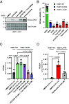
Screening hits reverse glycosphingolipid substrate accumulation in H4 cells. (A and B) HiBiT-GCase-L444P H4 cells were treated (starting at 50 to 60% confluence) with vehicle (DMSO, 0.3% v/v), NCGC326 (25 μM), trans-ISRIB (1.25 μM), or the combination of NCGC326 (25 μM) and trans-ISRIB (1.25 μM) for 3 d, and (starting at 90 to 100% confluence) with pladienolide B (100 nM) for 24 h. (A) GCase protein levels in cell lysates were visualized on western blot using the 2E2 antibody, with total protein as the loading control. (B) GCase activity in cell lysates of HiBiT-GCase H4 cells (WT, N370S, or L444P) treated with DMSO or compounds. Relative GCase activity was calculated by adjusting for protein concentration, correcting for GBA1-KO H4 cell background, and normalizing to HiBiT-WT+DMSO signal. (Error bars: SD [n = 8 technical replicates]). (C and D) Levels of glucosylsphingosine (GluSph) in HiBiT-GCase-WT and HiBiT-GCase-L444P H4 reporter lines were evaluated by supercritical fluid chromatography (SFC) separation coupled with tandem mass spectrometry (MS/MS) detection and normalized to total cellular inorganic phosphate (Pi) levels. Cells were treated with vehicle (0.3% v/v DMSO) or hit compounds for 3 d (C), except for the cytotoxic hit compound pladienolide B, for which treatment lasted 48 h (D). (Error bars: SD [n = 3 biological replicates]).

References

    1. Sidransky E., et al. , Multicenter analysis of glucocerebrosidase mutations in Parkinson’s disease. N. Engl. J. Med. 361, 1651–1661 (2009). - PMC - PubMed
    1. Nalls M. A., et al. , A multicenter study of glucocerebrosidase mutations in dementia with Lewy bodies. JAMA Neurol. 70, 727–735 (2013). - PMC - PubMed
    1. Richter F., et al. , A GCase chaperone improves motor function in a mouse model of synucleinopathy. Neurotherapeutics 11, 840–856 (2014). - PMC - PubMed
    1. Sardi S. P., et al. , Augmenting CNS glucocerebrosidase activity as a therapeutic strategy for Parkinsonism and other Gaucher-related synucleinopathies. Proc. Natl. Acad. Sci. U.S.A. 110, 3537–3542 (2013). - PMC - PubMed
    1. Maple-Grødem J., et al. , Association of GBA genotype with motor and functional decline in patients with newly diagnosed Parkinson disease. Neurology 96, e1036–e1044 (2021). - PMC - PubMed

MeSH terms

Substances