

Radiographic findings in dogs with 360 degrees gastric dilatation and volvulus
- PMID: 39388661
- PMCID: PMC11617607
- DOI: 10.1111/vru.13445
Radiographic findings in dogs with 360 degrees gastric dilatation and volvulus
Abstract
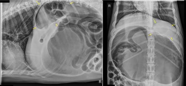
Gastric dilatation and volvulus (GDV) is a life-threatening emergency that requires urgent intervention. Radiographic features associated with 360-GDV in dogs have not been investigated. The aim of this retrospective observational study is to describe radiographic features and clinical variables in dogs affected with 360-GDV and to report agreement rates between different radiologists. We also report the sensitivity and specificity of radiographs to diagnose 360-GDV in dogs. Confirmed 360-GDV cases were retrieved, and the radiographic findings were compared with dogs presenting with gastric dilatation (GD) and 180-GDV. Images were reviewed and graded by three blinded board-certified radiologists. A total of 16 dogs with confirmed 360-GDV were identified. The median age was 10 years old (2-13 years). The sensitivity for detection of 360-GDV ranged between 43.7% and 50%, and the specificity between 84.6% and 92.1%. Interobserver agreement on final diagnosis was substantial (Kappa = 0.623; 0.487-0.760, 95% CI). The highest agreement rate was in cases of 180-GDV (87%), followed by the GD cases (72%) and 360-GDV (46%). Severe esophageal distension and absence of small intestinal dilation were the only radiographic features specifically associated with 360-GDV. A similar pyloric position was found between GD and 360-GDV. Additional radiographic variables that could help differentiate GD from 360-GDV include the degree of gastric distension and the peritoneal serosal contrast. Two cases with 360-GDV were misdiagnosed by the three radiologists as GD. In conclusion, radiographically, 360-GDV cases can reassemble GD and vice versa. Radiologists and clinicians should be aware of the low sensitivity of radiographs for the detection of 360-GDV.
Keywords: 360; dogs; gastric volvulus; radiographs.
© 2024 The Author(s). Veterinary Radiology & Ultrasound published by Wiley Periodicals LLC on behalf of American College of Veterinary Radiology.
Conflict of interest statement
The authors declare no conflict of interest.
Figures


References
-
- Kare T, Spencer JAA, Veterinary Surgery: Small Animal. Stomach. 2nd ed. 2017;1730‐1760.
-
- Ullmann B, Seehaus N, Hungerbühler S, Meyer‐Lindenberg A. Gastric dilatation volvulus: a retrospective study of 203 dogs with ventral midline gastropexy. J Small Anim Pract. 2016;57:18‐22. - PubMed
-
- Rosselli D, Updated Information on gastric dilatation and volvulus and gastropexy in dogs. Vet Clin North Am Small Anim Pract [Internet]. 2022; 52:317‐337. Available from: https://pubmed.ncbi.nlm.nih.gov/35082096/ - PubMed
-
- Sharp CR, Rozanski EA. Cardiovascular and systemic effects of gastric dilatation and volvulus in dogs. Top Companion Anim Med. 2014;29:67‐70. - PubMed
-
- Broome C, Walsh V. Gastric dilatation‐volvulus in dogs. N Z Vet J. 2003;51:275‐283. - PubMed
