Prognosis of Proximal and Distal Vertebrobasilar Artery Stent Placement
- PMID: 39389775
- PMCID: PMC11543063
- DOI: 10.3174/ajnr.A8389
Prognosis of Proximal and Distal Vertebrobasilar Artery Stent Placement
Abstract
Background and purpose: Vertebrobasilar artery stent placement (VBS) is potentially effective in preventing recurrent posterior circulation strokes; however, the incidences of in-stent restenosis and stented-territory ischemic events based on the location of stent placement have rarely been investigated. We aimed to investigate the characteristics and prognosis of VBS between intracranial and extracranial.
Materials and methods: This study was single-center retrospective cohort study, and we obtained medical records of patients who underwent VBS. We compared clinical and periprocedural factors between extracranial and intracranial VBS. The primary outcomes included the incidence of in-stent restenosis (>50% reduction in lumen diameter) and stented-territory ischemic events. We compared the incidence of in-stent restenosis and stented-territory ischemic events by using Kaplan-Meier curves.
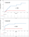
Results: Of the 105 patients, 41 (39.0%) underwent extracranial VBS, and 64 (61.0%) underwent intracranial VBS. During the follow-up, the incidences of in-stent restenosis and stented-territory ischemic events were 15.2% and 22.9%, respectively. The procedure time was longer (47.7 ± 19.5 minutes versus 74.5 ± 35.2 minutes, P < .001), and the rate of residual stenosis (≥30%) just after VBS was higher (2 [4.9%] versus 24 [37.5%], P < .001) in intracranial VBS than in extracranial VBS. Also, the incidences of in-stent restenosis were significantly higher in intracranial VBS than in extracranial VBS (4.9% versus 21.9%, P = .037). On the other hand, the incidences of stented-territory ischemic events (7.3% versus 32.8%, P < .001) were significantly higher in intracranial VBS than in extracranial VBS. The main mechanisms of stroke were artery-to-artery embolism (2 [66.7%]) in extracranial VBS, and artery-to-artery embolism (9 [42.9%]) and branch atheromatous disease (8 [38.1%]) in intracranial VBS. The Kaplan-Meier curve demonstrated a higher incidence of in-stent restenosis and stented-territory ischemic events in intracranial VBS than in extracranial VBS (P = .008 and P = .002, respectively).
Conclusions: During the follow-up, the incidence of in-stent restenosis and stented-territory ischemic events was higher in patients with intracranial VBS than in those with extracranial VBS. The higher rates of postprocedural residual stenosis might have contributed to the increased risk of in-stent restenosis. Furthermore, prolonged procedure time and additional stroke mechanism, including branch atheromatous disease, might be associated with a higher risk of stented-territory ischemic events in intracranial VBS.
© 2024 by American Journal of Neuroradiology.
Figures


References
-
- Labropoulos N, Nandivada P, Bekelis K. Stroke of the posterior cerebral circulation. Int Angiol 2011;30:105–14 - PubMed
MeSH terms
LinkOut - more resources
Full Text Sources
Medical
