

Synergistic effect of zinc oxide-cinnamic acid nanoparticles for wound healing management: in vitro and zebrafish model studies
- PMID: 39390421
- PMCID: PMC11468080
- DOI: 10.1186/s12896-024-00906-w
Synergistic effect of zinc oxide-cinnamic acid nanoparticles for wound healing management: in vitro and zebrafish model studies
Abstract
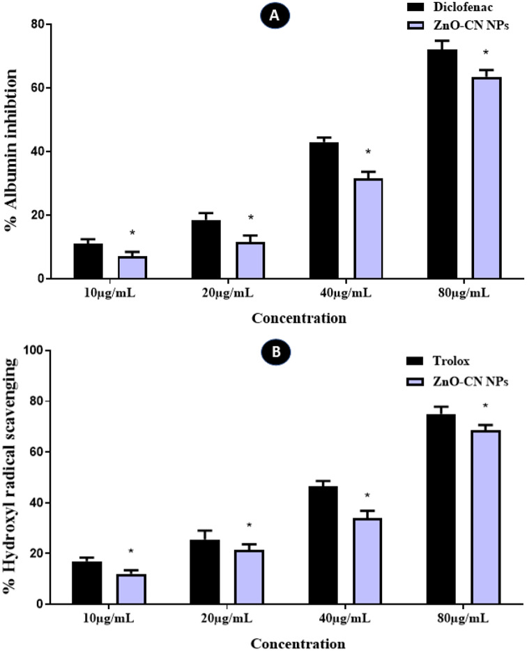
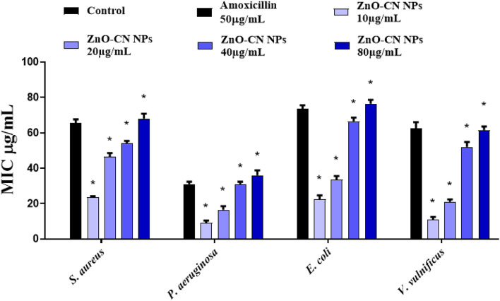
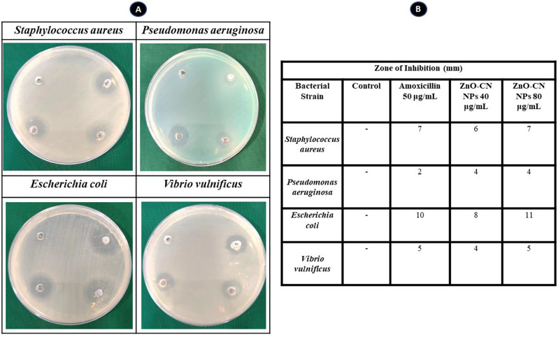
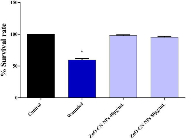
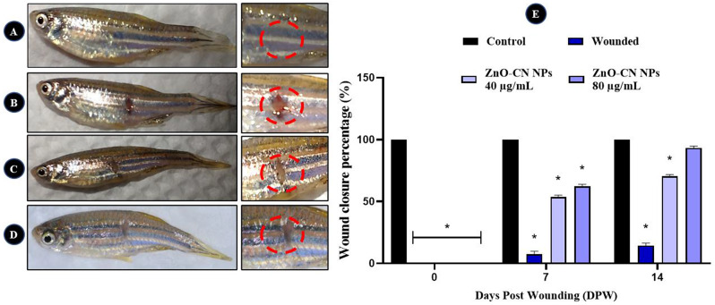
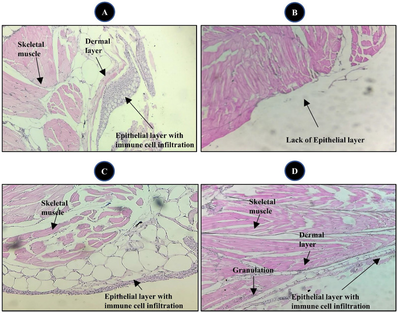
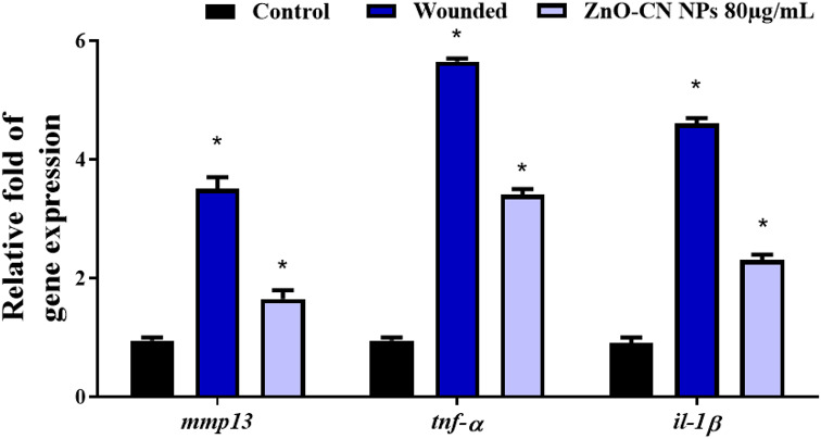
Wound infections resulting from pathogen infiltration pose a significant challenge in healthcare settings and everyday life. When the skin barrier is compromised due to injuries, surgeries, or chronic conditions, pathogens such as bacteria, fungi, and viruses can enter the body, leading to infections. These infections can range from mild to severe, causing discomfort, delayed healing, and, in some cases, life-threatening complications. Zinc oxide (ZnO) nanoparticles (NPs) have been widely recognized for their antimicrobial and wound healing properties, while cinnamic acid is known for its antioxidant and anti-inflammatory activities. Based on these properties, the combination of ZnO NPs with cinnamic acid (CA) was hypothesized to have enhanced efficacy in addressing wound infections and promoting healing. This study aimed to synthesize and evaluate the potential of ZnO-CN NPs as a multifunctional agent for wound treatment. ZnO-CN NPs were synthesized and characterized using key techniques to confirm their structure and composition. The antioxidant and anti-inflammatory potential of ZnO-CN NPs was evaluated through standard in vitro assays, demonstrating strong free radical scavenging and inhibition of protein denaturation. The antimicrobial activity of the nanoparticles was tested against common wound pathogens, revealing effective inhibition at a minimal concentration. A zebrafish wound healing model was employed to assess both the safety and therapeutic efficacy of the nanoparticles, showing no toxicity at tested concentrations and facilitating faster wound closure. Additionally, pro-inflammatory cytokine gene expression was analyzed to understand the role of ZnO-CN NPs in wound healing mechanisms. In conclusion, ZnO-CN NPs demonstrate potent antioxidant, anti-inflammatory, and antimicrobial properties, making them promising candidates for wound treatment. Given their multifunctional properties and non-toxicity at tested concentrations, ZnO-CN NPs hold significant potential as a therapeutic agent for clinical wound management, warranting further investigation in human models.
Keywords: Cinnamic acid; Nanomedicine; Wound healing; Wound infection; Zebrafish model; Zinc oxide nanoparticle.
© 2024. The Author(s).
Conflict of interest statement
The authors declare no competing interests.
Figures

References
-
- Makeri D, Odoki M, Eilu E, Agwu E. Update on prevalence and antimicrobial resistance of Staphylococcus aureus and Pseudomonas aeruginosa isolated from diabetic foot ulcers in Africa: a systematic review and meta-analysis. Bull Natl Res Cent. 2023;47:145.
-
- Mundhada A, Tenpe S. A study of organisms causing surgical site infections and their antimicrobial susceptibility in a tertiary care Government Hospital. Indian J Pathol Microbiol. 2015;58:195. - PubMed
-
- Kizilates F, Yakupogullari Y, Berk H, Oztoprak N, Otlu B. Risk factors for fecal carriage of extended-spectrum beta-lactamase-producing and carbapenem-resistant Escherichia coli and Klebsiella pneumoniae strains among patients at hospital admission. Am J Infect Control. 2021;49:333–9. - PubMed
-
- Bobrov AG, Getnet D, Swierczewski B, Jacobs A, Medina-Rojas M, Tyner S, et al. Evaluation of Pseudomonas aeruginosa pathogenesis and therapeutics in military‐relevant animal infection models. APMIS. 2022;130:436–57. - PubMed
MeSH terms
Substances
LinkOut - more resources
Full Text Sources
