

Changes and associations between synovial fluid and magnetic resonance imaging markers of osteoarthritis after high tibial osteotomy
- PMID: 39390512
- PMCID: PMC11465750
- DOI: 10.1186/s13075-024-03409-3
Changes and associations between synovial fluid and magnetic resonance imaging markers of osteoarthritis after high tibial osteotomy
Abstract
Background: Mechanobiological mechanisms of osteoarthritis (OA) are unclear. Our objectives were to explore: 1) changes in knee joint physiology using a large panel of synovial fluid biomarkers from before to one year after high tibial osteotomy (HTO) surgery, and 2) the association of changes in the synovial fluid biomarkers with the changes in MRI measures of knee effusion-synovitis and articular cartilage composition.
Methods: Twenty-six patients with symptomatic knee OA and varus alignment underwent synovial fluid aspirations and 3 T MRI before and one year after medial opening wedge HTO. Cytokine and growth factor levels in synovial fluid were measured with multiplex assays. Ontology and pathway enrichment was assessed using data protein sets with gene set enrichment analysis (GSEA), and analyzed using linear mixed effects models. MRIs were analyzed for effusion-synovitis and T2 cartilage relaxation time using manual segmentations. Changes in biomarker concentrations were correlated to changes in MRI effusion-synovitis volume and articular cartilage T2 relaxation times.
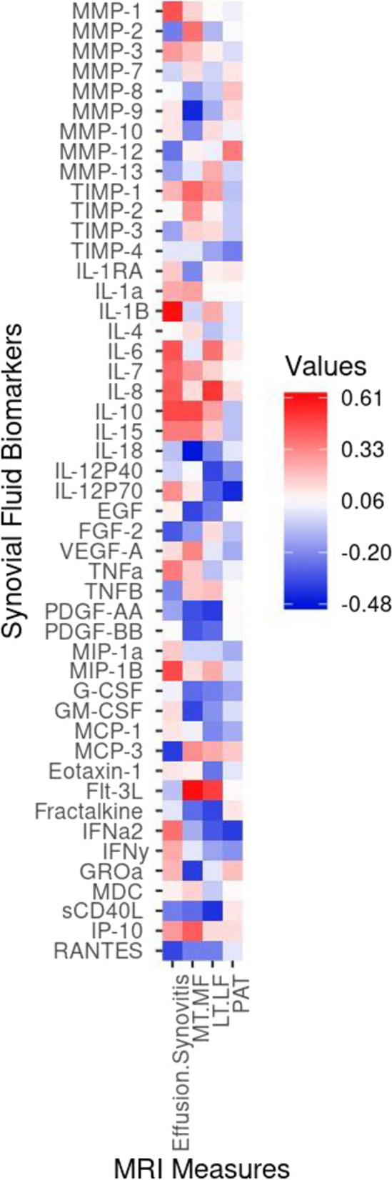
Results: Decreased enrichment in Toll-like receptor and TNF-α signalling was detected one year after HTO. The leading contributors to this reduction included IL-6, TNF-α and IL-1β, whereas the highest contributors to positive enrichment were EGF, PDGF-BB and FGF-2. Effusion-synovitis volume decreased (mean [95%CI]) one year after HTO (-2811.58 [-5094.40, -528.76mm3]). Effusion-synovitis volume was moderately correlated (r [95% CI]) with decreased MMP-1 (0.44 [0.05; 0.71]), IL-7 (0.41 [0.00; 0.69]) and IL-1β (0.59 [0.25; 0.80]) and increased MIP-1β (0.47 [0.10; 0.73]). Medial tibiofemoral articular cartilage T2 relaxation time decreased (mean [95% CI]) one year after HTO (-0.33 [-2.69; 2.05]ms). Decreased T2 relaxation time was moderately correlated to decreased Flt-3L (0.61 [0.28; 0.81]), IL-10 (0.47 [0.09; 0.73]), IP-10 (0.42; 0.03-0.70) and increased MMP-9 (-0.41 [-0.7; -0.03]) and IL-18 (-0.48 [-0.73; -0.10]).
Conclusions: Decreased aberrant knee mechanical loading in patients with OA is associated with decreased biological and imaging measures of inflammation (measured in synovial fluid and on MRI) and increased anabolic processes. These exploratory findings suggest that improvement in knee loading can produce long-term (one year) improvement in joint physiology.
Keywords: Biomarkers; Effusion-synovitis; High tibial osteotomy; Inflammation; Magnetic resonance imaging; Osteoarthritis.
© 2024. The Author(s).
Conflict of interest statement
CTA is a consultant for Abbvie, Amgen, Bristol Myers Squibb, Celgene, Fresenius Kabi, Gilead, Janssen, Merck, Novartis, Pfizer, Hoffmaan LaRoche, Sandoz, Sanofi-Genzyme, and UCB.
Figures


References
-
- Aigner T, Kim HA, Roach HI. Apoptosis in osteoarthritis. Rheum Dis Clin North Am. 2004;30:639–53. - PubMed
-
- Berenbaum F. Osteoarthritis as an inflammatory disease (osteoarthritis is not osteoarthrosis!). Osteoarthritis Cartilage. 2013;21:16–21. - PubMed
-
- Sellam J, Berenbaum F. The role of synovitis in pathophysiology and clinical symptoms of osteoarthritis. Nat Rev Rheumatol. 2010;6:625–35. - PubMed
MeSH terms
Substances
LinkOut - more resources
Full Text Sources
Medical
Miscellaneous
