. 2024 Oct 10;21(1):257.
doi: 10.1186/s12974-024-03249-7.

Selective neuronal expression of progranulin is sufficient to provide neuroprotective and anti-inflammatory effects after traumatic brain injury

Affiliations

Selective neuronal expression of progranulin is sufficient to provide neuroprotective and anti-inflammatory effects after traumatic brain injury

Sudena Wang et al. J Neuroinflammation. .

Abstract

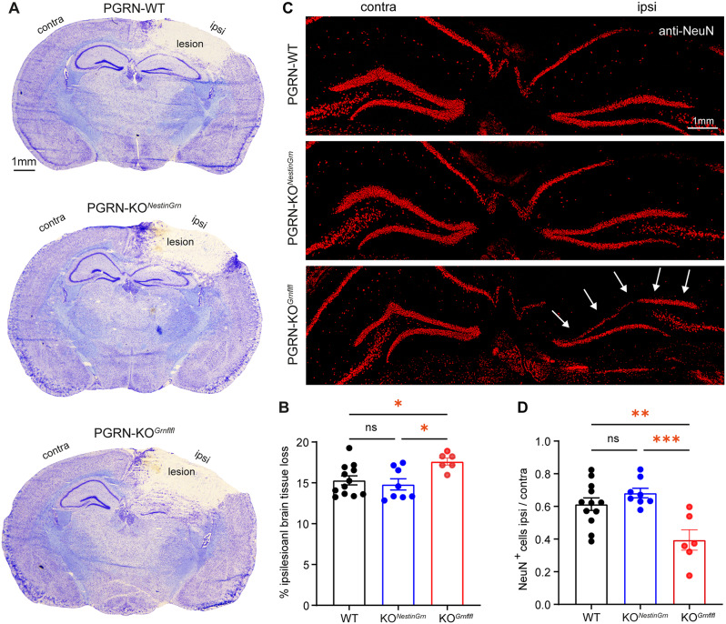
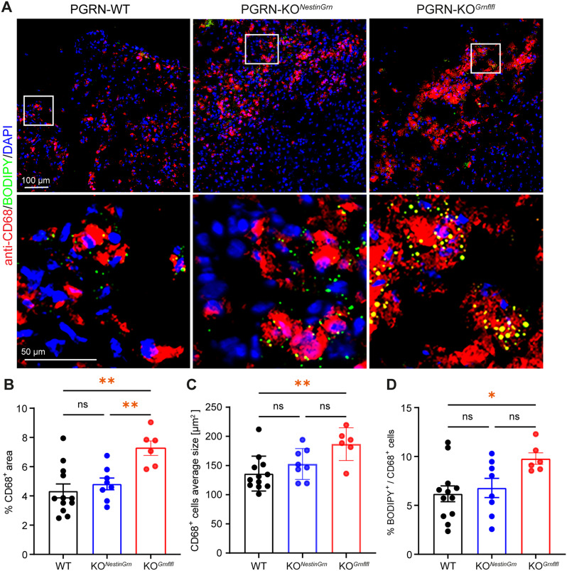
Progranulin (PGRN), which is produced in neurons and microglia, is a neurotrophic and anti-inflammatory glycoprotein. Human loss-of-function mutations cause frontotemporal dementia, and PGRN knockout (KO) mice are a model for dementia. In addition, PGRN KO mice exhibit severe phenotypes in models of traumatic or ischemic central nervous system (CNS) disorders, including traumatic brain injury (TBI). It is unknown whether restoration of progranulin expression in neurons (and not in microglia) might be sufficient to prevent excessive TBI-evoked brain damage. To address this question, we generated mice with Nestin-Cre-driven murine PGRN expression in a PGRN KO line (PGRN-KONestinGrn) to rescue PGRN in neurons. PGRN expression analysis in primary CNS cell cultures from naïve mice and in (non-) injured brain tissue from PGRN-KONestinGrn revealed expression of PGRN in neurons but not in microglia. After experimental TBI, examination of the structural brain damage at 5 days post-injury (dpi) showed that the TBI-induced loss of brain tissue and hippocampal neurons was exacerbated in PGRN-KOGrnflfl mice (PGRN knockout with the mGrn fl-STOP-fl allele, Cre-negative), as expected, whereas the tissue damage in PGRN-KONestinGrn mice was similar to that in PGRN-WT mice. Analysis of CD68+ immunofluorescent microglia and Cd68 mRNA expression showed that excessive microglial activation was rescued in PGRN-KONestinGrn mice, and the correlation of brain injury with Cd68 expression suggested that Cd68 was a surrogate marker for excessive brain injury caused by PGRN deficiency. The results show that restoring neuronal PGRN expression was sufficient to rescue the exacerbated neuropathology of TBI caused by PGRN deficiency, even in the absence of microglial PGRN. Hence, endogenous microglial PGRN expression was not essential for the neuroprotective or anti-inflammatory effects of PGRN after TBI in this study.

Keywords: CD68; Microglia; Neuroinflammation; Neuropathology; Neuroprotection; Progranulin; Therapy; Traumatic brain injury.


Conflict of interest statement

The authors declare no competing interests.

Figures

Fig. 1
Fig. 1
Progranulin expression in PGRN-KONestinGrn mice. (A, B) Grn mRNA expression in cell cultures from adult brains enriched for non-microglial neural cells (neurons and astrocytes) and microglia. The data are presented as box/scatter plots. The line is the median, the box shows the interquartile range, the whiskers show the minimum to maximum, and the scatters show the individual results of 7–9 mice per genotype, measured in duplicate. (C) Images of the contralesional hippocampal GCL (Bregma − 1.86 mm) showing anti-PGRN immunostaining and nuclear counterstaining by DAPI in PGRN-WT, PGRN-KONestinGrn, and PGRN-KOGrnflfl mice. Genotype-dependent PGRN expression in cells with microglial or neuronal morphology indicates PRGN expression in GCL neurons but not in microglia in PGRN-KONestinGrn mice. No specific signal was detected in PGRN-KOGrnflfl mice. (D) Triple fluorescence staining of the ipsilesional cortex (Bregma − 1.86 mm) at 5 dpi using anti-PGRN/anti-CD68/DAPI revealed PGRN expression in CD68+ microglia in PGRN-WT mice but not in PGRN-KONestinGrn or PGRN-KOGrnflfl mice. Arrows point to cells shown at higher magnification
Fig. 2
Fig. 2
Exacerbated structural brain damage in PGRN-KOGrnflfl mice is rescued in PGRN-KONestinGrn mice. (A) Representative images of cresyl violet-stained coronal brain sections from PGRN-WT, PGRN-KONestinGrn, and PGRN-KOGrnflfl mice at 5 dpi (Bregma − 1.86 mm). (B) Relative brain tissue loss (% of ipsilesional hemisphere) calculated from 16 consecutive sections (Bregma + 3.14 mm to − 4.36 mm). PGRN-WT and PGRN-KONestinGrn mice exhibit attenuated brain tissue loss compared to PGRN-KOGrnflfl mice. (C) Images of coronal brain sections (Bregma − 1.86 mm) showing anti-NeuN immunostaining in the hippocampal dentate gyrus of the contra- and ipsilesional hemispheres at 5 dpi. Arrows indicate exacerbated loss of GCL neurons in the ipisilesional suprapyramidal blade of PGRN-KOGrnflfl mice. (D) The number of NeuN+ neurons in the GCL was higher in PGRN-WT and PGRN-KONestinGrn mice than in PGRN-KOGrnflfl mice. The data points represent individual mice, PGRN-WT (n = 12), PGRN-KONestinGrn (n = 8) and PGRN-KOGrnflfl (n = 6), and the data are expressed as the mean ± SEM. One-way ANOVA with Holm–Šidák post hoc correction, *p < 0.05**p < 0.01, ***p < 0.001, ns = not significant
Fig. 3
Fig. 3
TBI-induced excessive Cd68 gene expression in PGRN-KOGrnflfl mice is attenuated in PGRN-KONestinGrn mice. (A-G) Gene expression analysis of inflammation-associated markers in ipsi- or contralesional brain tissues (Bregma + 0.64 mm to − 2.86 mm) was performed via RT‒qPCR. (A) Grn expression is highest in PGRN-WT mice and is reduced in PGRN-KONestinGrn and PGRN-KOGrnflfl mice. Grn expression in ipsilesional brain tissue was mildly greater in PGRN-KONestinGrn mice than in PGRN-KOGrnflfl mice but was substantially lower than that in WT mice because PGRN upregulation after TBI mainly occurs in microglia. (B-G) Column charts showing the mRNA expression of inflammation-associated markers (Aif1, Gfap, Cd68, Lyz2, Spp1, and Tnf). (D) Augmented ipsilesional Cd68 gene expression in PGRN-KOGrnflfl mice was partially rescued in PGRN-KONestinGrn mice. Two outliers were identified by Rout’s test and excluded (F). The data points represent individual mice, PGRN-WT (n = 12), PGRN-KONestinGrn (n = 8) and PGRN-KOGrnflfl (n = 6), and the data are expressed as the mean ± SEM. One-way ANOVA with Holm–Šidák post hoc correction, *p < 0.05, **p < 0.01, ***p < 0.001, **** p < 0.0001, ns = not significant
Fig. 4
Fig. 4
Cd68 gene expression is a surrogate marker of microglial PGRN deficiency and associated brain damage. Linear regression analyses to assess the relationship between Cd68 expression and ipsilesional brain tissue loss in PGRN-WT (n = 12), PGRN-KONestinGrn (n = 8), and PGRN-KOGrnflfl (n = 6) mice. Scatter plots with regression lines, 95% confidence intervals, correlation coefficients (r2), and p values are shown. The data points represent individual mice
Fig. 5
Fig. 5
Iba1+ microglial infiltration of the injured brain in PGRN-KOGrnflfl mice is reduced in PGRN-KONestinGrn mice. (A) Scheme illustrating the positions of the imaged brain regions. (B) Double immunostaining of the ipsi- and contralesional cortex at 5 dpi (Bregma − 1.86 mm) using anti-Iba1 and anti-GFAP antibodies showing TBI-evoked activation of microglia and astrocytes. (C) Higher magnification images of the boxed regions. (D, E) Column plots showing Iba1+ and GFAP+ counts in the ipsi- and contralesional cortices. The number of ipsilesional Iba1+ microglia was lower in PGRN-KONestinGrn mice than in PGRN-KOGrnflfl mice, whereas the number of contralesional GFAP+ astrocytes was greater in PGRN-KONestinGrn and PGRN-KOGrnflfl mice than in PGRN-WT mice. (F, G) Column plots showing the total anti-Iba1+ or anti- GFAP+ immunostained areas in the ipsi- and contralesional cortices. The area of ipsilesional Iba1+ immunostaining was smaller in PGRN-WT mice and PGRN-KONestinGrn mice than in PGRN-KOGrnflfl mice, whereas the immunostaining area of GFAP+ astrocytes was not different between genotypes. The data are expressed as the mean ± SEM, and the values are shown for individual mice, PGRN-WT (n = 12), PGRN-KONestinGrn (n = 8) and PGRN-KOGrnflfl (n = 6). One-way ANOVA (D, contra, E, ipsi), Brown-Forsythe ANOVA test (D, ipsi) and Kruskal‒Wallis test (E, contra) and post hoc Holm–Šidák, Dunnett T3 or Dunn’s corrections were used to calculate p values (*p < 0.05, **p < 0.01, ns = not significant)
Fig. 6
Fig. 6
Excessive CD68+ microglial infiltration of the injured brain in PGRN-KOGrnflfl mice is reduced in PGRN-KONestinGrn mice. (A) Triple-fluorescence staining of ipsilesional cortex at 5 dpi (Bregma − 1.86 mm) with anti-CD68/BODIPY/DAPI showed fewer CD68+ microglia in PGRN-WT and PGRN-KONestinGrn than in PGRN-KOGrnflfl mice and partial overlap of the BODIPY signal with that in CD68+ microglia. (B) Column plots showing reduced area occupancy by CD68+ microglia in PGRN-WT and PGRN-KONestinGrn mice compared to PGRN-KOGrnflfl mice. (C) Column plots showing reduced average size of CD68+ microglia in PGRN-WT mice compared to PGRN-KOGrnflfl mice. Differences between PGRN-KONestinGrn mice and PGRN-KOGrnflfl mice were statistically not significant (p = 0.07). (D) Column plots showing that the percentage of CD68+ microglia colabeled with BODIPY had the highest mean percentage in PGRN-KOGrnflfl mice, a reduced mean percentage in PGRN-KONestinGrn and a significant reduction in PGRN-WT mice. The data are expressed as the mean ± SEM, and the values from individual mice are shown, PGRN-WT (n = 12), PGRN-KONestinGrn (n = 8) and PGRN-KOGrnflfl (n = 6). One-way ANOVA and post hoc Holm–Šidák corrections were used to calculate p values (*p < 0.05, **p < 0.01, ns = not significant)

References

    1. Petkau TL, Neal SJ, Orban PC, MacDonald JL, Hill AM, Lu G, et al. Progranulin expression in the developing and adult murine brain. J Comp Neurol. 2010;518(19):3931–47. - PubMed
    1. Neill T, Buraschi S, Goyal A, Sharpe C, Natkanski E, Schaefer L, et al. EphA2 is a functional receptor for the growth factor progranulin. J Cell Biol. 2016;215(5):687–703. - PMC - PubMed
    1. Thomasen PB, Salasova A, Kjaer-Sorensen K, Woloszczuková L, Lavický J, Login H, et al. SorCS2 binds progranulin to regulate motor neuron development. Cell Rep. 2023;42(11):113333. - PubMed
    1. Altmann C, Hardt S, Fischer C, Heidler J, Lim H-Y, Häussler A, et al. Progranulin overexpression in sensory neurons attenuates neuropathic pain in mice: role of autophagy. Neurobiol Dis. 2016;96:294–311. - PubMed
    1. Tang W, Lu Y, Tian QY, Zhang Y, Guo FJ, Liu GY, et al. The growth factor progranulin binds to TNF receptors and is therapeutic against inflammatory arthritis in mice. Science. 2011;332(6028):478–84. - PMC - PubMed