. 2024 Oct 10;15(1):359.
doi: 10.1186/s13287-024-03932-9.

Connexin 43 regulates intercellular mitochondrial transfer from human mesenchymal stromal cells to chondrocytes

Affiliations

Connexin 43 regulates intercellular mitochondrial transfer from human mesenchymal stromal cells to chondrocytes

Rebecca M Irwin et al. Stem Cell Res Ther. .

Abstract

Background: The phenomenon of intercellular mitochondrial transfer from mesenchymal stromal cells (MSCs) has shown promise for improving tissue healing after injury and has potential for treating degenerative diseases like osteoarthritis (OA). Recently MSC to chondrocyte mitochondrial transfer has been documented, but the mechanism of transfer is unknown. Full-length connexin 43 (Cx43, encoded by GJA1) and the truncated, internally translated isoform GJA1-20k have been implicated in mitochondrial transfer between highly oxidative cells, but have not been explored in orthopaedic tissues. Here, our goal was to investigate the role of Cx43 in MSC to chondrocyte mitochondrial transfer. In this study, we tested the hypotheses that (a) mitochondrial transfer from MSCs to chondrocytes is increased when chondrocytes are under oxidative stress and (b) MSC Cx43 expression mediates mitochondrial transfer to chondrocytes.

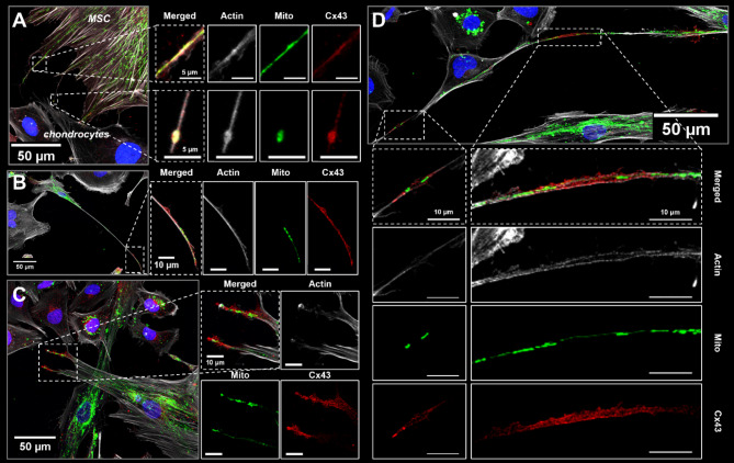
Methods: Oxidative stress was induced in immortalized human chondrocytes using tert-Butyl hydroperoxide (t-BHP) and cells were evaluated for mitochondrial membrane depolarization and reactive oxygen species (ROS) production. Human bone-marrow derived MSCs were transduced for mitochondrial fluorescence using lentiviral vectors. MSC Cx43 expression was knocked down using siRNA or overexpressed (GJA1 + and GJA1-20k+) using lentiviral transduction. Chondrocytes and MSCs were co-cultured for 24 h in direct contact or separated using transwells. Mitochondrial transfer was quantified using flow cytometry. Co-cultures were fixed and stained for actin and Cx43 to visualize cell-cell interactions during transfer.

Results: Mitochondrial transfer was significantly higher in t-BHP-stressed chondrocytes. Contact co-cultures had significantly higher mitochondrial transfer compared to transwell co-cultures. Confocal images showed direct cell contacts between MSCs and chondrocytes where Cx43 staining was enriched at the terminal ends of actin cellular extensions containing mitochondria in MSCs. MSC Cx43 expression was associated with the magnitude of mitochondrial transfer to chondrocytes; knocking down Cx43 significantly decreased transfer while Cx43 overexpression significantly increased transfer. Interestingly, GJA1-20k expression was highly correlated with incidence of mitochondrial transfer from MSCs to chondrocytes.

Conclusions: Overexpression of GJA1-20k in MSCs increases mitochondrial transfer to chondrocytes, highlighting GJA1-20k as a potential target for promoting mitochondrial transfer from MSCs as a regenerative therapy for cartilage tissue repair in OA.

Keywords: Arthritis; Cx43; GJA1; GJA1-20k; Gap junctions; MSCs; Osteoarthritis; Regenerative medicine.


Conflict of interest statement

The authors declare that they have no competing interests.

Figures

Fig. 1
Fig. 1
t-BHP-induced oxidative stress in chondrocytes increases incidence of mitochondrial transfer from MSCs. (A) Chondrocytes were cultured alone with or without t-BHP (12 µM, 24 h) to quantify mitochondrial polarization and ROS production (i) and prior to co-culture with MSCs (ii). (B) Ratio of depolarized to polarized mitochondria in chondrocytes after t-BHP or FCCP stimulation (n = 5–6). (C) Histograms represent ROS quantification for chondrocytes stimulated with t-BHP (pooled across n = 5 replicates). (D) Quantification of percentage of chondrocytes that were positive for CellROX green staining on flow cytometry (n = 5). (E) Z-projection of co-culture with chondrocytes and MSCs (red: chondrocytes; green: MSC mitochondria; blue: nuclei). Quadrant gate showing representative flow data for single color controls (F), co-cultures with unstimulated chondrocytes (G), and co-cultures with t-BHP stimulated chondrocytes (H). (I) Quantification of mitochondrial transfer from co-cultures (n = 7). *p < 0.05, **p < 0.01, ***p < 0.001, ****p < 0.0001
Fig. 2
Fig. 2
Direct cell-cell contacts are critical for MSC-Chondrocyte mitochondrial transfer. (A) Chondrocytes (GFP mitochondrial fluorescence) and MSCs (mCherry mitochondrial fluorescence) were co-cultured for 24 h in either 2D contact on cell culture plates or with no direct contact using transwell inserts. (B) Single color controls were used to establish gating strategy for identifying transfer events. Representative data for co-cultures from contact (C) and transwell (D) co-cultures. (E) Quantification of mitochondrial transfer from flow cytometry (n = 3, groups not sharing a letter are significantly different, p < 0.01). (F-L) Z-projections from 2D contact co-cultures captured using confocal imaging with MSCs (GFP mitochondria) and unlabeled chondrocytes (green: MSC mitochondria; grey: actin; blue: nuclei). MSC actin-positive filapodial extensions contain mitochondria (white asterisks) and MSC mitochondria localized in adjacent chondrocytes (white arrowheads)
Fig. 3
Fig. 3
Cx43 staining is enriched along actin cellular extensions containing mitochondria in MSCs. MSC actin cellular extensions were observed in contact co-cultures with chondrocytes after 24 h (mtGFP MSCs; unstained chondrocytes). Cells were stained for Cx43 and actin (phalloidin). Merged z-projections and individual channels are shown (green: MSC mitochondria; red: Cx43; grey: actin; blue: nuclei). (A) MSC mitochondria and Cx43 staining are found within MSC filapodial extensions. (B, C) Cx43 staining is enriched at the terminal end of MSC filapodial extensions that are actin-positive, and (D) Cx43 localizes to apparent cell-cell junction between a MSC and chondrocyte
Fig. 4
Fig. 4
GJA1 siRNA knocks down Cx43 expression in MSCs and decreases incidence of mitochondrial transfer. (A) Western blot analysis of full length Cx43 (GJA1-43k) and the truncated isoform GJA1-20k in MSCs after GJA1 siRNA treatment. Cx43 (B) and GJA1-20k (C) expression were normalized to α-tubulin (n = 3). (D) Relative isoform expression was calculated as the ratio of GJA1-20k to Cx43 (n = 3). (E-F) Representative images of Cx43 immunofluorescence for control (Stealth RNAi) and GJA1 siRNA treated MSCs. (G-H) Representative data from flow cytometry analyses of co-cultures. (I) Quantification of mitochondrial transfer events from co-cultures using flow cytometry (n = 8). Full-length western blot for quantification is presented in Supplementary Fig. 2. *p < 0.05, **p < 0.01, ***p < 0.001, ****p < 0.0001
Fig. 5
Fig. 5
GJA1-20k overexpression in MSCs increases mitochondrial transfer and MSC GJA1-20k expression correlates with transfer incidence. Western blot analysis of full length Cx43 (GJA1-43k) and the truncated isoform GJA1-20k in MSCs after lentiviral transduction (LacZ (control), GJA1+, GJA1-20k+). Cx43 (B) and GJA1-20k (C) expression were normalized to α-tubulin (n = 3). (D) Relative isoform expression was calculated as the ratio of GJA1-20k to Cx43 (n = 3). (E-G) Representative images of Cx43 immunofluorescence for LacZ (control), GJA1+, and GJA1-20k + MSCs. (H-J) Representative data from flow cytometry analyses of co-cultures. (K) Quantification of mitochondrial transfer events from co-cultures using flow cytometry (n = 8). (L-M) Linear correlations for normalized protein expression of full-length Cx43 (GJA1-43k) and GJA1-20k with normalized incidence of mitochondrial transfer (groups normalized to their respective controls). Groups not sharing a letter are significantly different (p < 0.05). Full-length western blot for quantification is presented in Supplementary Fig. 3. Uncropped western blot in (A) can be found in Supplementary Figs. 8 and 9
Fig. 6
Fig. 6
Hypothesized role of Cx43 and GJA1-20k in MSC to chondrocyte mitochondrial transfer. Our previous work implicated gap junction communication in MSC to chondrocyte mitochondrial transfer (i, Fahey + Scientific Reports 2022). Results presented here confirm the importance of Cx43 in mediating this phenomenon and reveal distinct roles for the full-length protein and the N-terminal truncated isoform (GJA1-20k). Given our imaging data and previous evidence that GJA1-20k increases trafficking of mitochondria along actin cytoskeletal elements (ii, iii) and recruits actin to organize cell trafficking pathways (iv), we propose that GJA1-20k likely aids in mitochondrial transfer by facilitating transport of mitochondria from MSCs to adjacent cells through mobilization of mitochondria via TNTs (ii), the formation of actin cellular extensions (iv), and/or packaging of mitochondria within microvesicles (iii). Future studies are warranted to further investigate these mechanisms

Update of

References

    1. GBD 2017 Disease and Injury Incidence and Prevalence Collaborators. Global, regional, and national incidence, prevalence, and years lived with disability for 354 diseases and injuries for 195 countries and territories, 1990–2017: a systematic analysis for the Global Burden of Disease Study 2017. Lancet [Internet]. 2018 Nov 10 [cited 2021 Sep 15];392(10159):1789. Available from: /pmc/articles/PMC6227754/. - PMC - PubMed
    1. DS C, PM R [Internet]. CJ V. Pharmaceutical therapy for osteoarthritis. 2012 May [cited 2021 Sep 15];4(5 Suppl). https://pubmed.ncbi.nlm.nih.gov/22632707/ - PubMed
    1. B G, FP T, MI H, RP G, MG C. KB F. Chondroprotection and the prevention of osteoarthritis progression of the knee: a systematic review of treatment agents. Am J Sports Med [Internet]. 2015 Mar 17 [cited 2021 Sep 15];43(3):734–44. https://pubmed.ncbi.nlm.nih.gov/24866892/ - PubMed
    1. Wei P, Bao R. Intra-Articular Mesenchymal Stem Cell Injection for Knee Osteoarthritis: Mechanisms and Clinical Evidence. Int J Mol Sci [Internet]. 2022 Jan 1 [cited 2024 Apr 24];24(1). https://pubmed.ncbi.nlm.nih.gov/36613502/ - PMC - PubMed
    1. Delco ML, Goodale M, Talts JF, Pownder SL, Koff MF, Miller AD, et al. Integrin α10β1-Selected mesenchymal stem cells mitigate the progression of Osteoarthritis in an equine Talar Impact Model. Am J Sports Med. 2020;48(3):612–23. - PubMed

LinkOut - more resources