. 2024 Oct 10;9(1):26.
doi: 10.1186/s40850-024-00214-y.

Regeneration of the caudal fin of the evolutionary ancient tropical gar Atractosteus tropicus

Affiliations

Regeneration of the caudal fin of the evolutionary ancient tropical gar Atractosteus tropicus

Alberto J Ríos-Flores et al. BMC Zool. .

Abstract

Background: The tropical gar (Atractosteus tropicus), a member of the Lepisosteidae family, is native to regions extending from southeastern Mexico to southern Costa Rica. This species serves as a unique bridge between tetrapods and teleosts due to its phylogenetic position, slow evolutionary rate, dense genetic map, gene similarities with humans, and ease of laboratory cultivation. As a taxonomic sister group to teleosts like the zebrafish (Danio rerio), known for its high regenerative capacity, it remains unclear whether the tropical gar shares a similar ability for regeneration.

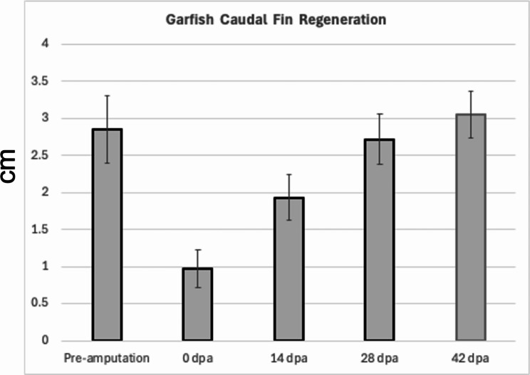
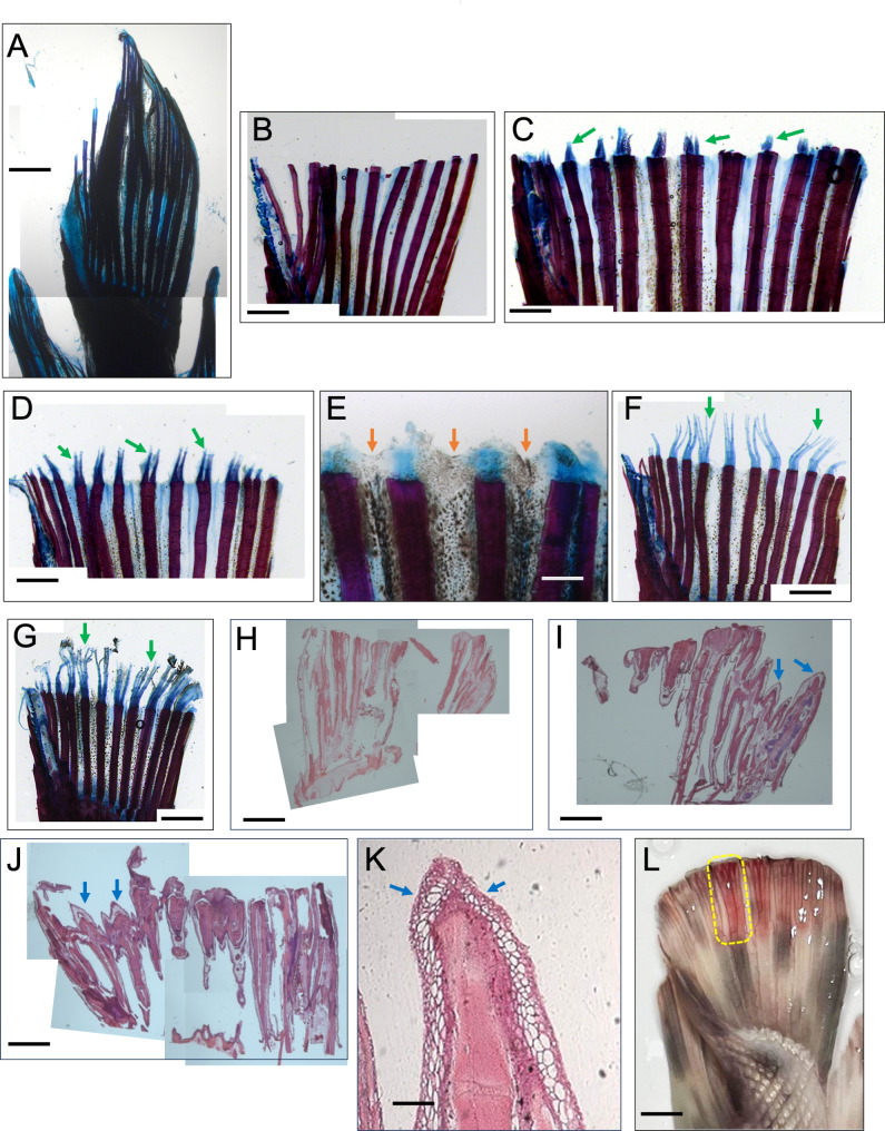
Results: This study aims to elucidate the caudal fin regeneration process in tropical gar through skeletal and histological staining methods. Juvenile specimens were observed over a two-month period, during which they were fed brine shrimp, and anesthetized with 1% eugenol for caudal fin amputation. Samples were collected at various days post-amputation (dpa). Alcian blue and alizarin red staining were used to highlight skeletal regeneration, particularly the formation of new cartilage, while histological staining with hematoxylin and eosin was performed to observe tissue regeneration at the amputation site.

Conclusions: The findings reveal a remarkable ability for caudal fin regeneration in juvenile tropical gar. Given the Garfish evolutionary relationship with teleosts, this opens new avenues for research into tissue regeneration across different groups of Actinopterygii.

Keywords: Caudal fin regeneration; EvoDevo; Garfish; Tropical gar.


Conflict of interest statement

The authors declare no competing interests.

Figures

Fig. 1
Fig. 1
The subject of study is the Garfish Atractosteus tropicus. (A) The geographical location of Atractosteus tropicus, it’s in the East and the South of Mexico in four states (underlined) and four different Central American countries. (B) here is shown one of the juvenile individuals use it in this study. (C) Phylogenetic tree using Vps18 protein sequences (highly conserved intracellular traffic protein) to show the position of the Garfish among several animals. (D) Phylogenetic tree using cytochrome b protein sequences to show the phylogenetic position of the Garfish Atractosteus tropicus in their taxonomically family Lepisosteidae. The map in (A) was obtained from Wikimedia commons according to the CC BY-SA 4.0 Attribution-sharealike 4.0 international (https://creativecommons.org/licenses/by-sa/4.0/#ref-appropriate-credit), and slightly modified for our purposes with Adobe Photoshop 2021
Fig. 2
Fig. 2
Comparison of caudal fin regeneration between zebrafish (Danio rerio) and the Garfish (Atractosteus tropicus). (A) adult individual of zebrafish. (B) juvenile Garfish. (CK) different time points pre- and post-amputation of the caudal fin. It takes about one month to regenerate a whole caudal fin in zebrafish. (KR) Caudal regeneration in Garfish at different time points for about two months. (MN) Initial steps of caudal regeneration in Garfish from 0 to 13 dpa (days post-amputation). At 4 dpa we detected the first visible signs of tissue regrowth that progress fast as could be seen at 13 dpa (see insets in white dotted boxes in O and P). Scale bars are (A) 5 mm (B) 20 mm (D) 5 mm (L) 10 mm (M) 2 mm. Figures MP are shown twice enlarged at the bottom of the figure
Fig. 3
Fig. 3
Caudal fin regeneration in 15 Garfishes. In this graph means values are shown for the Pre-amputation caudal fin and the regrowth velocity for the caudal fin after amputation as measured at 0, 14, 28 and 42 dpa. Error bars show Standard Deviation
Fig. 4
Fig. 4
Garfish skeletal bone regeneration. Cartilage and Bone staining’s in control fish (A) and at different post-amputation times (BG). (A and B) control and 0 dpa respectively. (C) At 4 dpa the regrowth of the cartilage is readily apparent (see green arrows). (D) At 6 dpa the rays cartilage is growing steadily and showing signs of branching (see green arrows). (E) In the close-up at 7 dpa we could observe that the tissue between growing between the rays (see orange arrows), pigment cells are clearly visible. (F and G) At 9 and 12 dpa respectively, the rays cartilage has grown considerable, and rays show several branches (see green arrows). We also used HE staining at (H) 2 dpa. (I) 5 dpa. (J) 8 dpa, and (K) close up at 9 dpa where new chondrocytes can be observed at the tip of the new formed rays. (L) At simple view on the stereomicroscope, at 16 dpa, a regenerated tail shows branched rays (See yellow dotted box). Scales bars are (A) 3 mm, (B, D, F, G - J) 3 mm, (C) 2 mm, (E) 0.5 mm, (K) 0,2 mm, and (L) 3 mm
Fig. 5
Fig. 5
Phylogenetic model showing the evolutionary position of Garfish between some other related animals. The Geological periods are represented by colored boxes at one side of the diagram from the Cambrian to the Quaternary period

References

    1. Wiley EO. The phylogeny and biogreography of fossil and recent Gars (Actinopterygii: Lepisosteidae). Misc. Publ Univ Kans Nat Hist Mus. 1976;64:1–111.
    1. Márquez-Couturier G, Vázquez-Navarrete CJ. (s.f.) Estado de arte de la biología y cultivo de pejelagarto (Atractosteus tropicus) AP AGRO Productividad. Vol. 8 Núm. 3 2015: Mayo-Junio.
    1. Instituto Mexicano de Investigación en Pesca y Acuacultura Sustentable. Subdelegaciones de Pesca-SAGARPA. Pejelagarto, distribución geográfica 2024. https://www.gob.mx/imipas/acciones-y-programas/acuacultura-pejelagarto
    1. Bussing W. Peces De las aguas continentales de Costa Rica. San José, Costa Rica: Editorial UCR; 2002.
    1. Espinosa-Perez H, Gaspar-Dilanar M, Fuentes-Mata P. Listados faunísticos de México III. Los peces dulceacuícolas mexicanos. México, D.F. Instituto De Biologia. Universidad Nacional Autónoma de México; 1993. p. 98.

LinkOut - more resources