. 2024 Sep;43(5):648-662.
doi: 10.23876/j.krcp.23.106. Epub 2024 Jan 2.

Maternal exposure to airborne particulate matter during pregnancy and lactation induces kidney injury in rat dams and their male offspring: the role of vitamin D in pregnancy and beyond

Affiliations

Maternal exposure to airborne particulate matter during pregnancy and lactation induces kidney injury in rat dams and their male offspring: the role of vitamin D in pregnancy and beyond

Min Hwa Son et al. Kidney Res Clin Pract. 2024 Sep.

Abstract

Background: Little is known about the transgenerational effects of maternal exposure to fine particulate matter (PM2.5) on offspring kidney health. This study investigated the effect of maternal administration of PM2.5 or PM2.5 with vitamin D during pregnancy and lactation on renal injury in rat dams and their offspring.

Methods: Nine pregnant Sprague-Dawley rats received oral administration of normal saline, airborne PM2.5, or PM2.5 with vitamin D from gestational day 11 to postpartum day 21. Kidneys of rat dams (n = 3 for each group) and their male offspring (n = 5 for each group) were taken for analysis on postpartum or postnatal day 21.

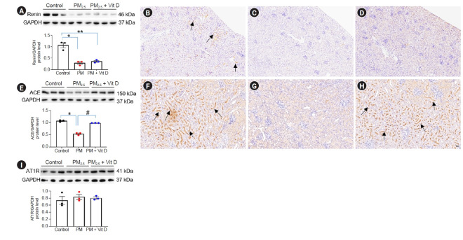
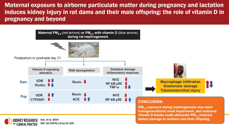
Results: Maternal PM2.5 exposure increased glomerular damage, tubulointerstitial injury, and cortical macrophage infiltration in both dams and pups; all increases were attenuated by vitamin D administration. In dam kidneys, PM2.5 increased the protein expression of vitamin D receptor (VDR), klotho, and tumor necrosis factor-α; vitamin D lessened these changes. The expressions of renin, nuclear factor erythroid 2-related factor 2 (Nrf2), and nuclear factor-kappa B (NF-κB) p50 decreased in rat dams exposed to PM2.5. In offspring kidneys, exposure to maternal PM2.5 reduced the expression of VDR, renin, angiotensin-converting enzyme (ACE), Nrf2, and NF-κB p50, but increased cytochrome P450 24A1 expression. Maternal vitamin D administration with PM2.5 enhanced VDR, ACE, and NF-κB p50 activities in pup kidneys.

Conclusion: PM2.5 exposure during nephrogenesis may exert transgenerational renal impairment, and maternal vitamin D intake could attenuate PM2.5-induced kidney damage in mothers and their offspring.

Keywords: Fetal development; Kidney diseases; Particulate matter; Renin-angiotensin system; Vitamin D.


Conflict of interest statement

Conflicts of interest

All authors have no conflicts of interest to declare.

Figures

Figure 1.
Figure 1.. Renal histological alterations in rat dams (A–E) and their male pups (F–J).
Dams and pups exposed to PM2.5 showed prominent renal structural changes (B, dams; G, pups) (arrows, sclerotic glomeruli; arrowheads, degenerated or dilated tubules) when compared to corresponding control kidneys (A, dams; F, pups). Maternal vitamin D (Vit D) supplementation ameliorated renal injury in both dams (C) and pups (H), as evidenced by the scores of glomerular damage (D, dams; I, pups) and tubulointerstitial injury (E, dams; J, pups). (A–C and F–H) Hematoxylin and eosin stain, ×100; bar = 100 µm (n = 3–5 for each group). PM, particulate matter. *p < 0.05, control vs. PM2.5 or PM2.5 + Vit D; #p < 0.05, PM2.5 vs. PM2.5 + Vit D.
Figure 2.
Figure 2.. Intrarenal vitamin D (Vit D) signaling in rat dams.
(A–D) Vitamin D receptor (VDR), (E–H) klotho, (I) CYP27B1, and (J) CYP24A1. Western blot analysis revealed increases in VDR (A) and klotho (E) activities in PM2.5-exposed dam kidneys as compared to the controls, which were reversed by Vit D treatment. Compared to the control rats (B), VDR was more strongly expressed in the dilated tubular epithelial cells and macula densa of the juxtaglomerular apparatus in PM2.5-exposed dam kidneys (C; arrows). Klotho was also highly expressed in almost all major tubular segments and glomeruli of PM2.5-exposed kidneys (F, controls; G, PM2.5-exposed kidneys, arrows). Maternal Vit D supplementation lessened both VDR (D) and klotho (H) expressions in the renal cortex. (B–D and F–H) Immunohistochemistry; ×200, bar = 50 µm (n = 3 for each group). No difference was observed in CYP27B1 (I) and CYP24A1 (J) expressions among the groups (*p < 0.05, control vs. PM2.5; #p < 0.05, PM2.5 vs. PM2.5 + Vit D). CYP, cytochrome P450 mixed-function oxidase; PM, particulate matter.
Figure 3.
Figure 3.. Intrarenal vitamin D (Vit D) signaling in rat pups.
(A–D) Vitamin D receptor (VDR), (E–H) CYP24A1, (I) klotho, and (J) CYP27B1. Western blot analysis revealed a decrease in VDR (A) and an increase in CYP24A1 (E) in pups from PM2.5-exposed dams as compared to the controls. VDR activity was restored by maternal Vit D supplementation. VDR expression was mainly observed in tubular epithelial cells of control pup kidneys (B, arrows), while it was markedly diminished in kidneys from pups exposed to maternal PM2.5 (C). Maternal Vit D intake restored intrarenal VDR expression in pups at the weaning (D, arrows). While CYP24A1 activity was rarely detected in control pup kidneys (F), it was highly expressed in the brush border of the tubules in pup kidneys exposed to maternal PM2.5 (G, arrows). Maternal Vit D intake did not reduce CYP24A1 expression (H, arrows). (B–D and F–H) Immunohistochemistry; ×200, bar = 50 µm (n = 3 for each group). No difference was observed in the intrarenal activities of klotho (I) and CYP27B1 (J) among the groups (*p < 0.05, control vs. PM2.5; #p < 0.05, PM2.5 vs. PM2.5 + Vit D). CYP, cytochrome P450 mixed-function oxidase; PM, particulate matter.
Figure 4.
Figure 4.. Intrarenal renin-angiotensin system signaling in rat dams.
(A–D) Renin, (E) angiotensin-converting enzyme (ACE), and (F) angiotensin II type 1 receptor (AT1R). Western blot analysis revealed a decrease in renin activity in PM2.5-exposed dam kidneys as compared to the controls (A). Immunohistochemically, renin expression was found in juxtaglomerular and interstitial cells in control kidneys (B, arrows). PM2.5 exposure reduced intrarenal renin expression (C), and vitamin D (Vit D) supplementation did not restore renin activity in the kidney (A and D). (B–D) Immunohistochemistry; ×200, bar = 50 µm (n = 3 for each group). No difference was observed in the intrarenal activities of ACE (E) and AT1R (F) among the groups (*p < 0.05, control vs. PM2.5 or PM2.5 + Vit D). PM, particulate matter.
Figure 5.
Figure 5.. Intrarenal renin-angiotensin system signaling in rat pups.
(A–D) Renin, (E–H) angiotensin-converting enzyme (ACE), and (I) angiotensin II type 1 receptor (AT1R). Western blot analysis revealed decreases in renin (A) and ACE (E) activities in pup kidneys exposed to maternal PM2.5 as compared to the controls. Intrarenal ACE activity was restored by maternal vitamin D (Vit D) supplementation (E). Renin expression was found in juxtaglomerular and interstitial cells in control kidneys (B, arrows) whereas it was rarely detected in pup kidneys of maternal PM2.5 exposure (C) and those of maternal Vit D intake with PM2.5 exposure (D). ACE was also widely expressed throughout the brush border of the proximal tubules in control pup kidneys (F, arrows). Maternal PM2.5 exposure remarkably reduced intrarenal ACE expression (G), while Vit D intake by dams restored ACE expression in the pup kidney (H, arrows). (B–D and F–H) Immunohistochemistry; ×200, bar = 50 µm (n = 3 for each group). No difference was observed in the intrarenal activity of AT1R among the groups (*p < 0.05, control vs. PM2.5; **p < 0.05, control vs. PM2.5 + Vit D; #p < 0.05, PM2.5 vs. PM2.5 + Vit D). PM, particulate matter.
Figure 6.
Figure 6.. Renal oxidative stress and inflammation in rat dams.
(A–D) Nuclear factor erythroid 2-related factor 2 (Nrf2), (E–H) nuclear factor-kappa B (NF-κB) p50, and (I–L) tumor necrosis factor alpha (TNF-α). Western blot analysis revealed decreases in Nrf2 (A) and NF-κB p50 (E) and an increase in TNF-α (I) in PM2.5-exposed dam kidneys as compared to the controls. Vitamin D (Vit D) supplementation reduced intrarenal TNF-α activity (I). While Nrf2 and NF-κB p50 expressions were well observed in control dam kidneys (B and F; arrows, respectively), they were weakly detected in PM2.5-exposed kidneys (C and G, respectively). Vit D intake did not restore their expressions (D and H). Intrarenal TNF-α expression was clearly detected throughout all tubular segments and glomeruli in the PM2.5 group (K, arrows) as compared to the control group (J) and the PM2.5 with Vit D group (L). (B–D, F–H, and J–L) Immunohistochemistry; ×200, bar = 50 µm (n = 3 for each group). *p < 0.05, control vs. PM2.5; **p < 0.05, control vs. PM2.5 + Vit D; #p < 0.05, PM2.5 vs. PM2.5 + Vit D. PM, particulate matter.
Figure 7.
Figure 7.. Renal oxidative stress and inflammation in rat pups.
(A–D) Nuclear factor erythroid 2-related factor 2 (Nrf2), (E–H) nuclear factor-kappa B (NF-κB) p50, (I) tumor necrosis factor alpha (TNF-α). Western blot analysis revealed decreases in Nrf2 (A) and NF-κB p50 (E) activities in pups from PM2.5-exposed dams as compared to the controls. Maternal vitamin D (Vit D) supplementation restored intrarenal NF-κB p50 activity in pup kidneys (E). While the Nrf2 and NF-κB p50 expressions were well observed in control pup kidneys (B and F; arrows, respectively), they were weakly detected in pup kidneys of maternal PM2.5 exposure (C and G, respectively). Maternal Vit D intake did not restore Nrf2 expression in pups (D) whereas it increased the intrarenal expression of NF-κB p50 (H, arrows). (B–D and F–H) Immunohistochemistry; ×200, bar = 50 µm (n = 5 for each group). No difference was observed in the intrarenal activity of TNF-α (I) among the groups (*p < 0.05, control vs. PM2.5; #p < 0.05, PM2.5 vs. PM2.5 + Vit D). PM, particulate matter.
Figure 8.
Figure 8.. Renal macrophage infiltration in dams (A–D) and pups (E–H).
Rat dams and their pups exposed to PM2.5 showed prominent cortical macrophage infiltration mainly in tubulointerstitium (B, dams; F, pups; arrows, respectively) as compared to the corresponding control rat kidneys (A, dams; E, pups). Vitamin D (Vit D) supplementation in both dams (C and D) and pups (G and H). (A–C and E–G) Immunohistochemistry; ×400, bar = 20 µm (n = 3–5 for each group). PM, particulate matter. *p < 0.05, control vs. PM2.5; #p < 0.05, PM2.5 vs. PM2.5 + Vit D.
None

Similar articles

Cited by

References

    1. Yin P, Brauer M, Cohen AJ, et al. The effect of air pollution on deaths, disease burden, and life expectancy across China and its provinces, 1990-2017: an analysis for the Global Burden of Disease Study 2017. Lancet Planet Health. 2020;4:e386–e398. - PMC - PubMed
    1. Bowe B, Xie Y, Li T, Yan Y, Xian H, Al-Aly Z. Particulate matter air pollution and the risk of incident CKD and progression to ESRD. J Am Soc Nephrol. 2018;29:218–230. - PMC - PubMed
    1. Oh J, Ye S, Kang DH, Ha E. Association between exposure to fine particulate matter and kidney function: results from the Korea National Health and Nutrition Examination Survey. Environ Res. 2022;212:113080. - PubMed
    1. Li X, Huang S, Jiao A, et al. Association between ambient fine particulate matter and preterm birth or term low birth weight: an updated systematic review and meta-analysis. Environ Pollut. 2017;227:596–605. - PubMed
    1. Morales-Rubio RA, Alvarado-Cruz I, Manzano-León N, et al. In utero exposure to ultrafine particles promotes placental stress-induced programming of renin-angiotensin system-related elements in the offspring results in altered blood pressure in adult mice. Part Fibre Toxicol. 2019;16:7. - PMC - PubMed

LinkOut - more resources