

Comparison of micro-flow imaging and contrast-enhanced ultrasonography in assessing segmental congestion after right living donor liver transplantation
- PMID: 39390717
- PMCID: PMC11532526
- DOI: 10.14366/usg.24114
Comparison of micro-flow imaging and contrast-enhanced ultrasonography in assessing segmental congestion after right living donor liver transplantation
Abstract
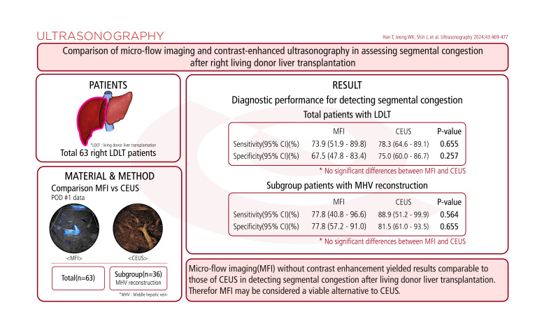
Purpose: This study aimed to determine whether micro-flow imaging (MFI) offers diagnostic performance comparable to that of contrast-enhanced ultrasonography (CEUS) in detecting segmental congestion among patients undergoing living donor liver transplantation (LDLT).
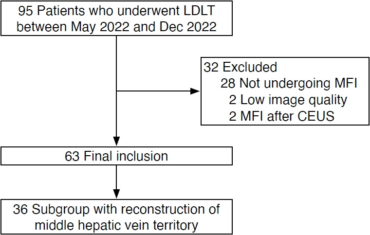
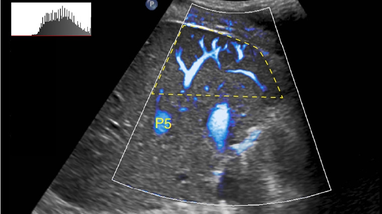
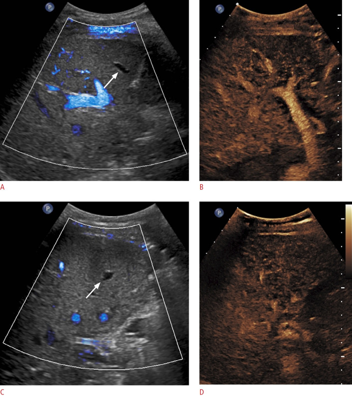
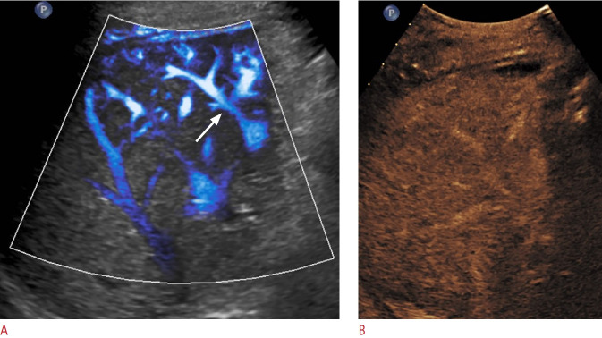
Methods: Data from 63 patients who underwent LDLT between May and December 2022 were retrospectively analyzed. MFI and CEUS data collected on the first postoperative day were quantified. Segmental congestion was assessed based on imaging findings and laboratory data, including liver enzymes and total bilirubin levels. The reference standard was a postoperative contrast-enhanced computed tomography scan performed within 2 weeks of surgery. Additionally, a subgroup analysis examined patients who underwent reconstruction of the middle hepatic vein territory.
Results: The sensitivity and specificity of MFI were 73.9% and 67.5%, respectively. In comparison, CEUS demonstrated a sensitivity of 78.3% and a specificity of 75.0%. These findings suggest comparable diagnostic performance, with no significant differences in sensitivity (P=0.655) or specificity (P=0.257) between the two modalities. Additionally, early postoperative laboratory values did not show significant differences between patients with and without congestion. The subgroup analysis also indicated similar diagnostic performance between MFI and CEUS.
Conclusion: MFI without contrast enhancement yielded results comparable to those of CEUS in detecting segmental congestion after LDLT. Therefore, MFI may be considered a viable alternative to CEUS.
Keywords: Contrast-enhanced ultrasound; Diagnostic accuracy; Hepatic congestion; Living donor liver transplant; Micro-flow imaging.
Conflict of interest statement
Woo Kyoung Jeong serves as Editor for the Ultrasonography, but has no role in the decision to publish this article. All remaining authors have declared no conflicts of interest.
Figures
Similar articles
-
The Preliminary Application of Simultaneous Display of Contrast-Enhanced Ultrasound and Micro-Flow Imaging Technology in the Diagnosis of Hepatic Tumors.J Ultrasound Med. 2023 Feb;42(3):729-737. doi: 10.1002/jum.16111. Epub 2022 Oct 11. J Ultrasound Med. 2023. PMID: 36217761
-
A preliminary study on the diagnostic value of contrast-enhanced ultrasound and micro-flow imaging for detecting blood flow signals in breast cancer patients.Gland Surg. 2024 Nov 30;13(11):2098-2106. doi: 10.21037/gs-24-264. Epub 2024 Nov 26. Gland Surg. 2024. PMID: 39678425 Free PMC article.
-
CEUS: a new imaging approach for postoperative vascular complications after right-lobe LDLT.World J Gastroenterol. 2009 Aug 7;15(29):3670-5. doi: 10.3748/wjg.15.3670. World J Gastroenterol. 2009. PMID: 19653347 Free PMC article.
-
Diagnostic performance of contrast-enhanced ultrasound in diagnosing hepatic artery occlusion after liver transplantation: A systematic review and meta-analysis.Clin Transplant. 2023 Nov;37(11):e15070. doi: 10.1111/ctr.15070. Epub 2023 Jul 3. Clin Transplant. 2023. PMID: 37398993
-
Diagnostic accuracy of contrast-enhanced ultrasound in assessing the therapeutic response to radio frequency ablation for liver tumors: systematic review and meta-analysis.Surg Endosc. 2018 Apr;32(4):2067-2075. doi: 10.1007/s00464-017-5903-4. Epub 2017 Dec 21. Surg Endosc. 2018. PMID: 29270801
References
-
- Lee SG, Park KM, Hwang S, Lee YJ, Kim KH, Ahn CS, et al. Adult-to-adult living donor liver transplantation at the Asan Medical Center, Korea. Asian J Surg. 2002;25:277–284. - PubMed
-
- Kim KW, Kim TK, Kim SY, Kim MJ, Park MS, Lee MG, et al. Doppler sonographic abnormalities suggestive of venous congestion in the right lobe graft of living donor liver transplant recipients. AJR Am J Roentgenol. 2007;188:W239–W245. - PubMed
-
- Lee S, Park K, Hwang S, Lee Y, Choi D, Kim K, et al. Congestion of right liver graft in living donor liver transplantation. Transplantation. 2001;71:812–814. - PubMed
-
- Inomata Y, Uemoto S, Asonuma K, Egawa H. Right lobe graft in living donor liver transplantation. Transplantation. 2000;69:258–264. - PubMed
-
- Ko GY, Sung KB, Yoon HK, Kim JH, Song HY, Seo TS, et al. Endovascular treatment of hepatic venous outflow obstruction after living-donor liver transplantation. J Vasc Interv Radiol. 2002;13:591–599. - PubMed
LinkOut - more resources
Full Text Sources
