

Huang Lian Jie Du decoction attenuated colitis via suppressing the macrophage Csf1r/Src pathway and modulating gut microbiota
- PMID: 39391314
- PMCID: PMC11464287
- DOI: 10.3389/fimmu.2024.1375781
Huang Lian Jie Du decoction attenuated colitis via suppressing the macrophage Csf1r/Src pathway and modulating gut microbiota
Abstract
Introduction: Ulcerative colitis, a subtype of chronic inflammatory bowel disease (IBD), is characterized by relapsing colonic inflammation and ulcers. The traditional Chinese herbal formulation Huang Lian Jie Du (HLJD) decoction is used clinically to treat diarrhea and colitis. However, the mechanisms associated with the effects of treatment remain unclear. This study aims to elucidate the molecular mechanistic effects of HLJD formulation on colitis.
Methods: Chronic colitis in mice was induced by adding 1% dextran sulfate sodium (DSS) to their drinking water continuously for 8 weeks, and HLJD decoction at the doses of 2 and 4 g/kg was administered orally to mice daily from the second week until experimental endpoint. Stool consistency scores, blood stool scores, and body weights were recorded weekly. Disease activity index (DAI) was determined before necropsy, where colon tissues were collected for biochemical analyses. In addition, the fecal microbiome of treated mice was characterized using 16S rRNA amplicon sequencing.
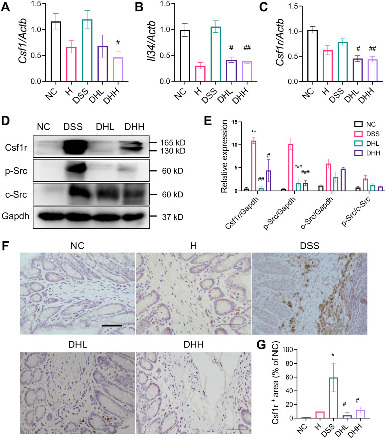
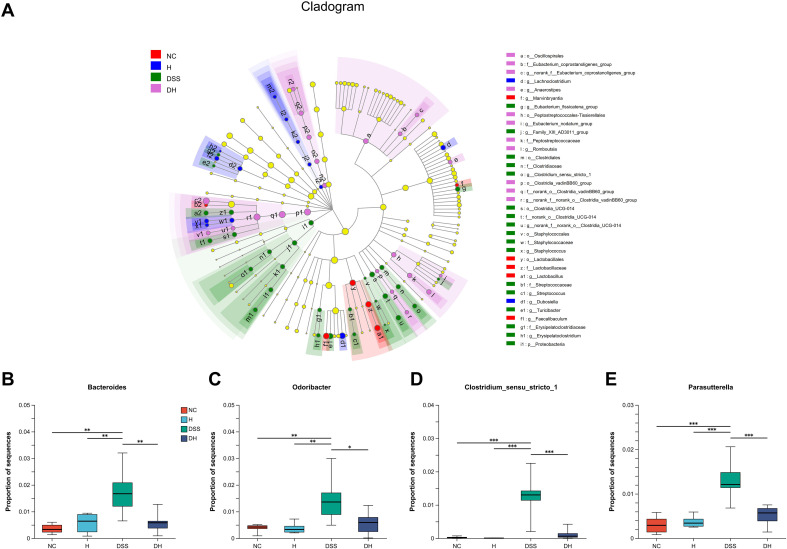
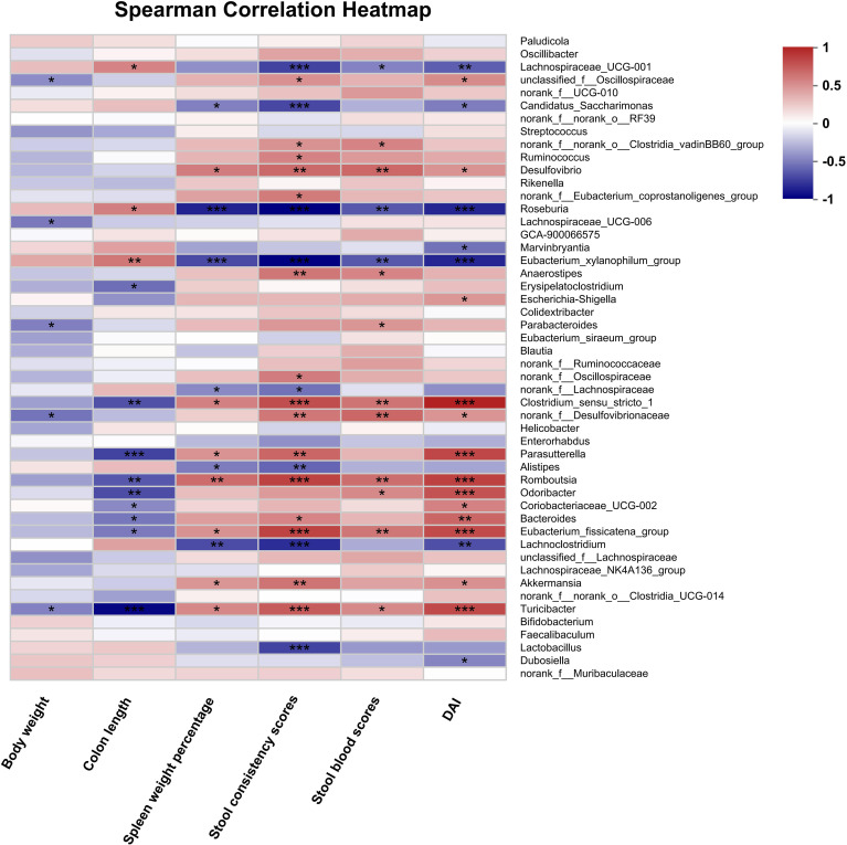
Results: HLJD decoction at doses of 2 and 4 g/kg relieved DSS-induced chronic colitis in mice by suppressing inflammation through compromised macrophage activity in colonic tissues associated with the colony-stimulating factor 1 receptor (Csf1r)/Src pathway. Furthermore, the HLJD formula could modify the gut microbiota profile by decreasing the abundance of Bacteroides, Odoribacter, Clostridium_sensu_stricto_1, and Parasutterella. In addition, close correlations between DAI, colon length, spleen weight, and gut microbiota were identified.
Discussion: Our findings revealed that the HLJD formula attenuated DSS-induced chronic colitis by reducing inflammation via Csf1r/Src-mediated macrophage infiltration, as well as modulating the gut microbiota profile.
Keywords: Csf1r; Src; colitis; gut microbiota; macrophage.
Copyright © 2024 Su, Liu, Zheng, Wu, Keng, Zhang and Li.
Conflict of interest statement
The authors declare that the research was conducted in the absence of any commercial or financial relationships that could be construed as a potential conflict of interest.
Figures


Similar articles
-
Huang-Lian-Jie-Du decoction alleviates depressive-like behaviors in dextran sulfate sodium-induced colitis mice via Trem2/Dap12 pathway.J Ethnopharmacol. 2023 Oct 28;315:116658. doi: 10.1016/j.jep.2023.116658. Epub 2023 May 30. J Ethnopharmacol. 2023. PMID: 37263316
-
Patchoulene epoxide mitigates colitis and hepatic damage induced by dextran sulfate sodium by regulating the colonic microbiota and purine metabolism.Front Immunol. 2025 Feb 14;16:1509114. doi: 10.3389/fimmu.2025.1509114. eCollection 2025. Front Immunol. 2025. PMID: 40028318 Free PMC article.
-
Qingchang Huashi Formula attenuates DSS-induced colitis in mice by restoring gut microbiota-metabolism homeostasis and goblet cell function.J Ethnopharmacol. 2021 Feb 10;266:113394. doi: 10.1016/j.jep.2020.113394. Epub 2020 Sep 15. J Ethnopharmacol. 2021. PMID: 32941971
-
Changdiqing decoction (CDQD) ameliorates colitis via suppressing inflammatory macrophage activation and modulating gut microbiota.Phytomedicine. 2025 Jul 25;143:156856. doi: 10.1016/j.phymed.2025.156856. Epub 2025 May 14. Phytomedicine. 2025. PMID: 40412060
-
Preventive Effects of Pyungwi-san against Dextran Sulfate Sodium- and Clostridium difficile-Induced Inflammatory Bowel Disease in Mice.Int J Mol Sci. 2019 Dec 16;20(24):6346. doi: 10.3390/ijms20246346. Int J Mol Sci. 2019. PMID: 31888274 Free PMC article.
Cited by
-
Huang-Lian-Jie-Du decoction alleviates cognitive deficits in Alzheimer's disease model 5xFAD mice by inhibiting Trem2/Dap12 signaling pathway.Chin Med. 2025 Apr 15;20(1):50. doi: 10.1186/s13020-025-01098-x. Chin Med. 2025. PMID: 40234956 Free PMC article.
-
Huang Lian Jie Du Decoction Prevents Chronic Alcoholic Encephalopathy and Improves Gut Microbiota Imbalance in Mice.Mol Neurobiol. 2025 Oct;62(10):13665-13677. doi: 10.1007/s12035-025-05151-6. Epub 2025 Jun 25. Mol Neurobiol. 2025. PMID: 40555894
-
The Role of Gut Microbiota on Intestinal Fibrosis in Inflammatory Bowel Disease and Traditional Chinese Medicine Intervention.J Inflamm Res. 2025 May 7;18:5951-5967. doi: 10.2147/JIR.S504827. eCollection 2025. J Inflamm Res. 2025. PMID: 40357383 Free PMC article. Review.
References
MeSH terms
Substances
LinkOut - more resources
Full Text Sources
Research Materials
Miscellaneous