Review
. 2024 Sep 9:5:xtae027.
doi: 10.1093/femsmc/xtae027. eCollection 2024.

Enterococcal-host interactions in the gastrointestinal tract and beyond

Affiliations
Review

Enterococcal-host interactions in the gastrointestinal tract and beyond

Wiam Abdalla Mo Madani et al. FEMS Microbes. .

Abstract

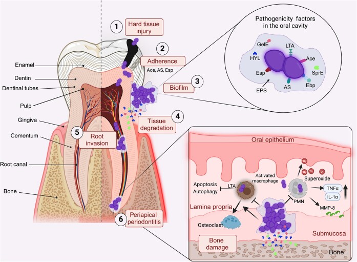
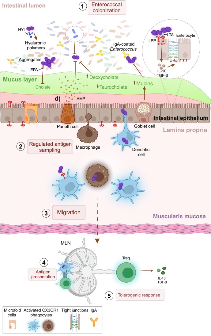
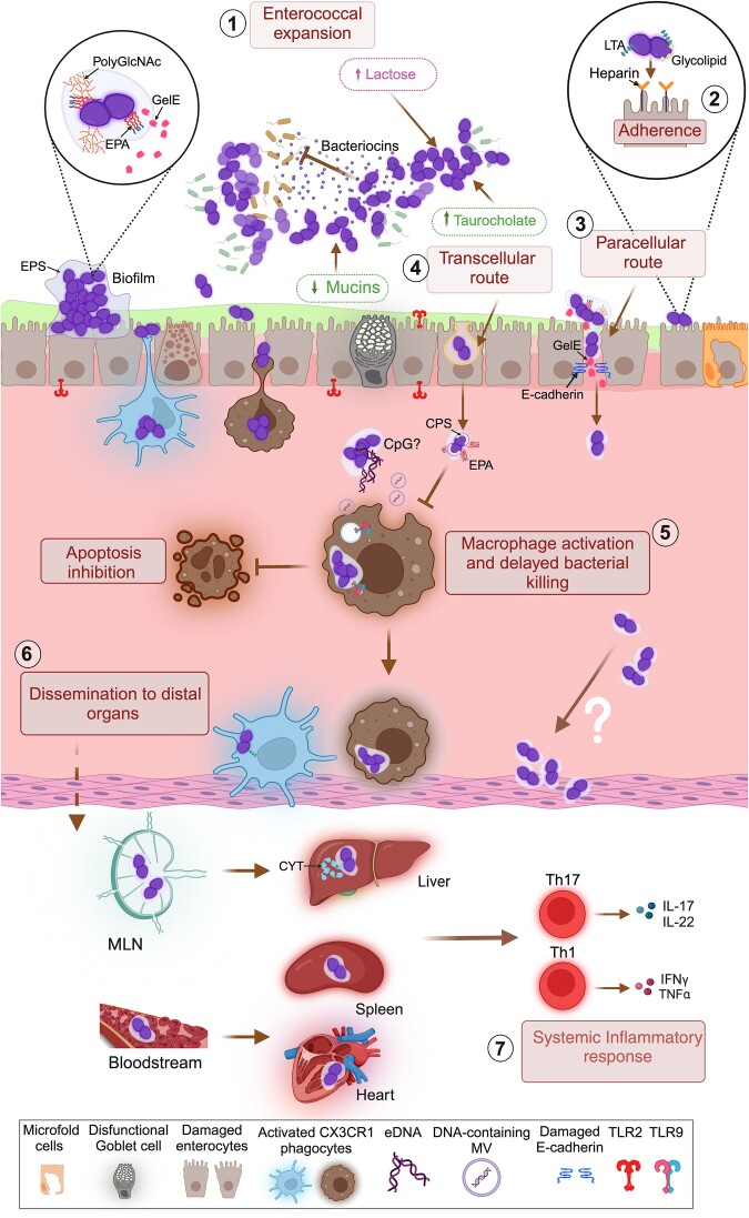
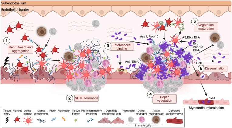
The gastrointestinal tract (GIT) is typically considered the natural niche of enterococci. However, these bacteria also inhabit extraintestinal tissues, where they can disrupt organ physiology and cause life-threatening infections. Here, we discuss how enterococci, primarily Enterococcus faecalis, interact with the intestine and other host anatomical locations such as the oral cavity, heart, liver, kidney, and vaginal tract. The metabolic flexibility of these bacteria allows them to quickly adapt to new environments, promoting their persistence in diverse tissues. In transitioning from commensals to pathogens, enterococci must overcome harsh conditions such as nutrient competition, exposure to antimicrobials, and immune pressure. Therefore, enterococci have evolved multiple mechanisms to adhere, colonize, persist, and endure these challenges in the host. This review provides a comprehensive overview of how enterococci interact with diverse host cells and tissues across multiple organ systems, highlighting the key molecular pathways that mediate enterococcal adaptation, persistence, and pathogenic behavior.

Keywords: commensal to pathogen transition; dysbiosis; enterococcal-host interactions; enterococci; gastrointestinal tract; inter-organ dissemination.


Conflict of interest statement

None declared.

Figures

Figure 1.
Figure 1.
Dynamic interactions of enterococci with oral tissues. The oral cavity includes various structures, with teeth being a prominent component. Each tooth comprises both hard and soft tissues; enamel is the calcified tissue covering the dentin in the crown of the tooth. Dentin, located just beneath the enamel, contains microscopic tubules called dentinal tubules. The cementum is a connective tissue covering the tooth root, attaching to the periodontal ligament. The soft tissues also include the pulp, which contains connective tissue, blood vessels, and nerves. (1) Hard tissue injury by mechanical and chemical insults allows enterococcal colonization in sensitive areas like the dentin, pulp tissue, and root canal system. (2) Enterococcus faecalis adheres to dentin via specific adhesins such as Ace (collagen-binding protein), Esp (enterococcal surface protein), and AS (aggregation substance), which is likely enhanced by salivary proteins. (3) Long-term colonization is promoted by biofilm formation on tooth surfaces and within the root canal system, where enterococci are encased by extracellular polymeric substances (EPS) comprised of proteins, fatty acids, and exopolysaccharides. Several enterococcal pathogenicity factors are produced within biofilms, including Esp, which promotes cell retention in this structure, Ebp (Endocarditis- and biofilm-associated pilus protein), and the cell-wall-anchored lipoteichoic acid (LTA). Moreover, enzymes like hyaluronidase (HYL), gelatinase (GelE), and sortase E (SprE) aid the dissolution of dentin’s mineral fraction (4), promoting calcified biofilm formation and penetration into dentinal tubules that facilitate further invasion and division within the root canals (5). The presence of E. faecalis in persistent apical periodontitis highlights its capacity to evade immune responses in the periapical region (6). Indeed, in this location, Enterococci can inhibit phagocytosis and autophagy in macrophages via LTA while enhancing macrophage differentiation into osteoclasts, resulting in increased bone resorption. Polymorphonuclear leukocytes (PMNs) can migrate into the root canal and respond to E. faecalis by producing extracellular superoxide, upregulating proinflammatory factors such as IL-1α and tumor necrosis factor-α (TNF-α), and releasing matrix metalloprotease (MMP-8), which collectively contributes to tissue damage. Enterococcal biofilms increase tolerance to antimicrobials and immune clearance by PMNs and macrophages, further promoting bone degradation. Hence, this sustained infection and inflammation in periapical tissues can lead to bone destruction and tooth loss.
Figure 2.
Figure 2.
Homeostatic interactions between enterococci and the intestine. Under eubiotic conditions, the long-term colonization (1) of enterococci in the gut lumen may be facilitated by multiple processes: the ability to utilize various gut nutrients, such as hyaluronic acid polymers, via hyaluronidases (HYL); the capacity to form biofilms/aggregates in the intestinal mucus layer, counteracting intestinal peristalsis; and the expression of EPA (enterococcal polysaccharide antigen) in the enterococcal cell wall that helps protect bacterial cells against bile acids like cholate. Transient expansion of enterococci in the intestine is limited by several factors, including the secretion of antimicrobial peptides (AMP) by Paneth cells, competition for nutrients with other commensal bacteria, elevated levels of deoxycholate bile acid, and active mucus secretion by Goblet cells. Enterococci on the luminal side can also be coated by IgA secreted by specialized gut plasma cells, preventing their binding to the mucus layer. If reaching the intestinal epithelium, E. faecalis lipoteichoic acid (LTA) and/or lipoproteins (LPP) may be recognized by Toll-like receptor (TLR)-2, which can trigger the production of anti-inflammatory cytokines, such as transforming growth factor β (TGF-β) and interleukin (IL)-10, while maintaining tight junction integrity between enterocytes. (2) Below the intestinal epithelium, lamina propria dendritic cells and macrophages constantly sample the gut lumen and phagocytose enterococci via TLR2 expressed on these myeloid cells. (3) Dendritic cells then migrate to the mesenteric lymph node (MLN), where they present enterococcal antigens to naïve T cells (4). This process can lead to the generation of regulatory T cells (Treg), which produce IL-10 and TGF-β, and thus orchestrate tolerance to these gut commensal bacteria (5).
Figure 3.
Figure 3.
Dysbiosis triggers enterococcal egress from the intestine. Disruption of intestinal homeostasis (dysbiosis) can lead to enterococcal overgrowth/dominance. (1) This expansion may be promoted by biofilm formation, where the extracellular polymeric substance (EPS), partly formed by poly N-acetylglucosamine (polyGlcNAc)-containing polymers, and the enterococcal polysaccharide antigen (EPA) enhance adherence to surfaces and resistance to antimicrobials and immune responses. Bacteriocin secretion and the ability to metabolize diverse carbon sources (lactose) may also provide a fitness advantage to E. faecalis. During dysbiosis, taurocholate levels increase (bile acid), which enterococci can tolerate. Reduced production of antimicrobial peptides, IgA, and mucus further allows E. faecalis to adhere to the epithelial layer. (2) Moreover, bacterial glycolipids and lipoteichoic acid (LTA), as well as colonic heparin/heparan sulfate receptors, may facilitate enterococcal attachment to epithelial cells. These conditions compromise the epithelial barrier, promoting enterococcal egress from the intestinal lumen (gut translocation) via two routes: paracellularly (3), where enterococcal cells adhered to the epithelial layer release gelatinases (GelE) that damage tight junction E-cadherin, allowing bacterial migration between intestinal epithelial cells, and transcellularly (4), where E. faecalis might pass across the barrier via direct endocytosis by enterocytes. (5) Translocated enterococci can be engulfed by lamina propria phagocytes, including CX3CR1 macrophages and dendritic cells. Enterococcal membrane vesicles (MV) containing DNA or extracellular DNA (eDNA) present in biofilms, which may contain CpG motifs, could be recognized by Toll-like receptor (TLR)-9 in lamina propria macrophages. Once phagocytosed, E. faecalis can persist/proliferate inside macrophages, especially when taken up as aggregates that inhibit apoptosis in these myeloid cells. Enterococcus faecalis can also inhibit phagocytosis through the expression of factors such as EPA and capsule (CPS), which may encase bacterial factors recognized by phagocytes. Enterococcal persistence within phagocytes leads to their activation and production of pro-inflammatory cytokines, aiding their dissemination (6) to mesenteric lymph nodes (MLN) and/or the bloodstream, facilitating spread to distal organs such as the liver, spleen, and heart. It is unknown whether extracellular E. faecalis can also egress the lamina propria or whether it needs phagocyte activity. Once in distal organs, such as the liver, exotoxins like cytolysin (CYT) may promote disease progression by lysing hepatocytes. (7) Enterococci can also trigger a systemic inflammatory response characterized by Th1 and Th17 cell polarization and the production of pro-inflammatory cytokines, such as interleukin (IL)-17, IL-22, interferon-gamma (IFNγ), and tumor necrosis factor-α (TNF-α).
Figure 4.
Figure 4.
Interactions between host cells and enterococci during IE. The endothelium, located in the inner layer of the heart chambers, valves, and blood vessels, becomes susceptible to IE when injured or perturbed, exposing subendothelial matrix components like fibrinogen, collagen, laminin, and fibronectin. (1) Platelets aggregate at the damaged endothelium, inducing cytokine production and upregulating tissue factor and fibrinogen. Active platelets produce fibrin, which stimulates further aggregation and serves as a scaffold for additional platelets and immune cells, promoting inflammation and forming a sterile NBTE vegetation (2). Bloodstream enterococci may bind to pre-existing NBTE or directly to damaged or inflamed cardiac surfaces, overcoming shear stress from high blood flow via different mechanisms (3): The von Willebrand Factor (vWF) mediates bacterial binding to the endocardium, acting as a bridge between bacteria and host cells; Ace and EfbA (fibronectin-binding protein) promote binding to subendothelial components such as collagen, laminin, and fibronectin; and enterococcal Asc-10 and Asa1 enhance attachment to fibrin by increasing cell hydrophobicity, all leading to enterococcal colonization and biofilm formation within the nascent septic vegetation (4). The maturation of the infected vegetation involves cycles of fibrin-platelet deposition, with bacteria stimulating platelet aggregation. Enterococcus faecalis interacts with platelets through envelope components such as AS, Ebp pili, and ElrA, promoting further aggregation and the extracellular release of adenosine diphosphate (ADP) from dense platelet δ-granules. Esp promotes enterococcal cell-cell aggregation by binding to cell envelope components like lipoteichoic acid (LTA) and, together with Asc-10 and EfbA, influences enterococcal biofilm maturation and growth. This septic vegetation growth and biofilm formation protect enterococci from antimicrobials to immune cells (5). GelE-mediated degradation of fibrin-rich matrices facilitates bacterial spread from vegetations to adjacent or distal sites (6). Enterococcus faecalis may invade endothelial cells via receptor (clathrin)-mediated endocytosis, further contributing to disease progression. Myocardial microlesions can result from the spread of infection, with the disulfide bond-forming protein A (DsbA) being necessary, enhancing cell death and suppressing the immune response.

Similar articles

Cited by

  • A new approach to osteoarthritis: gut microbiota.
    Aydin M, Avci GA, Yilmaz UI, Avci E. Aydin M, et al. Rev Assoc Med Bras (1992). 2025 May 2;71(3):e20241528. doi: 10.1590/1806-9282.20241528. eCollection 2025. Rev Assoc Med Bras (1992). 2025. PMID: 40332266 Free PMC article.
  • Cross-organ metabolite production and consumption in healthy and atherogenic conditions.
    Bae H, Jung S, Le J, Tamburini I, Kim J, Wang E, Song WS, Choi W, Jang KH, Kang T, Lopez ML, Ramirez C, Mohanty I, Kelly ME, Kim J, Kim R, Park SH, Baek J, Mendez B, Petrus P, de Aguiar Vallim TQ, Nicholas DA, Smith Q, Lee G, Seldin M, Jang C. Bae H, et al. Cell. 2025 Aug 7;188(16):4441-4455.e16. doi: 10.1016/j.cell.2025.05.001. Epub 2025 May 27. Cell. 2025. PMID: 40436017

References

    1. Aas JA, Paster BJ, Stokes LN et al. Defining the normal bacterial flora of the oral cavity. J Clin Microbiol. 2005;43:5721–32. - PMC - PubMed
    1. Agudelo Higuita NI, Huycke MM. Enterococcal disease, epidemiology, and implications for treatment. In: Gilmore MS, Clewell DB, Ike Y, Shankar N (eds), Enterococci: From Commensals to Leading Causes of Drug Resistant Infection. Boston: Massachusetts Eye and Ear Infirmary, 2014.
    1. Akbari Aghdam M, Soroush Barhaghi MH, Aghazadeh M et al. Virulence genes in biofilm producer Enterococcus faecalis isolates from root canal infections. Cell Mol Biol (Noisy-le-grand). 2017;63:55–59. - PubMed
    1. Al-Ahmad A, Muller N, Wiedmann-Al-Ahmad M et al. Endodontic and salivary isolates of Enterococcus faecalis integrate into biofilm from human salivary bacteria cultivated in vitro. J Endod. 2009;35:986–91. - PubMed
    1. Alghamdi F, Shakir M. The influence of Enterococcus faecalis as a dental root canal pathogen on endodontic treatment: a systematic review. Cureus. 2020;12:e7257. - PMC - PubMed

LinkOut - more resources