. 2024 Oct 2;15(42):17691-17701.
doi: 10.1039/d4sc03555k. Online ahead of print.

Dual-ligand PROTACS mediate superior target protein degradation in vitro and therapeutic efficacy in vivo

Affiliations

Dual-ligand PROTACS mediate superior target protein degradation in vitro and therapeutic efficacy in vivo

Yong Chen et al. Chem Sci. .

Abstract

Proteolysis targeting chimeras (PROTACs) are revolutionizing the drug development landscape due to their unique ability to selectively degrade disease-associated proteins. Conventional PROTACs are bivalent entities that induce ubiquitination and subsequent proteolysis of a chosen protein of interest (POI) by forming a ternary complex with an E3 ligase. We hypothesized that dual-ligand PROTACs, featuring two copies each of a POI ligand and an E3 ligase ligand, would facilitate the formation of high-avidity, long-lived ternary complexes inside cells, thereby increasing POI degradation potency. To this end, we developed a convergent synthesis route, using l-aspartic acid as a building block for homodimer synthesis, followed by copper-catalyzed azide-alkyne cycloaddition (CuAAC) to conjugate both dimers through a flexible linker. Dual-ligand PROTACs achieved up to a tenfold increase in degradation efficiency and a hundredfold increase in cytotoxicity in vitro across various cancer cell lines compared to their single-ligand counterparts. Furthermore, dual-ligand PROTACs sustain prolonged protein degradation, up to 60 hours after pulsing and washout. In vivo, in a mouse tumor model, the superior therapeutic activity of dual ligand PROTACs was observed.


Conflict of interest statement

The authors declare no competing financial interest.

Figures

Fig. 1
Fig. 1. Rationale for the design of dual-ligand PROTACs. (A) Conceptual representation of conventional single-ligand PROTACs and dual-ligand PROTACs. (B) The fate of ternary complex mediated by single-ligand PROTACs and dual-ligand PROTACs. According to our hypothesis, dual-ligand PROTACs can increase both the half-life and conformational landscape of the ternary complex. In the context of dual-ligand PROTACs-mediated ternary complex dynamic equilibrium, a protein that dissociates from ligand 2a possesses the potential to be promptly re-engaged by an adjacent identical ligand 2b. This interaction facilitates the formation of a novel ternary complex or enables the re-establishment of the initial ternary complex configuration. Conversely, in the scenario of single-ligand PROTACs systems, the disengagement of the protein from its ligand typically results in its subsequent diffusion away from the complex, resulting in the collapse of the original ternary complex structure.
Fig. 2
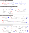
Fig. 2. Convergent synthesis of dual ligand BET PROTACs. (A) Chemical structure of the ligands used for the design of BET PROTACs. (B) General synthetic strategy to assemble the dual-ligand BET PROTACs. (C) Chemical structure of BET PROTACs based on pomalidomide for recruitment of the CRBN E3 ligase: dBET1 (single-ligand PROTACs), trivalent PROTACs (2J1P, 1J2P) and dual-ligand PROTACs (2J2P) (D) chemical structure of BET PROTACs based on VHL-ligand for recruitment of the VLH E3 ligase: MZ1 (single-ligand PROTACs), trivalent PROTACs (2J1V, 1J2V) and dual-ligand PROTACs (2J2V).
Fig. 3
Fig. 3. Detailed synthesis of dual-ligand and trivalent BET PROTACs. (A) Synthesis of 2J-N3 and 1J-N3. (B) Synthesis of 2P-alkyne and 1P-alkyne. (C) Synthesis of 2V-alkyne and 1V-alkyne. (D) Final PROTAC assembly by CuAAC conjugation.
Fig. 4
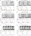
Fig. 4. Dual-ligand PROTACs induce robust and prolonged target protein degradation in vitro. HEK293 cells were treated with PROTACs recruiting the VHL E3 ligase (panels 1) and the CRBN E3 ligase (panels 2) for 4 h. Subsequently, cells were either lysed (A and B) or washed and cultured in the fresh medium until the desired time point of lysis (C). BRD2, BRD3 and BRD4 protein levels were measured by western blot (panels a for a representative image) and quantification was done by optical density integration of the protein bands (panels b). (A) BET protein degradation was screened for all synthesized PROTACs and commercial single-ligand PROTACs at a concentration of 0.1, 1, 10 μM, respectively with DMSO as the control vehicle. Experiments were repeated as independent replicates. Statistical analysis by one-way ANOVA. (**: p < 0.01, ***: p < 0.001, ****: p < 0.0001) (B) BET protein degradation by single- and dual-ligand PROTACs was tested over a 1 pM–10 μM concentration range (vehicle-only control contained DMSO). EC50 values were calculated by curve fitting of three independent experiments. (C) BET protein degradation by single- and dual-ligand PROTACs over time after wash-out. Experiments were performed as three independent replicates.
Fig. 5
Fig. 5. Dual-ligand PROTACs inhibit cancer cell growth in 2D and 3D in vitro cell cultures. (A) 2D-cultured cell lines were treated with an escalating dose of PROTACs for 48 h. Metabolic activity was assessed by CellTiter-Glo 2D assay. EC50 values were calculated from fitted curves. (B) A549 spheroids were treated with an escalating dose of PROTACs for 72 h. (Ba) Representative transmitted microscopy images. (Bb) Metabolic activity was assessed by CellTiter-Glo 3D assay. Both experiments were performed as three independent replicates.
Fig. 6
Fig. 6. Dual-ligand PROTACs 2J2V reduce tumor growth in vivo. A549 lung tumor cells were xenografted in Swiss nude immunodeficient (Crl:NU(Ico)-Foxn1nu) mice. Mice were treated with 2J2V and MZ1 through i.p. administration (A) treatment timeline. (B) Tumor growth curves. The tumor growth at day X is defined as the value of (tumor volume at day X) – (tumor volume at day 0)/(tumor volume at day 0) × 100%. (C) Body weight change curves. The body weight change at day X is defined as the value of (body weight at day X) – (body weight at day 0)/(body weight at day 0) × 100%. Statistical analysis was performed by two-way ANOVA. (**: p < 0.01).
Fig. 7
Fig. 7. Effects of MZ1 and 2J2V treatment on the proteome of A549 cells. A549 cells were treated with 10 nM of MZ1, 2J2V or an equal volume of DMSO for 4 hours and then the cell was lysed and the protein lysate was digested and prepared for Liquid chromatography-tandem mass spectrometry analysis. Further details are in the ESI data Section 4.8. Data are plotted as the log 2 of the normalized fold change in abundance against −log 10 of the P value per protein from three independent experiments. All t-tests performed were two-tailed assuming equal variances. Quantification of representative proteins can be found in ESI Fig. S8.

References

    1. Békés M. Langley D. R. Crews C. M. PROTAC targeted protein degraders: the past is prologue. Nat. Rev. Drug Discovery. 2022;21(3):181–200. doi: 10.1038/s41573-021-00371-6. - DOI - PMC - PubMed
    1. Sakamoto K. M. Kim K. B. Kumagai A. Mercurio F. Crews C. M. Deshaies R. J. Protacs: chimeric molecules that target proteins to the Skp1-Cullin-F box complex for ubiquitination and degradation. Proc. Natl. Acad. Sci. U. S. A. 2001;98(15):8554–8559. doi: 10.1073/pnas.141230798. - DOI - PMC - PubMed
    1. Bondeson D. P. Mares A. Smith I. E. Ko E. Campos S. Miah A. H. Mulholland K. E. Routly N. Buckley D. L. Gustafson J. L. Zinn N. Grandi P. Shimamura S. Bergamini G. Faelth-Savitski M. Bantscheff M. Cox C. Gordon D. A. Willard R. R. Flanagan J. J. Casillas L. N. Votta B. J. den Besten W. Famm K. Kruidenier L. Carter P. S. Harling J. D. Churcher I. Crews C. M. Catalytic in vivo protein knockdown by small-molecule PROTACs. Nat. Chem. Biol. 2015;11(8):611–617. doi: 10.1038/nchembio.1858. - DOI - PMC - PubMed
    1. Winter G. E. Buckley D. L. Paulk J. Roberts J. M. Souza A. Dhe-Paganon S. Bradner J. E. Phthalimide conjugation as a strategy for in vivo target protein degradation. Science. 2015;348(6241):1376–1381. doi: 10.1126/science.aab1433. - DOI - PMC - PubMed
    1. Zengerle M. Chan K.-H. Ciulli A. Selective Small Molecule Induced Degradation of the BET Bromodomain Protein BRD4. ACS Chem. Biol. 2015;10(8):1770–1777. doi: 10.1021/acschembio.5b00216. - DOI - PMC - PubMed

LinkOut - more resources