. 2024 Sep 17:5:1436855.
doi: 10.3389/falgy.2024.1436855. eCollection 2024.

A novel assay of excess plasma kallikrein-kinin system activation in hereditary angioedema

Affiliations

A novel assay of excess plasma kallikrein-kinin system activation in hereditary angioedema

Dan Sexton et al. Front Allergy. .

Erratum in

Abstract

Background: Cleaved high-molecular-weight kininogen (HKa) is a disease state biomarker of kallikrein-kinin system (KKS) activation in patients with hereditary angioedema due to C1 inhibitor deficiency (HAE-C1INH), the endogenous inhibitor of plasma kallikrein (PKa).

Objective: Develop an HKa-specific enzyme-linked immunosorbent assay (ELISA) to monitor KKS activation in the plasma of HAE-C1INH patients.

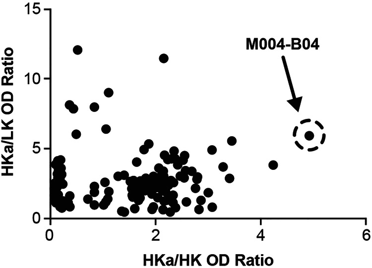
Methods: A novel HKa-specific antibody was discovered by antibody phage display and used as a capture reagent to develop an HKa-specific ELISA.

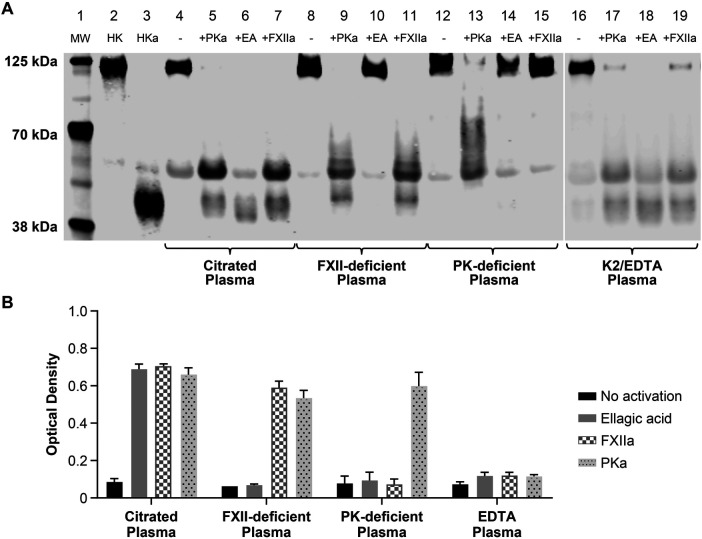
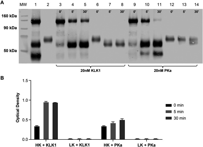
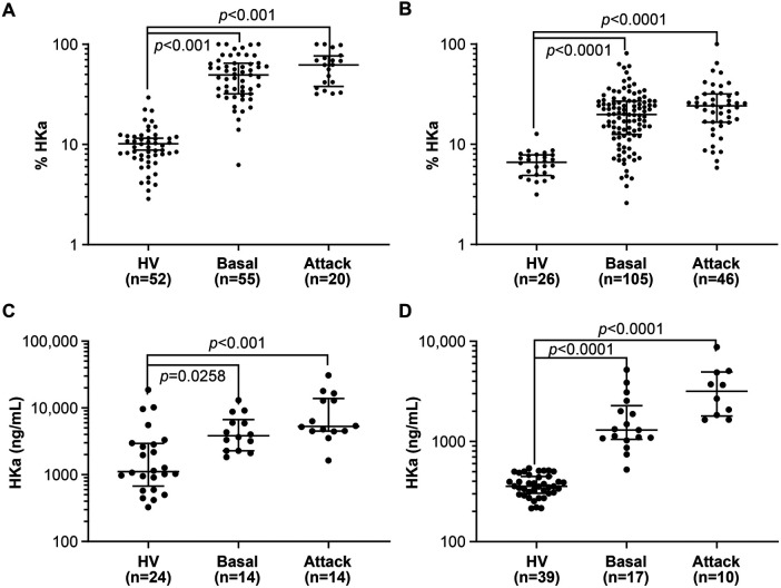
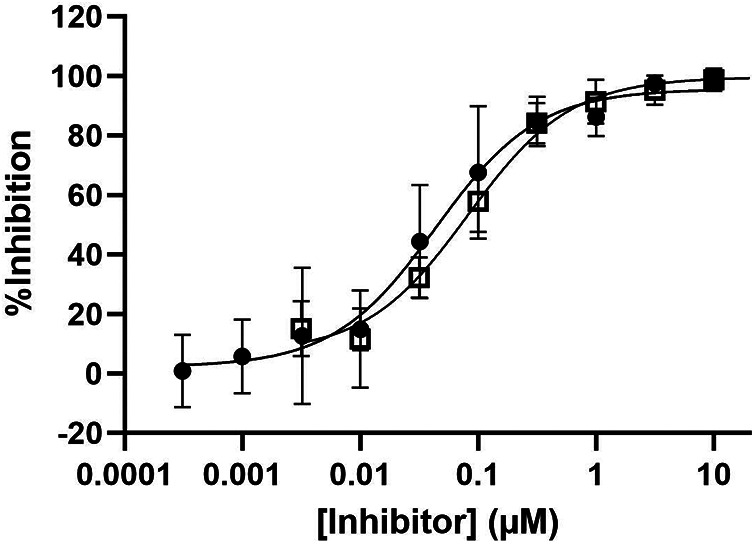
Results: Specific HKa detection following KKS activation was observed in plasma from healthy controls but not in prekallikrein-, high-molecular-weight kininogen-, or coagulation factor XII (FXII)-deficient plasma. HKa levels in plasma collected from HAE-C1INH patients in a disease quiescent state were higher than in plasma from healthy controls and increased further in HAE-C1INH plasma collected during an angioedema attack. The specificity of the assay for PKa-mediated HKa generation in minimally diluted plasma activated with exogenous FXIIa was demonstrated using a specific monoclonal antibody inhibitor (lanadelumab, IC50 = 0.044 µM).

Conclusions: An ELISA was developed for the specific and quantitative detection of HKa in human plasma to support HAE-C1INH drug development. Improved quantification of the HKa biomarker may facilitate further pathophysiologic insight into HAE-C1INH and other diseases mediated by a dysregulated KKS and may enable the design of highly potent inhibitors targeting this pathway.

Keywords: biomarkers; bradykinin; hereditary angioedema; phage display; plasma kallikrein.


Conflict of interest statement

JC, KK, CB, and DY are employees of Takeda Development Center Americas, Inc., and hold stock/stock options in Takeda Pharmaceuticals Company Limited. SJ is an employee of Takeda Pharmaceuticals USA, Inc., and holds stock/stock options in Takeda Pharmaceuticals Company Limited. DS is an employee of Sexton Bio Consulting, LLC, and a former employee of Takeda Development Center Americas, Inc., and holds Takeda Pharmaceuticals Company Limited stock or stock options. NP, JK, and MR-H are former employees of Takeda Development Center Americas, Inc., and hold stock/stock options in Takeda Pharmaceuticals Company Limited. RF is a former employee of Shire, a Takeda company. GS is an employee of Charles River Laboratories. The authors declare that this study received funding from Takeda. The funder had the following involvement in the study: research and publication of this article.

Figures

Figure 1
Figure 1
Discovery of an HKa-specific antibody by phage display selection and screening. Recombinant Fab fragments were passively immobilized onto 384-well plates before the addition of either biotinylated HKa, intact HK, or LK followed by detection using streptavidin-HRP and TMB as described in the Methods section. Fab, fragment antigen-binding; HK, high-molecular-weight kininogen; HKa, cleaved high-molecular-weight kininogen; HRP, horseradish peroxidase; LK, low-molecular-weight kininogen; OD, optical density; TMB, 3,3′,5,5′-tetramethylbenzidine.
Figure 2
Figure 2
Characterization of M4-B4 reactivity in human plasma. Sodium citrate and EDTA plasma from healthy volunteers, FXII-deficient sodium citrate plasma, and PK-deficient sodium citrate plasma were either untreated or activated for 20 min at 37°C with either 100 nM PKa, ellagic acid reagent, or 10 nM FXIIa before analysis by western blotting (A) using an anti-HK monoclonal antibody (11H05) or by an ELISA (B) using M4-B4 as a capture antibody and a 1:1 mixture of 11H05 and 13B12 anti-HK monoclonal antibodies as detection antibodies. Lane composition are as follows: molecular-weight markers (MW) (lane 1), purified HK (lane 2), purified HKa (lane 3), citrate plasma (lane 4), citrate plasma + PKa (lane 5), citrate plasma + ellagic acid (lane 6), citrate plasma + FXIIa (lane 7), FXII-deficient plasma (lane 8), FXII-deficient plasma + PKa (lane 9), FXII-deficient plasma + ellagic acid (lane 10), FXII-deficient plasma + FXIIa (lane 11), prekallikrein-deficient plasma (lane 12), prekallikrein-deficient plasma + PKa (lane 13), prekallikrein-deficient plasma + ellagic acid (lane 14), prekallikrein-deficient plasma + FXIIa (lane 15), EDTA plasma (lane 16), EDTA-deficient plasma + PKa (lane 17), EDTA-deficient plasma + ellagic acid (lane 18), and EDTA-deficient plasma + FXIIa (lane 19). EA, ellagic acid; FXIIa, coagulation factor XIIa; HK, high-molecular-weight kininogen; HKa, cleaved high-molecular-weight kininogen; K2/EDTA, dipotassium ethylenediaminetetraacetic acid; LK, low-molecular-weight kininogen; MW, molecular weight; PK, prekallikrein; PKa, plasma kallikrein.
Figure 3
Figure 3
Comparative kininogen digestion by tissue vs. plasma kallikrein. Purified HK and LK were analyzed by western blotting (A) or an ELISA (B) using M4-B4 as a capture antibody and 11H05 and 13B12 as detection antibodies following incubation with either KLK1 or PKa. The western blot analysis was performed using a polyclonal sheep anti-kininogen antibody for detection that cross-reacted with LK as well as HK. The lane compositions are as follows: molecular-weight markers (MW), a mixture of purified intact HK and HKa (1.5 µM each) (lane 1); purified LK (1.5 µM) (lane 2); HK (1.5 µM) incubated with 20 nM KLK1 for 0, 5, and 30 min (lanes 3–5); LK (1.5 µM) incubated with 20 nM KLK1 for 0, 5, and 30 min (lanes 6–8); HK (1.5 µM) incubated with 20 nM PKa for 0, 5, and 30 min (lanes 9–11); and LK (1.5 µM) incubated with 20 nM PKa for 0, 5, and 30 min (lanes 12–14). HK, high-molecular-weight kininogen; HKa, cleaved high-molecular-weight kininogen; KLK1, tissue kallikrein 1; LK, low-molecular-weight kininogen; MW, molecular weight; PK, prekallikrein; PKa, plasma kallikrein.
Figure 4
Figure 4
Comparison of HKa levels in healthy volunteers and HAE-C1INH patients by western blotting and an ELISA. The statistical analysis of the data in (A–D) is summarized in Supplementary Table S3. (A) Comparison of the percentage of HKa measured by western blotting in the citrated plasma of healthy volunteers with that from HAE-C1INH patients collected between (basal) and during an attack. (B) Comparison of the percentage of HKa measured by western blotting in the SCAT169 plasma of healthy volunteers with that from HAE-C1INH patients collected between (basal) and during an attack. (C) Comparison of HKa levels measured by an ELISA in the citrated plasma of healthy volunteers with those from HAE-C1INH patients collected between (basal) and during an attack. (D) Comparison of HKa levels measured by an ELISA in the SCAT169 plasma of healthy volunteers with those from HAE-C1INH patients collected between (basal) and during an attack. Group means and 95% confidence intervals are shown. Groups were compared with each other by one-way ANOVA. HAE-C1INH, hereditary angioedema due to a deficiency in total (type I) or functional C1 inhibitor protein (type II); HKa, cleaved high-molecular-weight kininogen; HV, healthy volunteer.
Figure 5
Figure 5
Inhibition of HKa generation in activated plasma by PKa inhibitors. Pooled sodium citrate human plasma samples were spiked with the antibody PKa inhibitor lanadelumab (closed circles) or a small-molecule PKa inhibitor BD-105294 (open squares). Plasma samples were activated for 30 min with 5 nM FXIIa. Post activation samples were analyzed at a 150-fold dilution. A dose-response relationship was fitted to averaged values from at least three experiments of duplicate measurements through non-linear regression (GraphPad Prism) using a three-parameter hyperbolic competitive equation {Y = Bottom + [Top-Bottom]/[1 + (X/IC50)]} to provide estimated IC50 values for lanadelumab [IC50 = 0.04 µM, with a 95% confidence interval (CI) from 0.030 to 0.064 µM] and BD-105294 (IC50 = 0.082 µM, with a 95% CI from 0.059 to 0.11 µM). FXIIa, coagulation factor XIIa; HKa, cleaved high-molecular-weight kininogen; IC50, half maximal inhibitory concentration; PKa, plasma kallikrein.

References

    1. Long AT, Kenne E, Jung R, Fuchs TA, Renne T. Contact system revisited: an interface between inflammation, coagulation, and innate immunity. J Thromb Haemost. (2016) 14(3):427–37. 10.1111/jth.13235 - DOI - PubMed
    1. Schmaier AH. The contact activation and kallikrein/kinin systems: pathophysiologic and physiologic activities. J Thromb Haemost. (2016) 14(1):28–39. 10.1111/jth.13194 - DOI - PubMed
    1. de Maat S, Joseph K, Maas C, Kaplan AP. Blood clotting and the pathogenesis of types I and II hereditary angioedema. Clin Rev Allergy Immunol. (2021) 60(3):348–56. 10.1007/s12016-021-08837-6 - DOI - PMC - PubMed
    1. Srivastava P, Gailani D. The rebirth of the contact pathway: a new therapeutic target. Curr Opin Hematol. (2020) 27(5):311–9. 10.1097/MOH.0000000000000603 - DOI - PMC - PubMed
    1. Marceau F, Bachelard H, Bouthillier J, Fortin JP, Morissette G, Bawolak MT, et al. Bradykinin receptors: agonists, antagonists, expression, signaling, and adaptation to sustained stimulation. Int Immunopharmacol. (2020) 82:106305. 10.1016/j.intimp.2020.106305 - DOI - PubMed

LinkOut - more resources