. 2024 Aug 22;27(10):110784.
doi: 10.1016/j.isci.2024.110784. eCollection 2024 Oct 18.

GATA-4 overexpressing BMSC-derived exosomes suppress H/R-induced cardiomyocyte ferroptosis

Affiliations

GATA-4 overexpressing BMSC-derived exosomes suppress H/R-induced cardiomyocyte ferroptosis

Zhiyuan Xiao et al. iScience. .

Abstract

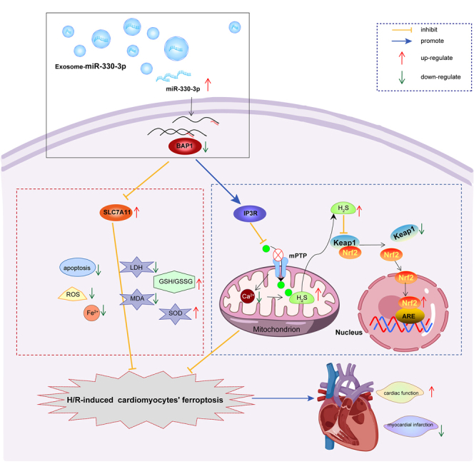
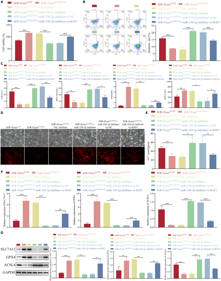
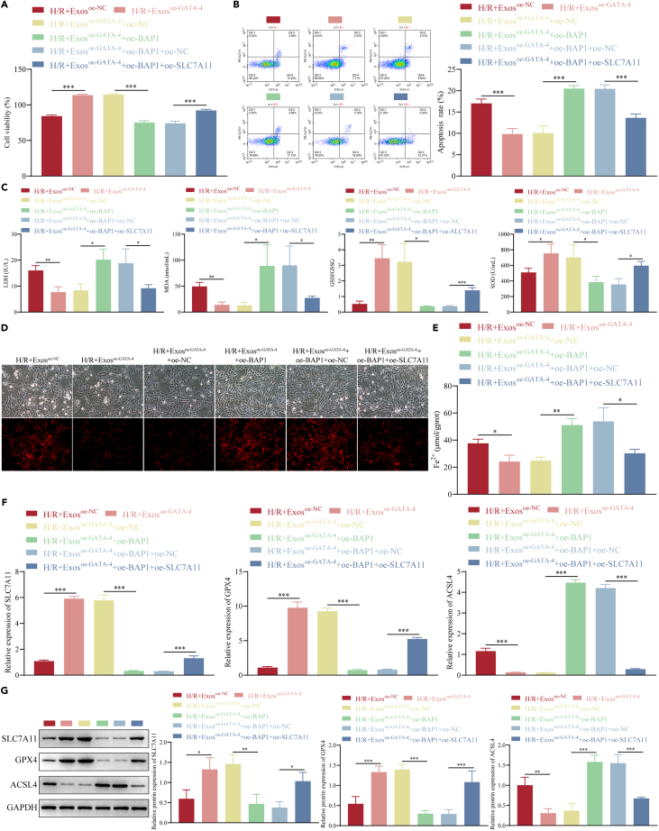
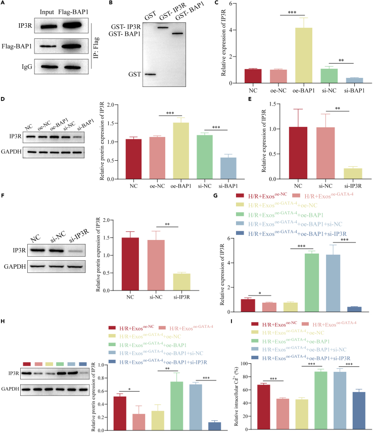
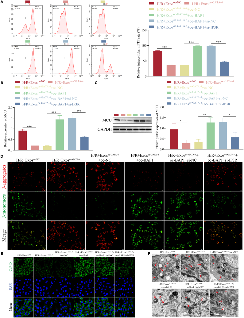
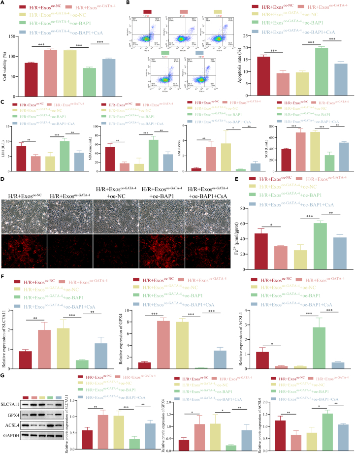
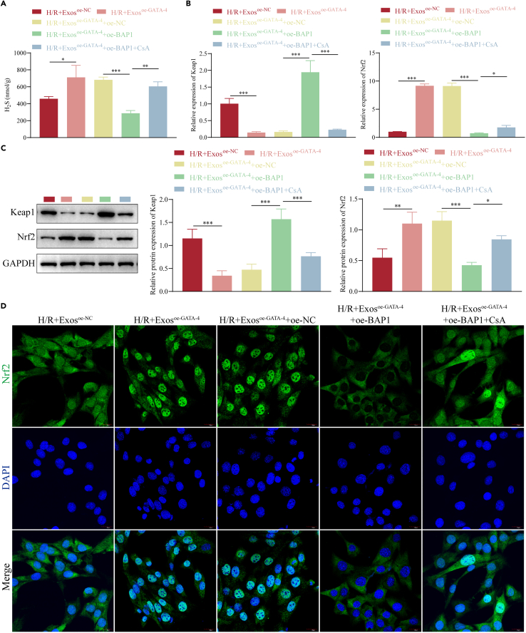
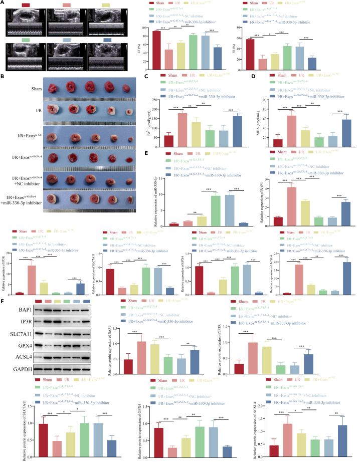
Bone marrow mesenchymal stem cell (BMSC)-derived exosomes overexpressing GATA-4 (Exosoe-GATA-4) can protect cardiac function. Mitochondrial permeability transition pore (mPTP) has a crucial role in ferroptosis. This study aimed to assess the mechanism of Exosoe-GATA-4 in myocardial ischemia/reperfusion (I/R) injury. Exos were successfully excreted, and 185 differential expression miRNAs were obtained using bioinformatics. The Exosoe-GATA-4 effectively suppressed hypoxia/reoxygenation (H/R)-induced cardiomyocytes' ferroptosis, while the effects were reversed by miR-330-3p inhibitor. miR-330-3p targeted negative regulated BAP1. The effects of miR-330-3p inhibitor were reversed by knock-down BAP1. Also, BAP1 reversed the effects of Exosoe-GATA-4 on H/R-induced cardiomyocytes' ferroptosis by downregulating SLC7A11. Mechanistically, BAP1 interacted with IP3R and increased cardiomyocytes' Ca2+ level, causing mPTP opening and mitochondrial dysfunction, promoting H/R-induced cardiomyocytes' ferroptosis. Moreover, hydrogen sulfide (H2S) content was increased and regulated the keap1/Nrf2 signaling pathway by Exosoe-GATA-4 treated. Exosoe-GATA-4 effectively suppresses H/R-induced cardiomyocytes' ferroptosis by upregulating miR-330-3p, which regulates the BAP1/SLC7A11/IP3R axis and inhibits mPTP opening.

Keywords: Cell biology; Molecular biology.


Conflict of interest statement

All authors declare no competing interests.

Figures

None
Graphical abstract
Figure 1
Figure 1
Differential expression miRNAs in BMSC-derived exosomes (Exos) overexpressing GATA-4 compared to BMSC-derived Exosoe−NC group (A) TEM image of Exos. (B) NanoSight analysis. (C) Western blot was used to evaluate exosome markers. (D and E) Volcano plot (D) and heatmap (E) of differential expression miRNAs. (F) RT-qPCR detected the expression levels of differential expression miRNAs. n = 3, ∗∗p < 0.01, ∗∗∗p < 0.001. Data are represented as mean ± SD.
Figure 2
Figure 2
BMSC-derived exosomes overexpressing GATA-4 inhibits H/R-induced ferroptosis by upregulating miR-330-3p (A and B) Transfection efficiency (A) and the expression of miR-330-3p (B) detected by RT-qPCR. (C) CCK-8 assay. (D) Flow cytometry assessing early and late cell apoptosis. (E) The content of LDH, MDA, and GSH was detected by ELISA. (F and G) The ROS (F) and Fe2+ (G) levels were detected by Kits assay. (H and I) The expression levels of SLC7A11, GPX4, and ACSL4 were detected by RT-qPCR (H) and western blot (I). n = 3, ∗p < 0.05, ∗∗p < 0.01, ∗∗∗p < 0.001. Data are represented as mean ± SD.
Figure 3
Figure 3
miR-330-3p target-regulating BAP1 (A) Cytoscape software visualization (only shows the 31 target genes). (B) The combined sequence between miR-330-3p and BAP1. (C) Dual-luciferase reporter assay. (D) AgO2-RIP assay. (E and F) Transfection efficiency of si-NC and si-BAP1 in cardiomyocytes was detected by (E) RT-qPCR and western blot (F). (G and H) The expression levels of BAP1 were detected by RT-qPCR (G) and western blot (H). n = 3, ∗∗p < 0.01, ∗∗∗p < 0.001. Data are represented as mean ± SD.
Figure 4
Figure 4
BMSC-derived exosomes overexpressing GATA-4 inhibits H/R-induced cardiomyocyte ferroptosis by miR-330-3p target-regulating BAP1 (A) CCK-8 assay. (B) Early and late cell apoptosis detected by flow cytometry. (C) The content of LDH, MDA, and GSH was detected by ELISA. (D and E) The ROS (D) and Fe2+ (E) levels were detected by Kits assay. (F and G) The expression levels of SLC7A11, GPX4 and ACSL4 were detected by RT-qPCR (F) and western blot (G). n = 3, ∗p < 0.05, ∗∗p < 0.01, ∗∗∗p < 0.001. Data are represented as mean ± SD.
Figure 5
Figure 5
BAP1 negative regulated the expression of SLC7A11 (A) Luciferase reporter assay. AgO2-RIP assay. (B and C) Transfection efficiency of oe-NC and oe-BAP1 in cardiomyocytes was detected by RT-qPCR (B) and western blot (C). (D) The expression levels of BAP1 and SLC7A11 were detected by western blot. (E and F) Transfection efficiency of oe-NC and oe-SLC7A11 in cardiomyocytes was detected by RT-qPCR (E) and western blot (F). (G and H) The expression levels of SLC7A11 were detected by RT-qPCR (G) and western blot (H). n = 3, ∗p < 0.05, ∗∗p < 0.01, ∗∗∗p < 0.001. Data are represented as mean ± SD.
Figure 6
Figure 6
BAP1 reverses the inhibiting role of BMSC-derived exosome overexpressing GATA-4 on H/R-induced cardiomyocyte ferroptosis by downregulating target gene SLC7A11 (A) CCK-8 assay. (B) Early and late cell apoptosis detected by flow cytometry. (C) The content of LDH, MDA, and GSH was detected by ELISA. (D and E) The ROS (D) and Fe2+ (E) levels were detected by Kits assay. (F and G) The expression levels of SLC7A11, GPX4, and ACSL4 were detected by RT-qPCR (F) and western blot (G). n = 3, ∗p < 0.05, ∗∗p < 0.01, ∗∗∗p < 0.001. Data are represented as mean ± SD.
Figure 7
Figure 7
BAP1 interacts with IP3R (A) CoIP assay. (B) GST pull-down assay. (C and D) The expression levels of IP3R were detected by (C) RT-qPCR and western blot (D). (E and F) Transfection efficiency of si-NC and si-IP3R in cardiomyocytes was detected by RT-qPCR (E) and western blot (F). (G and H) The expression levels of IP3R were detected by RT-qPCR (G) and western blot (H). (I) The Ca2+ levels were detected by Kits assay. n = 3, ∗p < 0.05, ∗∗p < 0.01, ∗∗∗p < 0.001. Data are represented as mean ± SD.
Figure 8
Figure 8
BAP1 induces cardiomyocytes Ca2+ overload, leading to mPTP activation and mitochondrial dysfunction through interaction with IP3R (A) mPTP assessment by flow cytometry. (B and C) The expression level of MCU was detected by RT-qPCR (B) and western blot (C). (D) Mitochondrial membrane potential assay (scale bar: 20 μm). (E) IF detected the fluorescence intensity of CyP-D (scale bar: 20 μm). (F) TEM was reviewed for mitochondrial morphology (scale bar: 1 μm). n = 3, ∗p < 0.05, ∗∗p < 0.01, ∗∗∗p < 0.001. Data are represented as mean ± SD.
Figure 9
Figure 9
BAP1 induces mPTP opening, which reverses the inhibiting role of BMSC-derived Exosoe−GATA−4 on H/R-induced cardiomyocyte ferroptosis through interaction with IP3R (A) CCK-8 assay. (B) Early and late cell apoptosis detected by flow cytometry. (C) Levels of LDH, MDA, and GSH were detected by ELISA. (D and E) The ROS (D) and Fe2+ (E) levels were assessed using Kits assay. (F and G) The expression levels of SLC7A11, GPX4, and ACSL4 were detected by RT-qPCR (F) and western blot (G). n = 3, ∗p < 0.05, ∗∗p < 0.01, ∗∗∗p < 0.001. Data are represented as mean ± SD.
Figure 10
Figure 10
BMSC-derived Exosoe−GATA−4 inhibits H/R-induced cardiomyocytes ferroptosis related to keap1/Nrf2 signaling pathway (A) The content of H2S was assessed using Kits assay. (B and C) The expression levels of keap1 and Nrf2 were detected using RT-qPCR (B) and western blot (C). (D) The expression of Nrf2 was detected using IF (scale bar: 20 μm). n = 3, ∗p < 0.05, ∗∗p < 0.01, ∗∗∗p < 0.001. Data are represented as mean ± SD.
Figure 11
Figure 11
BMSC-derived Exosoe−GATA−4 inhibits I/R-induced cardiomyocyte ferroptosis by upregulated miR-330-3p in vivo (A) An ultrasonic cardiogram estimated cardiac function. (B) The TTC staining. (C and D) The Fe2+ (C) and MDA (D) levels were assessed using Kits assay. (E) The mRNA expression levels of miR-330-3p, BAP1, IP3R, SLC7A11, GPX4, and ACSL4 were detected by RT-qPCR. (F) The protein expressions of BAP1, IP3R, SLC7A11, GPX4, and ACSL4 were detected by western blot. n = 6, ∗p < 0.05, ∗∗p < 0.01, ∗∗∗p < 0.001. Data are represented as mean ± SD.

Similar articles

Cited by

References

    1. Agüero F., Marrugat J., Elosua R., Sala J., Masiá R., Ramos R., Grau M., REGICOR Investigators New myocardial infarction definition affects incidence, mortality, hospitalization rates and prognosis. Eur. J. Prev. Cardiol. 2015;22:1272–1280. doi: 10.1177/2047487314546988. - DOI - PubMed
    1. Chi H.J., Chen M.L., Yang X.C., Lin X.M., Sun H., Zhao W.S., Qi D., Dong J.L., Cai J. Progress in Therapies for Myocardial Ischemia Reperfusion Injury. Curr. Drug Targets. 2017;18:1712–1721. doi: 10.2174/1389450117666160401120308. - DOI - PubMed
    1. Braunwald E. The treatment of acute myocardial infarction: the Past, the Present, and the Future. Eur. Heart J. Acute Cardiovasc. Care. 2012;1:9–12. doi: 10.1177/2048872612438026. - DOI - PMC - PubMed
    1. Shen S., Wang Z., Sun H., Ma L. Role of NLRP3 Inflammasome in Myocardial Ischemia-Reperfusion Injury and Ventricular Remodeling. Med. Sci. Mon. Int. Med. J. Exp. Clin. Res. 2022;28 doi: 10.12659/msm.934255. - DOI - PMC - PubMed
    1. Li J., Cao F., Yin H.L., Huang Z.J., Lin Z.T., Mao N., Sun B., Wang G. Ferroptosis: past, present and future. Cell Death Dis. 2020;11:88. doi: 10.1038/s41419-020-2298-2. - DOI - PMC - PubMed

LinkOut - more resources