. 2025 Jan 7;151(1):31-44.
doi: 10.1161/CIRCULATIONAHA.124.069041. Epub 2024 Oct 11.

A Rare Noncoding Enhancer Variant in SCN5A Contributes to the High Prevalence of Brugada Syndrome in Thailand

Affiliations

A Rare Noncoding Enhancer Variant in SCN5A Contributes to the High Prevalence of Brugada Syndrome in Thailand

Roddy Walsh et al. Circulation. .

Abstract

Background: Brugada syndrome (BrS) is a cardiac arrhythmia disorder that causes sudden death in young adults. Rare genetic variants in the SCN5A gene encoding the Nav1.5 sodium channel and common noncoding variants at this locus are robustly associated with the condition. BrS is particularly prevalent in Southeast Asia but the underlying ancestry-specific factors remain largely unknown.

Methods: Genome sequencing of BrS probands and population-matched controls from Thailand was performed to identify rare noncoding variants at the SCN5A-SCN10A locus that were enriched in patients with BrS. A likely causal variant was prioritized by computational methods and introduced into human induced pluripotent stem cell (hiPSC) lines using CRISPR-Cas9. The effect of the variant on SCN5A expression and Nav1.5 sodium channel current was then assessed in hiPSC-derived cardiomyocytes (hiPSC-CMs).

Results: A rare noncoding variant in an SCN5A intronic enhancer region was highly enriched in patients with BrS (detected in 3.9% of cases with a case-control odds ratio of 45.2). The variant affects a nucleotide conserved across all mammalian species and predicted to disrupt a Mef2 transcription factor binding site. Heterozygous introduction of the enhancer variant in hiPSC-CMs caused significantly reduced SCN5A expression from the variant-containing allele and a 30% reduction in Nav1.5-mediated sodium current density compared with isogenic controls, confirming its pathogenicity. Patients with the variant had severe phenotypes, with 89% experiencing cardiac arrest.

Conclusions: This is the first example of a functionally validated rare noncoding variant at the SCN5A locus and highlights how genome sequencing in understudied populations can identify novel disease mechanisms. The variant partly explains the increased prevalence of BrS in this region and enables the identification of at-risk variant carriers to reduce the burden of sudden cardiac death in Thailand.

Keywords: Asia, Southeastern; Brugada syndrome; genetics; noncoding genetic variant.


Conflict of interest statement

None.

Figures
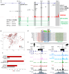
Figure 1.
Figure 1.
Identification of a noncoding enhancer variant in SCN5A associated with BrS. A, Overview of the coding sequence and regulatory elements at the SCN10ASCN5A locus, highlighting single nucleotide polymorphisms (SNPs) from Brugada syndrome (BrS) genome-wide association study (GWAS; lead SNP and SNPs with r2>0.5 in European populations), the EMERGE track of cardiac-specific epigenetic markers, the location of the 4 rare and low-frequency noncoding variants associated with BrS in Thai patients, and odds ratios (ORs) and P values for each haplotype. B, Principal component analysis for patients with BrS and controls from Thailand. C, BrS case and control frequencies for SCN5A coding variants: ultrarare variants (gnomAD filtering allele frequency <0.00001), the low-frequency variant p.Arg965Cys, and other low-frequency variants (gnomAD filtering allele frequency <0.001). D, Sequence alignment of 6 mammalian species, including human and mouse, of the hs2177 enhancer core that encompasses the GRCh38:3-38580380-A-C variant (indicated by box). The variant is located in and disrupts a predicted and conserved Mef2 motif site (indicated by blue shading) and is flanked by T-box (red shading) and Gata (green shading) recognition sites as predicted by Homer. E, UCSC browser views of the hs2177-SCN5A RE5 enhancer in human (hg38) and mouse (mm10). Relevant cardiac transcription factor chromatin immunoprecipitation sequencing and assay for transposase-accessible chromatin with sequencing traces from the literature were plotted (see Table S1 for data set references). ACM indicates atrial cardiomyocytes; CM, cardiomyocytes; and VCM, ventricular cardiomyocytes.
Figure 2.
Figure 2.
Experimental characterization of the RE5 variant. A, Overview of experimental validation: generation of human induced pluripotent stem cell–derived cardiomyocytes (hiPSC-CMs) from PGP1 cell line, isogenic control (red), and RE5 variant line (blue), where the variant was introduced in the heterozygous state by CRISPR-Cas9 editing (graphic produced with BioRender). B, Allelic balance of SCN5A expression in the 2 lines was assessed using the heterozygous synonymous coding variant c.3183A>G present in the PGP1 line (596 bases upstream of the RE5 variant in exon 17), with the C exonic allele demonstrated to be in phase with the RE5 variant. C, Allelic balance of SCN5A expression in the control and RE5 variant lines as determined by RNA sequencing (4 biological replicates each), where P value refers to the Z test differences in allelic expression ratios. D, Sodium current (INa) characterization in control hiPSC-CMs (PGP1; 26 cells from 6 differentiations) and hiPSC-CMs of the 2 RE5 variant lines (A7, 21 cells from 6 differentiations; and C8, 21 cells from 5 differentiations), showing representative traces of INa activated during the first depolarizing pulses of the voltage clamp protocol shown in the inset. E, Average current–voltage (I-V) relationships of peak INa (left) and boxplots depicting INa densities (median and boxes represent interquartile range), determined at −20 mV (right). *Significance compared with controls (P<0.05; Kruskal-Wallis test, followed by Dunn comparisons).
Figure 3.
Figure 3.
Clinical aspects of carriers of the RE5 variant. A, Family pedigree of one of the patients with Brugada syndrome (BrS) carrying the RE5 variant with ages indicated (the proband is II-7, as indicated). Three family members with the variant tested positive for BrS after ajmaline challenge (II-1, III-1, III-3; black fill). Two other family members with the variant did not consent to ajmaline challenge: II-4 displayed a borderline baseline Brugada type I pattern and III-6 displayed a baseline Brugada type III pattern. One individual also tested positive for BrS after ajmaline challenge despite not carrying the RE5 variant (III-5), which is not atypical for SCN5A pathogenic variants in BrS families and reflects the complex genetic pathogenesis of the disease. No other noncarriers display the Brugada marker on ECG. B, Epicardial substrate ablation areas (after ajmaline challenge) for patients with BrS carrying the RE5 variant (n=3; substrate sizes 40.0, 29.2, and 17.2 cm2; mean±SD, 28.7±11.5 cm2) compared with data from Ciconte et al for carriers of pathogenic coding variants in SCN5A (SCN5A-positive; n=49; mean±SD, 18.8±5.7 cm2) and patients with BrS without SCN5A variants (SCN5A-negative; n=146; mean±SD, 11.9±4.8 cm2; 1-way ANOVA, P<0.0001; Tukey honestly significant difference post hoc for RE5 vs SCN5A-negative, P<0.0001). C, Baseline (above) and postajmaline (below) epicardial mapping for the proband of the pedigree in A, with the color bar representing local activation time from early (red) to late (blue) activation (scale in milliseconds). The latest activation was localized at the right ventricular outflow tract (RVOT), the target for radiofrequency ablation, before and after ajmaline administration. However, the latest activation at the RVOT was markedly prolonged after ajmaline, increasing from 130 ms before to 200 ms after. AP indicates anterior-posterior; INF, inferior; LL, left lateral; LV, left ventricle; and RV, right ventricle.
Figure 4.
Figure 4.
Overview of BrS genetic architecture in Thailand and European-ancestry populations. Top, Graphical representation of the genetic contribution of rare and low-frequency variants to Brugada syndrome (BrS) in Thailand (left) and European ancestry populations (right), using a likely conservative estimate of a 4-fold higher prevalence of BrS in Thailand., The absolute frequency of ultrarare loss-of-function coding variants in SCN5A is likely to be broadly similar in both ancestries (represented by the dark blue squares), but the diagnostic yield of such variants in BrS cohorts from Thailand is relatively lower (≈5% vs ≈20%) because of the different disease prevalence between ancestries (highlighted by the overall square sizes). A proportion of the additional susceptibility in Thai patients with BrS is explained by the low-frequency SCN5A coding variant p.Arg965Cys (≈6.5% of cases) and the noncoding RE5 enhancer variant described in this study (≈4% of cases). Bottom, Population frequencies of SCN5A p.Arg965Cys and RE5 variants in South and East Asian countries (see Table S8 for details of data sets used and exact variant frequencies; gray = data not available).

References

    1. Behr ER, Ben-Haim Y, Ackerman MJ, Krahn AD, Wilde AAM. Brugada syndrome and reduced right ventricular outflow tract conduction reserve: a final common pathway? Eur Heart J. 2021;42:1073–1081. doi: 10.1093/eurheartj/ehaa1051 - PubMed
    1. Walsh R, Lahrouchi N, Tadros R, Kyndt F, Glinge C, Postema PG, Amin AS, Nannenberg EA, Ware JS, Whiffin N, et al. ; Nantes Referral Center for inherited cardiac arrhythmia. Enhancing rare variant interpretation in inherited arrhythmias through quantitative analysis of consortium disease cohorts and population controls. Genet Med. 2021;23:47–58. doi: 10.1038/s41436-020-00946-5 - PMC - PubMed
    1. Hosseini SM, Kim R, Udupa S, Costain G, Jobling R, Liston E, Jamal SM, Szybowska M, Morel CF, Bowdin S, et al. ; National Institutes of Health Clinical Genome Resource Consortium. Reappraisal of reported genes for sudden arrhythmic death. Circulation. 2018;138:1195–1205. doi: 10.1161/CIRCULATIONAHA.118.035070 - PMC - PubMed
    1. Wilde AAM, Semsarian C, Márquez MF, Shamloo AS, Ackerman MJ, Ashley EA, Sternick EB, Barajas-Martinez H, Behr ER, Bezzina CR, et al. ; Document Reviewers. European Heart Rhythm Association (EHRA)/Heart Rhythm Society (HRS)/Asia Pacific Heart Rhythm Society (APHRS)/Latin American Heart Rhythm Society (LAHRS) expert consensus statement on the state of genetic testing for cardiac diseases. Europace. 2022;24:1307–1367. doi: 10.1093/europace/euac030 - PMC - PubMed
    1. Barc J, Tadros R, Glinge C, Chiang DY, Jouni M, Simonet F, Jurgens SJ, Baudic M, Nicastro M, Potet F, et al. ; KORA-Study Group. Genome-wide association analyses identify new Brugada syndrome risk loci and highlight a new mechanism of sodium channel regulation in disease susceptibility. Nat Genet. 2022;54:232–239. doi: 10.1038/s41588-021-01007-6 - PMC - PubMed

Substances