. 2024 Nov 15;213(10):1452-1466.
doi: 10.4049/jimmunol.2400395.

NK Cell and Monocyte Dysfunction in Multisystem Inflammatory Syndrome in Children

Affiliations

NK Cell and Monocyte Dysfunction in Multisystem Inflammatory Syndrome in Children

Jenna K Dick et al. J Immunol. .

Abstract

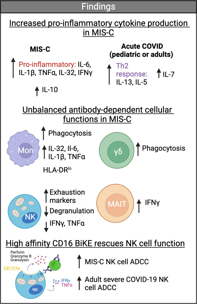
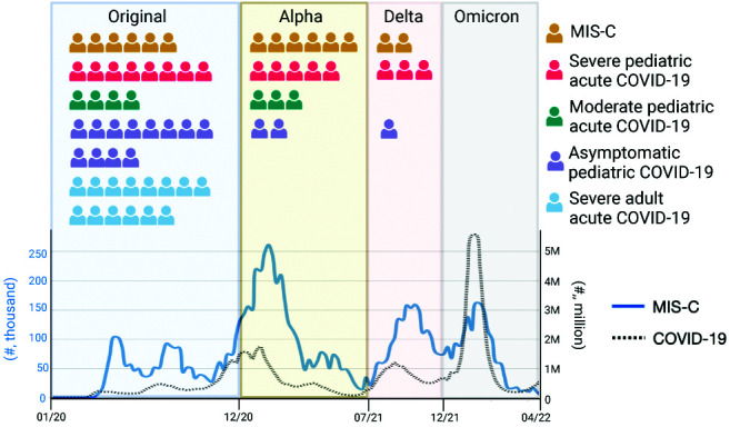
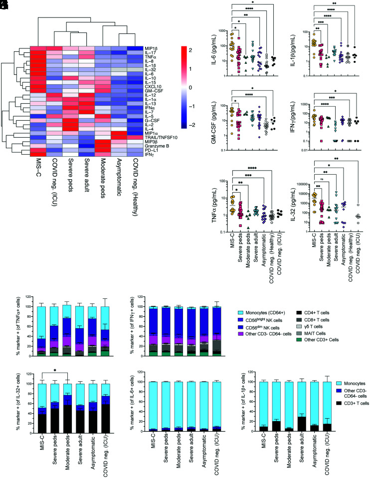
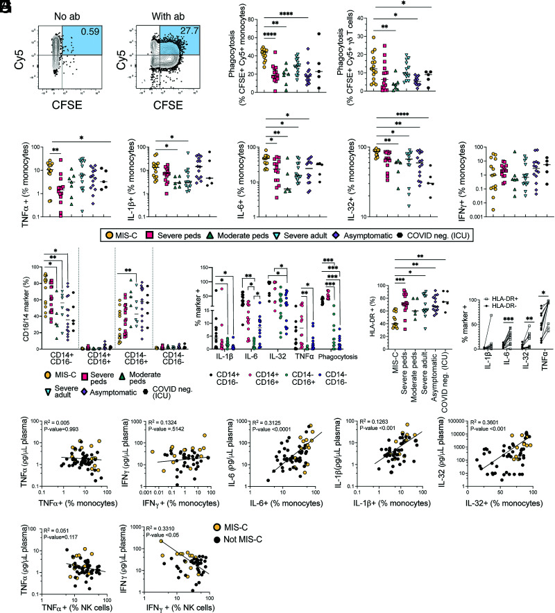
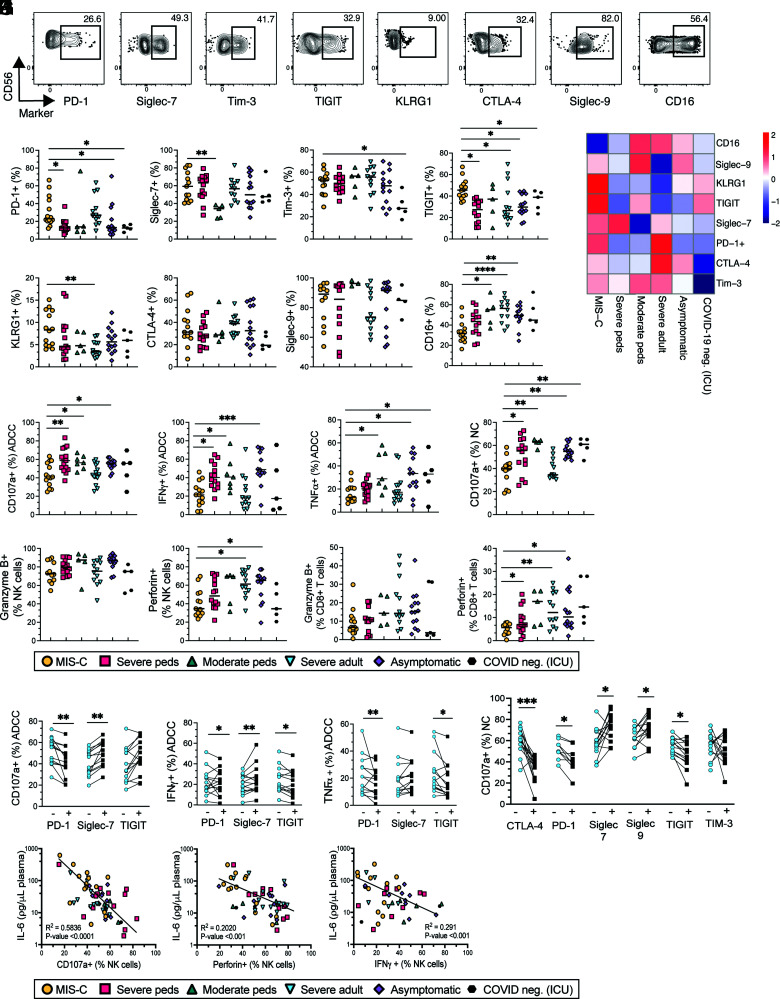
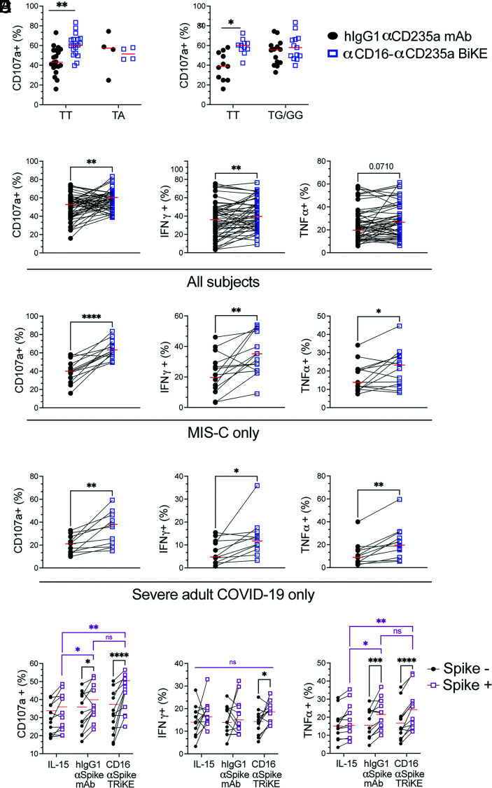
Multisystem inflammatory syndrome in children (MIS-C) is a severe complication of SARS-CoV-2 infection characterized by multiorgan involvement and inflammation. Testing of cellular function ex vivo to understand the aberrant immune response in MIS-C is limited. Despite strong Ab production in MIS-C, SARS-CoV-2 nucleic acid testing can remain positive for 4-6 wk postinfection. Therefore, we hypothesized that dysfunctional cell-mediated Ab responses downstream of Ab production may be responsible for delayed clearance of viral products in MIS-C. In MIS-C, monocytes were hyperfunctional for phagocytosis and cytokine production, whereas NK cells were hypofunctional for both killing and cytokine production. The decreased NK cell cytotoxicity correlated with an NK exhaustion marker signature and systemic IL-6 levels. Potentially providing a therapeutic option, cellular engagers of CD16 and SARS-CoV-2 proteins were found to rescue NK cell function in vitro. Taken together, our results reveal dysregulation in Ab-mediated cellular responses of myeloid and NK cells that likely contribute to the immune pathology of this disease.


Conflict of interest statement

The authors have no financial conflicts of interest.

Figures

None
Graphical abstract
FIGURE 1.
FIGURE 1.
Study schematic of enrollment of each subject between March 2020 and December 2021 during the SARS-CoV-2 pandemic. Visual representation is shown of each COVID-19+ subject enrolled and the predominant circulating variant when they were infected. The timing of acute COVID-19 infection in MIS-C patients is unknown and was presumed to be 1 mo prior to MIS-C presentation (5). The weekly number of MIS-C cases is depicted in blue (left y-axis), and the number of weekly COVID-19 cases overall is depicted in a black dotted line (right y-axis). The case data were sourced from the Centers for Disease Control and Prevention. The figure is based on Rybinka et al. (43).
FIGURE 2.
FIGURE 2.
Distinct cytokine and chemokine profiles between MIS-C and acute COVID-19 control groups. (A) Profiles of cytokines and chemokines in plasma shown as a heatmap and stratified by group: MIS-C (n = 14), severe pediatric (n = 13), moderate pediatric (n = 5), severe adult (n = 14), asymptomatic (n = 15), COVID-19 negative healthy (n = 15), COVID-19 negative ICU (n = 6). Patients who received convalescent plasma were removed from this analysis. Color intensity of each cell represents the average Z score of cytokine measures within each group. (B–G) Analyte IL-6, IL-1β, GM-CSF, IFN-γ, TNF-α, and IL-32 plasma levels in MIS-C patients compared with each control group. Statistical analyses were performed using a Kruskal–Wallis test with a Dunn multiple comparison test. Black lines indicate median. *p <0.05, **p <0.01, ***p <0.001, ****p <0.0001. (HL) Intracellular cytokine production of IFN-γ, TNF-α, IL-32, IL-6, and IL-1β in an Ab-dependent cellular cytotoxicity/Ab-dependent cellular phagocytosis assay. (H) and (I) are from one flow panel, and (J)–(L) are from a different myeloid cell flow cytometry panel. Statistical analyses were performed using a Kruskal–Wallis test with a Dunn multiple comparison test. *p < 0.05.
FIGURE 3.
FIGURE 3.
In MIS-C, monocytes and γδ T cells have increased phagocytosis whereas monocytes have increased cytokine production. (A) Combined flow cytometry files from all subjects of CD64+ monocytes showing CFSE by Cy5 on monocytes incubated with RBCs with or without anti-CD235a and labeled with CFSE and cholesterol-oligo-Cy5 probe (n = 72). Phagocytosis is defined as percent of cells that are both CFSE+ and Cy5+. (B and C) Proportion of monocytes (B) and γδ T cells (C) performing phagocytosis, as defined by the flow plots in (A), stratified by group. Black lines indicate median. (C) Intracellular cytokine staining of cells after incubation with Ab-coated RBCs. Proportion of monocytes that produced TNF-α, IFN-γ, IL-1β, IL-6, and IL-32, stratified by group. Black lines indicate median. (D) Proportion of CD16 and CD14 monocyte subsets by group. Black lines indicate median. (E) Intracellular cytokine staining and measurement of phagocytosis on monocytes after incubation with Ab-coated RBCs stratified based on expression of CD14 and CD16 in MIS-C subjects. (F) Proportion of HLA-DR+ cells for monocytes, stratified by group. Black lines indicate median. (G) Intracellular cytokine staining and measurement of phagocytosis on monocytes after incubation with Ab-coated RBCs stratified based on expression of HLA-DR in MIS-C subjects. For (A)–(G), statistical analyses were performed using a Kruskal–Wallis test with a Dunn multiple comparison test. *p < 0.05, **p < 0.01. ***p < 0.001, ****p < 0.0001. (H and I) Proportion of proinflammatory cytokines produced by monocytes (H) and NK cells (I) compared with the plasma levels of respective cytokines by ELISA. R2 goodness-of-fit analysis on nonlinear regression line (log transformed) is shown. Yellow dots indicate MIS-C subjects. Black dots indicate non–MIS-C subjects from all other groups for which samples have paired ELISA and flow cytometry data (not all subjects included, n varies depending on flow cytometry test run). The p values were determined by a Pearson correlation.
FIGURE 4.
FIGURE 4.
In MIS-C, CD56dim NK cells exhibit an exhausted phenotype and have decreased cytotoxicity. (A) Combined flow cytometry plots of all subjects for CD56dim NK cells (gated on singlets, live cells, CD3CD64) by expression of exhaustion markers (n = 72). (B) Proportion of expression of selected exhaustion markers on CD56dim NK cells stratified by group: MIS-C (n = 14), severe peds (pediatrics) (n = 21), moderate peds (pediatrics) (n = 7), severe adults (n = 14), asymptomatic (n = 15), COVID-19 negative ICU (n = 6). Black lines indicate median. (C) Profiles of exhaustion markers on CD56dim NK cells shown as a heatmap and stratified by group. Color intensity represents average of marker expression divided the average expression from every subject. (D and E) Proportion of CD56dim NK cells degranulated (CD107a) or produced IFNγ-, or TNF-α in ADCC (D) or natural cytotoxicity assay (E). Black lines depict median. (F and G) Proportion of CD56dim NK cells (F) or CD8 T cells (G) expressing granzyme B and perforin. For (B)–(F), statistical analyses were performed using a Kruskal–Wallis test with a Dunn multiple comparison test. For (G), a paired t test was performed. Black lines depict median. *p < 0.05, **p < 0.01, ***p < 0.001, ****p < 0.0001. (H and I) Proportion of CD56dim NK cell degranulation, IFN-γ and TNF-α production in an ADCC (H) or NC (I) assay based on expression of exhaustion markers. Statistical analyses were performed using a paired t test. *p < 0.05, **p < 0.01, ***p < 0.001. (J–L) Plasma levels of IL-6 compared with the proportion of degranulating (J), perforin-positive (K), and IFN-γ–producing (L) NK cells. R2 goodness-of-fit analysis on log-transformed regression line is shown. Statistical testing was done with a Pearson correlation test.
FIGURE 5.
FIGURE 5.
Cellular engagers that target CD16 with high affinity improve NK cell function compared with a human IgG1 Ab. (A) Proportion of degranulation in response to an RBC target opsonized with an hIgG1 anti-CD235a Ab (black dots) or an anti-CD16/anti-CD235a BiKE (open squares) based on FcγR3A SNP (rs10127939 or c.230T>A on the left panel and rs396991 or c.559T>G on the right panel) genotypes. (B–D) Proportion of degranulation as measured by CD107a, IFN-γ, and TNF-α production comparing an hIgG1 anti-CD235a Ab (black dots) to anti-CD16/anti-CD235a BiKE (blue dots) in all subjects (B) in MIS-C alone (C) or in severe adult subjects alone (D). (E) Proportion of NK cells degranulating or producing cytokines as measured by CD107a, IFN-γ, and TNF-α comparing HEK 293 cells (Spike −) with a HEK 293 Spike-expressing cell line (Spike +) when cultured with IL-15 alone, an hIgG1 anti–SARS-CoV-2 Spike mAb (10 µg/ml), or anti–CD16-IL-15-αSARS-CoV-2 Spike TRiKE (10 µg/ml). E:T ratio = 3:1. Statistical analyses were performed using a paired t test. *p < 0.05, **p < 0.01. ***p < 0.001, ****p < 0.0001. Red lines depict median.

References

    1. Patel, J. M. 2022. Multisystem inflammatory syndrome in children (MIS-C). Curr. Allergy Asthma Rep. 22: 53–60. - PMC - PubMed
    1. Vogel, T. P., Top K. A., Karatzios C., Hilmers D. C., Tapia L. I., Moceri P., Giovannini-Chami L., Wood N., Chandler R. E., Klein N. P., et al. . 2021. Multisystem inflammatory syndrome in children and adults (MIS-C/A): case definition & guidelines for data collection, analysis, and presentation of immunization safety data. Vaccine 39: 3037–3049. - PMC - PubMed
    1. Cui, X., Zhao Z., Zhang T., Guo W., Guo W., Zheng J., Zhang J., Dong C., Na R., Zheng L., et al. . 2021. A systematic review and meta-analysis of children with coronavirus disease 2019 (COVID-19). J. Med. Virol. 93: 1057–1069. - PMC - PubMed
    1. Whittaker, E., Bamford A., Kenny J., Kaforou M., Jones C. E., Shah P., Ramnarayan P., Fraisse A., Miller O., Davies P., et al. .; PIMS-TS Study Group and EUCLIDS and PERFORM Consortia . 2020. Clinical characteristics of 58 children with a pediatric inflammatory multisystem syndrome temporally associated with SARS-CoV-2. JAMA 324: 259–269. - PMC - PubMed
    1. Feldstein, L. R., Rose E. B., Horwitz S. M., Collins J. P., Newhams M. M., Son M. B. F., Newburger J. W., Kleinman L. C., Heidemann S. M., Martin A. A., et al. .; CDC COVID-19 Response Team . 2020. Multisystem inflammatory syndrome in U.S. children and adolescents. N. Engl. J. Med. 383: 334–346. - PMC - PubMed

Supplementary concepts