. 2024 Oct 11;10(41):eado6492.
doi: 10.1126/sciadv.ado6492. Epub 2024 Oct 11.

Mechanism of degrader-targeted protein ubiquitinability

Affiliations

Mechanism of degrader-targeted protein ubiquitinability

Charlotte Crowe et al. Sci Adv. .

Abstract

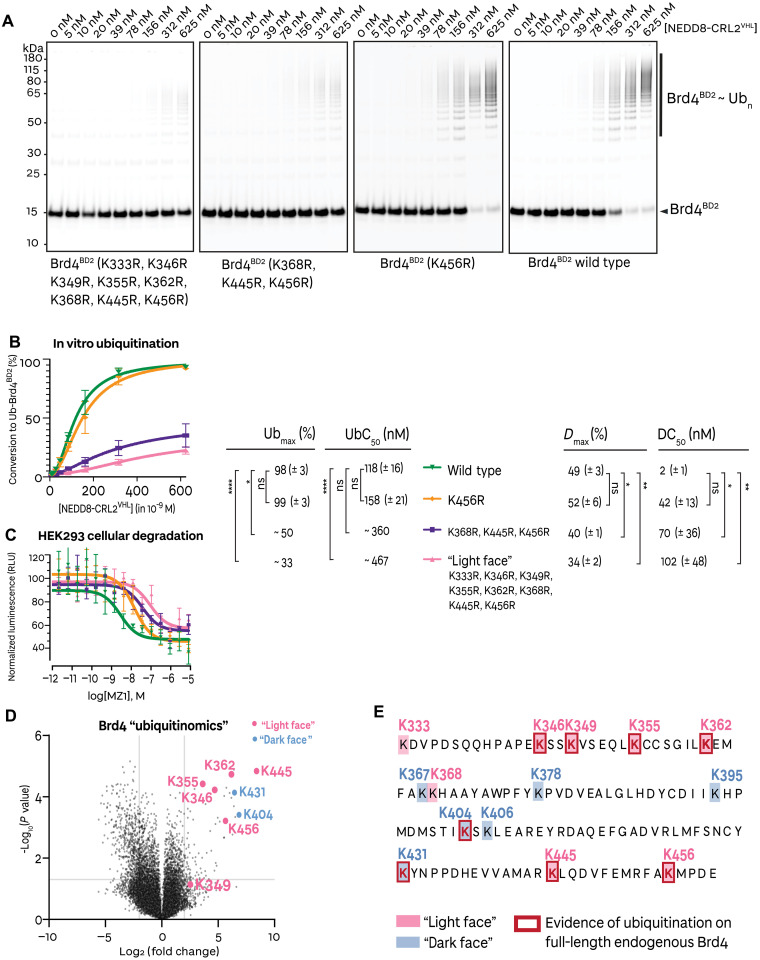
Small-molecule degraders of disease-driving proteins offer a clinically proven modality with enhanced therapeutic efficacy and potential to tackle previously undrugged targets. Stable and long-lived degrader-mediated ternary complexes drive fast and profound target degradation; however, the mechanisms by which they affect target ubiquitination remain elusive. Here, we show cryo-EM structures of the VHL Cullin 2 RING E3 ligase with the degrader MZ1 directing target protein Brd4BD2 toward UBE2R1-ubiquitin, and Lys456 at optimal positioning for nucleophilic attack. In vitro ubiquitination and mass spectrometry illuminate a patch of favorably ubiquitinable lysines on one face of Brd4BD2, with cellular degradation and ubiquitinomics confirming the importance of Lys456 and nearby Lys368/Lys445, identifying the "ubiquitination zone." Our results demonstrate the proficiency of MZ1 in positioning the substrate for catalysis, the favorability of Brd4BD2 for ubiquitination by UBE2R1, and the flexibility of CRL2 for capturing suboptimal lysines. We propose a model for ubiquitinability of degrader-recruited targets, providing a mechanistic blueprint for further rational drug design.


Figures

Fig. 1.
Fig. 1.. MZ1 orients the Brd4 bromodomain 2 neo-substrate toward Rbx1.
(A) Chemical structure of the bivalent PROTAC MZ1 (in purple: the Brd4BD2 ligand; in green: the VHL ligand). (B) SDS-PAGE of the assembled Brd4BD2-MZ1-(NEDD8)-CRL2VHL-UBE2R1(C93K)-Ub complex applied to cryo-EM grids. (C) Mass photometry histogram of the fully assembled Brd4BD2-MZ1-(NEDD8)-CRL2VHL-UBE2R1(C93K)-Ub complex (purple) applied to cryo-EM grids, versus (NEDD8)-CRL2VHL (green). The peaks have been fitted with Gaussian curves. (D) The ~4.0-Å-resolution cryo-EM reconstruction of Brd4BD2-MZ1-(NEDD8)-CRL2VHL. The E3 ligase NEDD8-CRL2VHL (green) recruits MZ1 (yellow), which, in turn, binds the neo-substrate Brd4BD2 (purple). A ribbon representation of the complex is modeled into the displayed cryo-EM volume (same color scheme). (E) The cryo-EM model (this study) of the VHL-MZ1-Brd4BD2 interface overlaid with the same interface from the crystal structure by Gadd et al. (13) (PDB: 5T35). Brd4BD2 displays a conserved interface proximal to the MZ1-VHL binding site, and a re-orientation distal to the MZ1-VHL binding site, toward the E3 ligase RING box domain.
Fig. 2.
Fig. 2.. Brd4 bromodomains are ubiquitinated at lysines accessible to the E3 ligase RING box domain.
(A) Coomassie-stained SDS-PAGE of in vitro Brd4BD2 ubiquitination in the presence of MZ1-CRL2VHL-UBE2R1-Ub (in purple: gel bands subjected to mass spectrometry analysis). (B) Cryo-EM volume of Brd4BD2 in complex with MZ1-(NEDD8)-CRL2VHL with ubiquitinated lysines on Brd4BD2 highlighted in pink and unmodified lysines highlighted in light blue as mapped by mass spectrometry following in vitro ubiquitination. (C) Identified mass spectrum for the Brd4BD2 K-GG–modified peptide at K456. “Score” refers to the Andromeda score. (D) Sequence alignments of the bromodomain 2 of Brd2, Brd3, Brd4, and BrdT. Ubiquitinated lysines on Brd4BD2 (as shown by mass spectrometry) are highlighted in pink and unmodified lysines are highlighted in light blue, with sequence-aligned lysines of other BD2s represented in the same color, suggesting which lysines may be modified in other BET BD2s.
Fig. 3.
Fig. 3.. Assembly of a Brd4BD2 ubiquitination structural mimetic.
(A) Schematic of the biochemical strategy used to trap MZ1-Brd4BD2-Ub(G76S, K48C)-UBE2R1(C93K, S138C, C191S, C223S)-Ub in complex with (NEDD8)-CRL2VHL. (B) SDS-PAGE of the assembled (NEDD8)-CRL2VHL-MZ1-Brd4BD2-Ub(G76S, K48C)-UBE2R1(C93K, S138C, C191S, C223S)-Ub complex applied to cryo-EM grids. (C) Mass photometry histogram of the fully assembled (NEDD8)-CRL2VHL-MZ1-Brd4BD2-Ub(G76S, K48C)-UBE2R1(C93K, S138C, C191S, C223S)-Ub complex (purple) applied to cryo-EM grids, versus (NEDD8)-CRL2VHL (green). The peaks have been fitted with Gaussian curves.
Fig. 4.
Fig. 4.. Structural snapshots of Brd4BD2 ubiquitination complex assembly.
(A) Fixed discrete states and continuous states are captured by cryo-EM corresponding to the dynamics of the cross-linked (NEDD8)-CRL2VHL-MZ1-Brd4BD2-Ub(G76S, K48C)-UBE2R1(C93K, S138C, C191S, C223S)-Ub polyubiquitination species. (B) The ~3.7-Å-resolution cryo-EM reconstruction of closed cross-linked (NEDD8)-CRL2VHL-MZ1-Brd4BD2-Ub(G76S, K48C)-UBE2R1(C93K, S138C, C191S, C223S)-Ub. The E3 ligase (NEDD8)-CRL2VHL (shades of green) recruits MZ1 (yellow), which, in turn, binds the neo-substrate Brd4BD2 (purple). UBE2R1 (red) is engaged by Rbx1, with donor ubiquitin (pale yellow) covalently bound to UBE2R1(C93K).
Fig. 5.
Fig. 5.. Brd4BD2 lysine mutants illustrate specificity in ubiquitinability and degradability.
(A) Representative in vitro ubiquitination assay with Alexa Fluor 647 (AF647)–labeled recombinantly expressed and purified Brd4BD2 mutants, performed in the presence of (NEDD8)-CRL2VHL, MZ1, and UBE2R1. The species were resolved by SDS-PAGE and scanned at 647 nm to visualize unmodified Brd4BD2 and ubiquitinated Brd4BD2 species. (B) Quantification of the percent conversion from unmodified Brd4BD2 to ubiquitinated Brd4BD2 species from the in vitro ubiquitination assay. The error bars represent the mean of N = 3 replicates with SEM. UbC50 and Ubmax values are calculated. (C) MZ1 dose-dependent cellular degradation of NanoLuc-Brd4BD2 wild-type and lysine mutants. The error bars represent the mean of N = 4 replicates with SEM. DC50 and Dmax values are calculated. (D) Global changes in the cellular “ubiquitinome,” as assessed by di-glycine remnant mass spectrometry profiling on HEK293 cells following treatment with 1 μM MZ1 for 15 min. The depicted volcano plot shows 25,843 ubiquitinated precursors presented as log2 fold change relative to DMSO controls. The positions of light face lysines, which showed evidence of modification in vitro, are highlighted in pink, while dark face lysines, which showed no evidence of modification in vitro but are found ubiquitinated in cells, are highlighted in blue. (E) Sequence of Brd4BD2 overlaying the results of in vitro ubiquitination and ubiquitinomics from this study. The legend denotes light face lysines (highlighted in pink; evidence of ubiquitination in vitro on recombinant Brd4BD2), dark face lysines (highlighted in blue; no evidence of ubiquitination in vitro on recombinant Brd4BD2), and lysines that showed evidence of modification from cellular ubiquitinomics experiments (red bold font and red box).
Fig. 6.
Fig. 6.. Schematic of structural and mechanistic feature of ubiquitinability of target proteins revealed from this study.

References

    1. Zhao L., Zhao J., Zhong K., Tong A., Jia D., Targeted protein degradation: Mechanisms, strategies and application. Signal Transduct. Target. Ther. 7, 113 (2022). - PMC - PubMed
    1. Chirnomas D., Hornberger K. R., Crews C. M., Protein degraders enter the clinic—A new approach to cancer therapy. Nat. Rev. Clin. Oncol. 20, 265–278 (2023). - PMC - PubMed
    1. Zheng N., Shabek N., Ubiquitin ligases: Structure, function, and regulation. Annu. Rev. Biochem. 86, 129–157 (2017). - PubMed
    1. Harper J. W., Schulman B. A., Cullin-RING ubiquitin ligase regulatory circuits: A quarter century beyond the F-box hypothesis. Annu. Rev. Biochem. 90, 403–429 (2021). - PMC - PubMed
    1. Cowan A. D., Ciulli A., Driving E3 ligase substrate specificity for targeted protein degradation: Lessons from nature and the laboratory. Annu. Rev. Biochem. 91, 295–319 (2022). - PubMed

MeSH terms