. 2024 Oct 22;18(42):29045-29058.
doi: 10.1021/acsnano.4c10154. Epub 2024 Oct 11.

Tetrahydropyrimidine Ionizable Lipids for Efficient mRNA Delivery

Affiliations

Tetrahydropyrimidine Ionizable Lipids for Efficient mRNA Delivery

Ivan Isaac et al. ACS Nano. .

Abstract

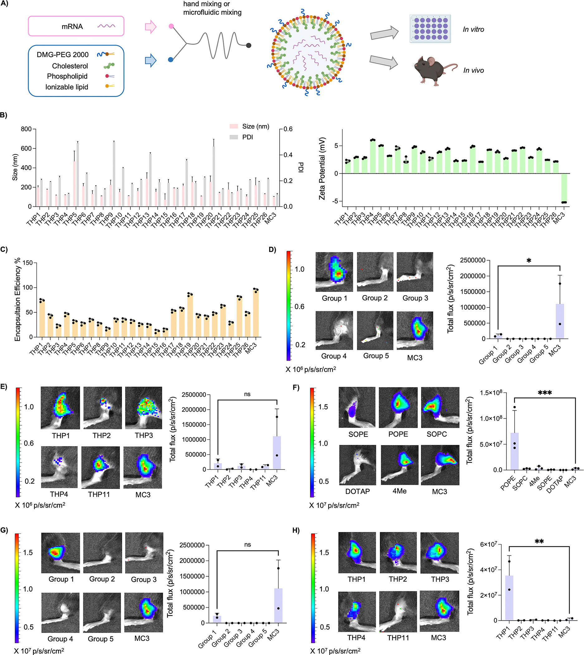
Lipid nanoparticles (LNPs) have emerged as an effective and promising technology for messenger RNA (mRNA) delivery, offering a potential solution to physiological barriers and providing an alternative approach to gene therapy without the drawbacks associated with viral delivery. However, efficiently delivering mRNA remains a significant challenge in nucleic acid-based therapies due to the limitations of current LNP platforms in achieving optimal endosomal escape and mRNA release, which largely relies on finding a suitable ionizable lipid. Additionally, the synthesis of these ionizable lipids involves multiple chemical reactions, often making the process time-consuming and difficult to translate. In this study, we employed a facile, catalyst-free, and versatile one-pot multicomponent reaction (MCR) to develop a library of ionizable lipids featuring a pharmacologically significant tetrahydropyrimidine (THP) backbone, tailored for enhanced mRNA delivery. A library of 26 THP ionizable lipids was systematically synthesized in just 3 h and formulated with luciferase mRNA for initial in vitro screening. The THP LNPs exhibited tunable particle sizes, favorable ζ-potentials, and high encapsulation efficiencies. Among them, THP1 demonstrated the highest transfection efficiency both in vitro and in vivo after intramuscular administration, comparable to DLin-MC3-DMA (MC3), a conventional benchmark. Further optimization of THP1 with phospholipids significantly enhanced intramuscular mRNA delivery and showed sustained protein expression in vivo for up to 5 days. More importantly, it demonstrated successful intravenous delivery in a dose-dependent manner with minimal toxicity, as indicated by hematological, histopathological, and proinflammatory cytokine assessments. Furthermore, THP1 LNPs also demonstrated the ability to edit genes in specific liver tissues in a tdTomato transgenic mouse model, highlighting their precision and utility in targeted therapeutic applications. These findings position THP1 LNPs as promising candidates for advancing mRNA-based therapies, with significant implications for clinical translation in vaccine delivery and CRISPR/Cas9-mediated gene editing in the liver.

Keywords: biomaterials; in vivo delivery; ionizable lipid; lipid nanoparticles; mRNA; multicomponent reaction; tetrahydropyrimidine.


Conflict of interest statement

The authors declare the following competing financial interest(s): I.I. and C.B. have filed a patent application based on this work.

Figures

Figure 1.
Figure 1.
Design and facile synthesis of THP ionizable lipids using THP MCR. (A) Schematic illustration of THP multicomponent reaction used for the synthesis of THP ionizable lipids. Full list of aliphatic amines (left) and heterocyclic amines (right) used to generate the library are shown. (B) Chemical structures of all the 26 THP ionizable lipids synthesized.
Figure 2.
Figure 2.
Physicochemical characterization and in vivo screening of THP LNPs. (A) Overview of the synthesis of LNPs for in vitro and in vivo evaluations. (B) The size (nm), polydispersity index (PDI), and ζ-potential (mV) of THP LNPs. (C) mRNA encapsulation efficiency of all THP LNPs. (D) IVIS images at 6h post injection and graphical representation of total flux of FLuc mRNA encapsulated with all THPs, batched based on chemical structures detailed in Figure S3. C57BL/6 mice were injected intramuscularly with 0.5 mg/kg of pooled THP nanoparticles (n = 2 biologically independent mice, initial screening, *P < 0.05, **P < 0.01, ***P < 0.001. NS, not significant, one-way ANOVA with Bonferroni post hoc analysis). (E) Individual intramuscular injections of all the THPs from Group 1. Representative IVIS images at 6h post injection and graphical representation of total flux of THP1, THP2, THP3, THP4, and THP11 LNPs at a dose of 0.5 mg/kg (n = 2 biologically independent mice, initial screening, *P < 0.05, **P < 0.01, ***P < 0.001. NS, not significant, one-way ANOVA with Bonferroni post hoc analysis). (F) IVIS Images at 6h post injection and graphical representation of total flux of THP1 formulated with phospholipids SOPE, POPE, SOPC, DOTAP, and 4Me injected intramuscularly in C57BL/6 mice at a dose of 0.5 mg/kg (n = 2 biologically independent mice, initial screening, *P < 0.05, **P < 0.01, ***P < 0.001. NS, not significant, one-way ANOVA with Bonferroni post hoc analysis). (G) Representative IVIS images at 6h post injection and graphical representation of total flux of all the THPs formulated with POPE, batched based on chemical structures detailed in Figure S3. C57BL/6 mice were injected intramuscularly with 0.5 mg/kg of pooled THP nanoparticles (n = 2 biologically independent mice, initial screening, *P < 0.05, **P < 0.01, ***P < 0.001. NS, not significant, one-way ANOVA with Bonferroni post hoc analysis). (H) Representative IVIS images at 6h post injection and graphical representation of total flux of all the THPs from Group 1 formulated with POPE and injected intramuscularly in C57BL/6 mice at a dose of 0.5 mg/kg (n = 2 biologically independent mice, initial screening, *P < 0.05, **P < 0.01, ***P < 0.001. NS, not significant, one-way ANOVA with Bonferroni post hoc analysis).
Figure 3.
Figure 3.
Validation and evaluation of intramuscular versus intravenous administration of the top ionizable lipid THP1. (A) Chemical structure of our top performing THP ionizable lipid, THP1. (B) Comparison of size, PDI, ζ-potential measurements, and mRNA encapsulation efficiency of THP1 formulated with POPE, THP1 formulated with DOPE, and MC3 formulated using hand mixing and microfluidic mixing (n = 3, ± SD, *P < 0.05, **P < 0.01, ***P < 0.001. NS, not significant, one-way ANOVA with Bonferroni post hoc analysis). (C) Representative IVIS images at 6h post injection and graphical representation of total flux of THP1 formulated with POPE using microfluidic mixing and injected intramuscularly in C57BL/6 mice at a dose of 0.5 mg/kg (n = 4 biologically independent mice, ± SD, *P < 0.05, **P < 0.01, ***P < 0.001. NS, not significant, two-tailed unpaired Student’s t test analysis). (D) In vivo kinetics of FLuc expression following intramuscular injection. THP1 LNPs formulated with Luc mRNA were injected intramuscularly into mice at a dose of 0.5 mg/kg (n = 3 biologically independent mice, ± SD). The luciferase expression was visualized at 12, 24, 48, 72, and 120 h after injection by IVIS.
Figure 4.
Figure 4.
Pharmacokinetics and toxicity of THP1 LNPs. (A) Representative IVIS images at 24h post injection and graphical representation of total flux of THP1 formulated with DOPE, THP1 formulated with POPE and MC3 LNPs injected intravenously at a dose of 0.5 mg/kg (n = 3 biologically independent mice, ± SD, *P < 0.05, **P < 0.01, ***P < 0.001. NS, not significant, one-way ANOVA with Bonferroni post hoc analysis). (B) Representative IVIS images at 24h post injection and graphical representation of total flux of different doses (0.25, 0.5, and 1 mg/kg) of THP1 LNPs administered intravenously in C57BL/6 mice (n = 3 biologically independent mice, ± SD, *P < 0.05, **P < 0.01, ***P < 0.001. NS, not significant, one-way ANOVA with Bonferroni post hoc analysis). (C) Serum levels of liver enzymes, aspartate aminotransferase (AST), alanine aminotransferase (ALT) and Alkaline Phosphatase after intravenous administration with PBS, THP1 and MC3 at a dose of 0.5 mg/kg (n = 3, ± SD, *P < 0.05, **P < 0.01, ***P < 0.001. NS, not significant, one-way ANOVA with Bonferroni post hoc analysis). (D) Levels of IL-1β, IL-6, and TNFα in mice intravenously treated with THP1 and MC3 LNPs at a dose of 0.5 mg/kg (n = 3 biologically independent mice, ± SD, *P < 0.05, **P < 0.01, ***P < 0.001; NS indicates no significance, one-way ANOVA with Bonferroni post hoc analysis). PBS-injected mice were kept as the control group. (E) Volcano plot of differential gene expression. X-axis: the logarithm of fold change; y-axis: False Discovery Rate (FDR).
Figure 5.
Figure 5.
THP1 enabled highly efficient mRNA delivery and tissue-specific tdTomato expression in the liver. (A) Schematic illustration of Cre mRNA delivery and Cre-mediated genetic deletion of the stop cassette to activate tdTomato expression in a cre-loxP mouse model (Ai14). LNPs formulated with Cre mRNA were injected intravenously in Ai14 mice, and isolated livers were imaged using the IVIS. (B) Representative tdTomato expression in the liver 48h post injection and graphical representation of total flux of THP1 and MC3 LNPs containing Cre mRNA, and PBS treated control injected intravenously to Ai14 mice at a dose of 0.5 mg/kg (n = 3 biologically independent mice, ± SD, *P < 0.05, **P < 0.01, ***P < 0.001. NS, not significant, one-way ANOVA with Bonferroni post hoc analysis). (C) (i) Immunohistochemistry of tdTomato in Ai14 liver: Representative images showing immunohistochemical staining of tdTomato in the liver 24h post injection. Ai14 mice treated intravenously with THP1 and MC3 LNPs encapsulating Cre mRNA, and with PBS at a dose of 0.5 mg/kg (n = 3 biologically independent mice, images captured at 20× magnification, ± SD). (ii) Immunofluorescence analysis of tdTomato in Ai14 liver: Representative immunofluorescent images of liver sections 24h post injection with THP1 and D-MC3 LNPs encapsulating Cre mRNA, and with PBS (control) via intravenous injection in Ai14 mice at a dose of 0.5 mg/kg. DAPI was used to label the nuclei. Images were captured at 20× magnification from three biologically independent mice. (iii) Histological examination of LNP treated Ai14 livers: Representative histology images of liver sections 24h post-treatment with THP1 and MC3 encapsulating Cre mRNA, and PBS (control) via intravenous injection in Ai14 mice at a dose of 0.5 mg/kg. Hematoxylin and Eosin (H&E) staining was performed, with images taken at 10× magnification. Scale bars: 200 μm (n = 3 biologically independent mice).

References

    1. Karikó K; Whitehead K; van der Meel R What Does the Success of mRNA Vaccines Tell Us about the Future of Biological Therapeutics? Cell Syst. 2021, 12 (8), 757–758. - PMC - PubMed
    1. Mukai H; Ogawa K; Kato N; Kawakami S Recent Advances in Lipid Nanoparticles for Delivery of Nucleic Acid, mRNA, and Gene Editing-Based Therapeutics. Drug Metab. Pharmacokinet. 2022, 44, No. 100450. - PMC - PubMed
    1. Qiu M; Li Y; Bloomer H; Xu Q Developing Biodegradable Lipid Nanoparticles for Intracellular mRNA Delivery and Genome Editing. Acc. Chem. Res. 2021, 54 (21), 4001–4011. - PubMed
    1. Holt CE; Bullock SL Subcellular mRNA Localization in Animal Cells and Why It Matters. Science 2009, 326 (5957), 1212–1216. - PMC - PubMed
    1. Olszewska M; Bujarski JJ; Kurpisz M P-Bodies and Their Functions during mRNA Cell Cycle: Mini-Review. Cell Biochem. Funct. 2012, 30 (3), 177–182. - PubMed

Publication types

LinkOut - more resources