Multicenter Study
. 2024 Dec;11(6):e200323.
doi: 10.1212/NXI.0000000000200323. Epub 2024 Oct 11.

Pediatric MOG-Ab-Associated Encephalitis: Supporting Early Recognition and Treatment

Affiliations
Multicenter Study

Pediatric MOG-Ab-Associated Encephalitis: Supporting Early Recognition and Treatment

Nee Na Kim et al. Neurol Neuroimmunol Neuroinflamm. 2024 Dec.

Erratum in

Abstract

Background and objectives: Antibodies to myelin oligodendrocyte glycoprotein (MOG-Ab) have recently been reported in patients with encephalitis who do not fulfill criteria for acute disseminated encephalomyelitis (ADEM). We evaluated a cohort of these children and compared them with children with ADEM.

Methods: This retrospective, multicenter cohort study comprised consecutive patients <18 years of age with MOG-Ab who fulfilled criteria for autoimmune encephalitis. These patients were stratified into (1) children not fulfilling criteria for ADEM (encephalitis phenotype) and (2) children with ADEM. Clinical/paraclinical data were extracted from the electronic records. Comparisons were made using the Mann-Whitney U test and χ2 Fisher exact test for statistical analysis.

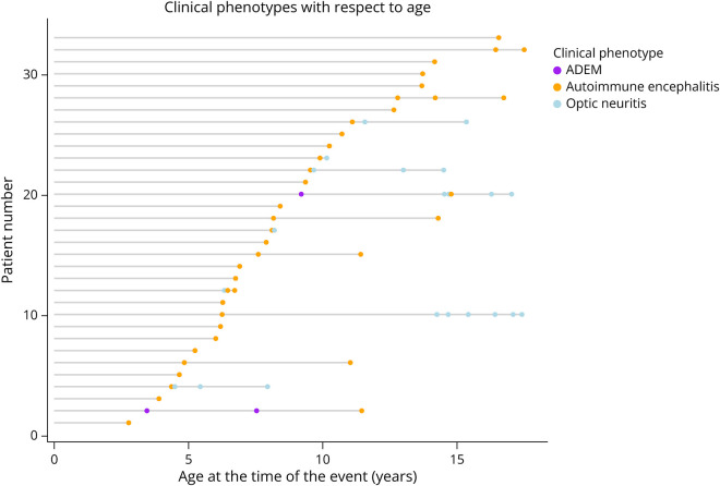
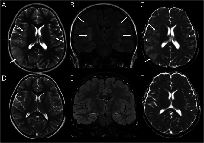
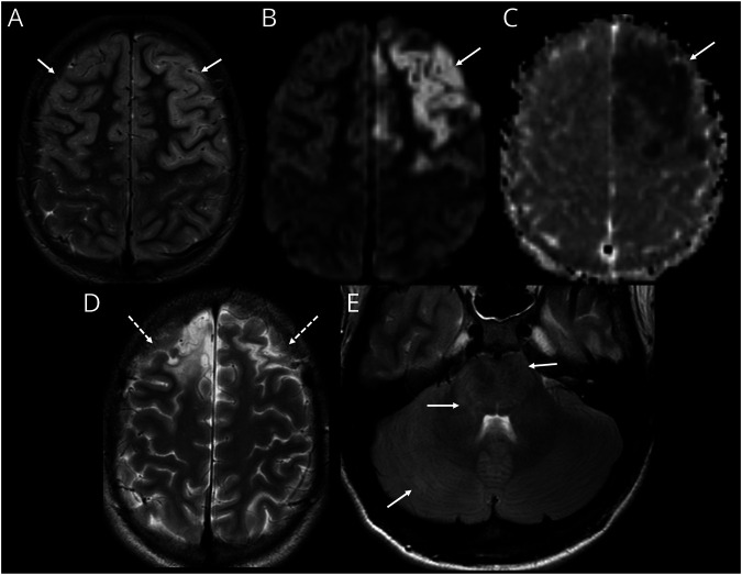
Results: From 235 patients with positive MOG-Ab, we identified 33 (14%) with encephalitis and 74 (31%) with ADEM. The most common presenting symptoms in children with encephalitis were headache (88%), seizures (73%), and fever (67%). Infective meningoencephalitis was the initial diagnosis in 67%. CSF pleocytosis was seen in 79%. Initial MRI brain was normal in 8/33 (24%) patients. When abnormal, multifocal cortical changes were seen in 66% and unilateral cortical changes in 18%. Restricted diffusion was demonstrated in 43%. Intra-attack new lesions were seen in 7/13 (54%). When comparing with children with ADEM, children with encephalitis were older (median 8.9 vs 5.7 years, p = 0.005), were more likely to be admitted to intensive care (14/34 vs 4/74, p < 0.0001), were given steroid later (median 16.6 vs 9.6 days, p = 0.04), and were more likely to be diagnosed with epilepsy at last follow-up (6/33 vs 1/74, p = 0.003).

Discussion: MOG-Ab should be tested in all patients with suspected encephalitis even in the context of initially normal brain MRI. Although exclusion of infections should be part of the diagnostic process of any child with encephalitis, in immunocompetent children, when herpes simplex virus CSF PCR and gram stains are negative, these features do not preclude the diagnosis of immune mediated disease and should not delay initiation of first-line immunosuppression (steroids, IVIG, plasma exchange), even while awaiting the antibody results.


Conflict of interest statement

O. Abdel-Mannan receives funding from the Association British Neurologists, MS Society and the Berkeley Foundation’s AW Pidgley Memorial Trust. O. Cicarelli is NIHR Research Professor (RP - 2017-08-ST2-004). She also receives funding from MRC, United Kingdom and National MS Society and, NIHR and Rosetrees Trust. She is a member of independent DSMB for Novartis, gave a teaching talk on McDonald criteria in a Merck local symposium, and contributed to an Advisory Board for Biogen; she is Deputy Editor of Neurology, for which she receives an honorarium. E. Flanagan has served on advisory boards for Alexion, Genentech, Horizon Therapeutics and UCB. He has received research support from UCB. He received royalties from UpToDate. Dr. Flanagan is a site principal investigator in a randomized clinical trial of Rozanolixizumab for relapsing myelin oligodendrocyte glycoprotein antibody-associated disease run by UCB; is a site principal investigator and a member of the steering committee for a clinical trial of satralizumab for relapsing myelin oligodendrocyte glycoprotein antibody-associated disease run by Roche/Genentech; has received funding from the NIH (R01NS113828); and is a member of the medical advisory board of the MOG project. Dr. Flanagan is an editorial board member of Neurology, Neuroimmunology and Neuroinflammation, The Journal of the Neurologic Sciences and Neuroimmunology Reports. A patent has been submitted on DACH1-IgG as a biomarker of paraneoplastic autoimmunity; C. Hemingway receives grant support from the MRC and MS Society. She has served as a consultant to Novartis, Roche, UCB and Sanofi. M. Lim receives personal fees from Octapharma, Roche, Novartis, Amgen; grants from the National Institute of Health Research, Action Medical Research, Boston Children's Hospital Research Fund, and the GOSH charity outside the submitted work; and being the United Kingdom clinical lead for the MR-MinMo study. R. Forsyth received grant funding from the NIHR Efficacy and Mechanism Evaluation Programme. J. Palace is partly funded by highly specialized services to run a national congenital myasthenia service and a neuromyelitis service, support for scientific meetings and honorariums for advisory work from Merck Serono, Biogen Idec, Novartis, Teva, Chugai Pharma, and Bayer Schering, Alexion, Roche, Genzyme, MedImmune, EuroImmun, MedDay, Abide ARGENX, UCB and Viela Bio and grants from Merck Serono, Novartis, Biogen Idec, Teva, Abide, MedImmune, Bayer Schering, Genzyme, Chugai, and Alexion. She has received grants from the MS society, Guthrie Jackson Foundation, NIHR, Oxford Health Services Research Committee, EDEN, MRC, GMSI, John Fell, and Myaware for research studies. E. Wassmer received grants from Action Medical Research and MS Society; Travel-Educational grants, consultancy and/or speaking fees from UCB, Shire, Merck Serona, Novartis, Bayer, Biogen Idec, Genzyme, Novartis, PTC Therapeutics, Alexion, GMP-Orphan and IGES U.K. Pharma Ltd. The other authors report no disclosures. Go to Neurology.org/NN for full disclosures.

Figures

Figure 1
Figure 1. Graphic Representation of Attack Phenotypes Over Time in the Autoimmune Encephalitis Study Population
X axis shows the age of the child at the time of clinical attack. Each horizontal line represents one patient, with dots corresponding to each clinical attack stratified to ADEM (purple), autoimmune encephalitis (yellow), and optic neuritis (blue). Of the 12 patients who relapsed, 3 (25%) have relapsed within 3 months of the first attack.
Figure 2
Figure 2. Intra-Attack Lesion Dynamic in Patient With MOG-Ab Autoimmune Encephalitis
(A–D) MRI on day 18 of illness. Axial T2-weighted image (A) shows diffuse cerebral edema with mild cortical hyperintensity and some effacement of the sulci and ventricles. Axial diffusion-weighted imaging (DWI) (B) and corresponding apparent diffusion coefficient (ADC) (C) images show cortical restricted diffusion which is asymmetric with greater involvement of the right cerebral hemisphere (arrows, B and C). Contrast-enhanced axial T1-weighted image shows leptomeningeal enhancement (arrows, D). (E–H) MRI on day 24 of illness. Axial T2-weighted image (E) shows improvement of cerebral edema evidenced by resolution of sulcal and ventricular effacement. The cortical T2 hyperintensity is now more conspicuous (arrows, E). Axial DWI (F) and corresponding ADC (G) images show evolution of changes with new involvement of the left posterior temporal and parietal cortices (arrow, F). The previously seen diffusion abnormalities in the right mesial frontal lobe and right precuneus have now resolved (dashed arrows, F and G). Contrast-enhanced axial T1-weighted image shows persistent leptomeningeal enhancement (arrows, H). MOG-Ab = antibodies to myelin oligodendrocyte glycoprotein.
Figure 3
Figure 3. Imaging Features Mimicking HSV Encephalitis in a Child With of MOG-Ab Autoimmune Encephalitis
(A–C) MRI on day 5 of illness. Axial T2-weighted image (A) shows swelling and hyperintense signal involving the right insula, operculum, and temporo-parietal regions. Subtle swelling and signal change is noted, involving the left operculum (arrows, A). Coronal fluid-attenuated inversion recovery (FLAIR) image (B) demonstrates bilateral, asymmetric swelling and hyperintensity involving the opercula and insular regions (arrows, B). Axial apparent diffusion coefficient (ADC) (C) image shows hyperintense signal in keeping with facilitated diffusion because of vasogenic edema (arrows, C). (D–F) Follow-up MRI after 3 months. Axial T2-weighted (E), coronal FLAIR (B), and axial ADC (C) images show resolution of the previously seen lesions, without evidence of scarring. HSV = herpes simplex virus; MOG-Ab = antibodies to myelin oligodendrocyte glycoprotein.
Figure 4
Figure 4. Cerebral Cortical Encephalitis in a Child With MOG-Ab
(A–C) MRI during first attack axial T2-weighted image (A) shows swelling and hyperintense signal involving the frontal lobe cortices bilaterally (arrows, A). Axial diffusion-weighted imaging (DWI) sequence (B) and ADC image (C) demonstrates restricted diffusion involving the left frontal cortex (arrows, B and C). (D and E) MRI after second episode 6 years later. Axial T2-weighted image (D) shows scarring and volume loss in the frontal lobes as a sequela of the first episode (dashed arrows, D). Axial fluid-attenuated inversion recovery image (E) at the level of the posterior fossa shows new swelling and hyperintensity involving the right cerebellar hemisphere, pons, and the middle cerebellar peduncles (arrows, E). MOG-Ab = antibodies to myelin oligodendrocyte glycoprotein.
Figure 5
Figure 5. Evolution of MRI Lesion Patterns During an Acute Attack and Subsequent Attacks in 2 Different Patients
(A–E) Case 1. Axial T2-weighted image performed during 1st acute attack shows patchy hyperintense lesions involving the deep gray nuclei (arrows, A), which resolved on the follow-up MRI 3 months later. (B) Second attack, 4 years later, T2-weighted sequence at this point showed diffuse cerebral edema evidenced by sulcal and ventricular effacement in addition to new thalamic and basal ganglia lesions (arrows, C). These changes resolved on 3-month follow-up MRI (D). MRI during 3rd attack (8 years from initial attack) showed bilateral cortical lesions in addition to the deep gray lesions (arrows, E). (F–I) Case 2. MRI at symptom onset (headache, lethargy, and ataxia) was normal, except for features of raised intracranial pressure, evidenced by bilateral posterior scleral flattening (arrows, F). Note normal appearance of the brainstem and cerebellum (G). CSF opening pressure was found to be 40 cm, and patient was treated as “idiopathic intracranial hypertension.” (A) Repeat MRI was performed 3 weeks later because of ongoing clinical symptomatology, which shows patchy T2/fluid-attenuated inversion recovery hyperintense lesions involving the brainstem, middle cerebellar peduncles, and cerebellar white matter (arrows, H) and the left hypothalamus and right thalamus (arrows, I).

References

    1. Wells E, Hacohen Y, Waldman A, et al. . Neuroimmune disorders of the central nervous system in children in the molecular era. Nat Rev Neurol. 2018;14(7):433-445. doi:10.1038/s41582-018-0024-9. - DOI - PubMed
    1. Graus F, Titulaer MJ, Balu R, et al. . A clinical approach to diagnosis of autoimmune encephalitis. Lancet Neurol. 2016;15(4):391-404. doi:10.1016/S1474-4422(15)00401-9. - DOI - PMC - PubMed
    1. Cellucci T, Van Mater H, Graus F, et al. . Clinical approach to the diagnosis of autoimmune encephalitis in the pediatric patient. Neurol Neuroimmunol Neuroinflamm. 2020;7(2):e663. doi:10.1212/NXI.0000000000000663. - DOI - PMC - PubMed
    1. Chen LW, Guasp M, Olivé-Cirera G, et al. . Antibody investigations in 2,750 children with suspected autoimmune encephalitis. Neurol Neuroimmunol Neuroinflamm. 2024;11(1):e200182. doi:10.1212/NXI.0000000000200182. - DOI - PMC - PubMed
    1. Hacohen Y, Wright S, Waters P, et al. . Paediatric autoimmune encephalopathies: clinical features, laboratory investigations and outcomes in patients with or without antibodies to known central nervous system autoantigens. J Neurol Neurosurg Psychiatry. 2013;84(7):748-755. doi:10.1136/jnnp-2012-303807. - DOI - PMC - PubMed

Publication types