. 2024 Nov;300(11):107868.
doi: 10.1016/j.jbc.2024.107868. Epub 2024 Oct 10.

Structure function analysis of ADP-dependent cyanobacterial phosphofructokinase reveals new phylogenetic grouping in the PFK-A family

Affiliations

Structure function analysis of ADP-dependent cyanobacterial phosphofructokinase reveals new phylogenetic grouping in the PFK-A family

Lu Shen et al. J Biol Chem. 2024 Nov.

Abstract

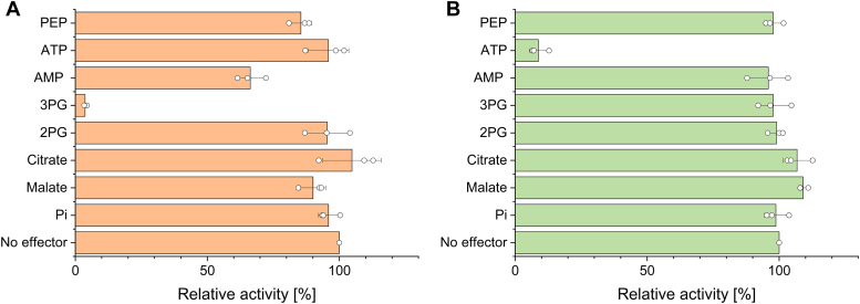
Depending on the light conditions, photosynthetic organisms switch between carbohydrate synthesis or breakdown, for which the reversibility of carbohydrate metabolism, including glycolysis, is essential. Kinetic regulation of phosphofructokinase (PFK), a key-control point in glycolysis, was studied in the cyanobacterium Synechocystis sp. PCC 6803. The two PFK iso-enzymes (PFK- A1, PFK-A2), were found to use ADP instead of ATP, and have similar kinetic characteristics, but differ in their allosteric regulation. PFK-A1 is inhibited by 3-phosphoglycerate, a product of the Calvin-Benson-Bassham cycle, while PFK-A2 is inhibited by ATP, which is provided by photosynthesis. This regulation enables cyanobacteria to switch PFK off in light, and on in darkness. ADP dependence has not been reported before for the PFK-A enzyme family and was thought to be restricted to the PFK-B ribokinase superfamily. Phosphate donor specificity within the PFK-A family could be related to specific binding motifs in ATP-, ADP-, and PPi-dependent PFK-As. Phylogenetic analysis revealed a distribution of ADP-PFK-As in cyanobacteria and in a few alphaproteobacteria, which was confirmed in enzymatic studies.

Keywords: ADP; ADP-dependent PFK-A; ATP; PFK-A superfamily; PPi signature binding motifs; allosteric regulation; cyanobacteria.


Conflict of interest statement

Conflict of interest The authors declare that they have no conflicts of interest with the contents of this article.

Figures

Figure 1
Figure 1
Overview of pathways involved in CO2-fixation and carbohydrate metabolism in Synechocystis sp. PCC6803. The key enzymes of the reversible EMP pathway, phosphofructokinase (PFK) and the bifunctional fructose-1,6-bisphosphatase/sedoheptulose-1,7-bisphosphatase (FBP/SBPase) are shown. Pathways are differentiated by arrow color: The EMP pathway is shown in green, the OPP pathway in cyan, the ED pathway (under discussion) in blue, the CBB cycle in purple, and the NOPP pathway in red. The PK pathway was omitted for clarity. 3PG, 3-phosphoglycerate; 6PG, 6-phosphogluconate; BPG, 1,3-bisphosphoglycerate; CBB, Calvin–Benson–Bassham cycle; DHAP, dihydroxyacetone phosphate; E4P, erythrose 4-phosphate; ED, Entner-Doudoroff pathway; EMP, Embden–Meyerhof–Parnas pathway; F6P, fructose 6-phosphate; FBP, fructose 1,6-bisphosphate; GAP, glyceraldehyde 3-phosphate; G6P, glucose 6-phosphate; KDPG, 2-keto-3-deoxy-6-phosphogluconate; OPP, oxidative pentose phosphate pathway; NOPP, non-oxidative pentose phosphate pathway; PYR, pyruvate; Ru5P, ribulose 5-phosphate; RuBP, ribulose 1,5-bisphophate; Ri5P, ribose 5-phosphate; S7P, sedoheptulose 7-phosphate; SBP, sedoheptulose 1,7-bisphosphate; X5P, xylulose 5-phosphate. The figure was created using BioRender.com.
Figure 2
Figure 2
Effector studies for PFK-A isoenzymes from Synechocystis sp. PCC 6803. The influence of different metabolites/effectors on ADP-PFK-A1 (A) and ADP_PFK-A2 is shown. The influence of different metabolites/effectors on ADP-PFK-A1 (A) and ADP-PFK-A2 (B) is shown. Assays were performed with subsaturating concentrations of F6P (0.08 mM for PFK-A1 and 0.07 mM for PFK-A2) and ADP (0.4 mM for PFK-A1 and 0.2 mM for PFK-A2). The different effectors were tested at a concentration of 1 mM. The relative activity (%) in comparison to the control without effector (100%) are shown. The specific activity without effector for ADP-PFK-A1 was 1.35 U/mg and for ADP-PFK-A2 2.77 U/mg. The means and standard deviations for three technical replicates (n = 3) are shown.
Figure 3
Figure 3
Characterization of the Synechocystis PFK-A isoenzymes. (A-C) PFK-A1, (sll1196) kinetics and the effect of 3PG. A, F6P saturation, with 3 mM ADP, and three different 3PG concentrations, (B) ADP saturation, with 3 mM F6P, and three different 3PG concentrations, (C) the effect of 3PG, with six different combinations of F6P and ADP concentrations; (DF) PFK-A2, (sll0745) kinetics and the effect of ATP. (D) F6P saturation with 3 mM ADP and three different ATP concentrations, (E) ADP saturation, with 0.8 mM F6P, and three different ATP concentrations, (F) ATP inhibition, with six different combinations of F6P and ADP concentrations. The MWC model (Equation 1) fit is shown with parameter values given in Table 1; colored bands indicate the 95% confidence intervals. Error bars indicate the standard deviation of the mean with n = 3 (technical replicates).
Figure 4
Figure 4
Structural comparison of the Synechocystis PFK-A1 with other PFK-As. Ribbon representations are shown for the monomers of Synechocystis ADP-dependent PFK-A1 (Sll1196, alphafold, brown) (A) and PFK-A2 (Sll0745, alphafold, grey) (B) in comparison with the ATP-dependent PFK-A from S. aureus (5xz9, crystal structure, blue) (30) (C) and PPi-dependent PFK-A from T. tenax (TTX 1277, alphafold, yellow) (D). The superimposition of all four monomers shown in (E) clearly shows that the cyanobacterial ADP-PFK-As adopt the typical PFK-A fold.
Figure 5
Figure 5
Phylogenetic analysis of the PFK-A family. The phylogenetic affiliations of the cyanobacterial and alphaproteobacterial ADP-PFK-As within the PFK-A superfamily are given (with uniprot accession number if available, otherwise Genbank or NCBI accessions). Enzymes characterized in this study are marked in boldface. The “40 kDa subgroup”, include ATP-PFK-As (previously named Group 1 according to (15, 27, 28), dark brown), as well as PPi- and ADP-PFK-As (previously designated as Group 3, shown in brown and light brown respectively). The ADP-PFK-As subcluster is further subdivided into ADP-PFK-A1 (Sll1196) and ADP-PFK-A2 (Sll0745) homologs. The “50 kDa subgroup” (group 2a) of ATP-PFK-As is shown in blue, and the “50 kDa” (group 2 “short”) and the “60 kDa subgroup” (group 2 “long”) of PPi-dependent PFK-As are shown in light and dark green, respectively. The evolutionary history was inferred by using the Maximum Likelihood method and the Le/Gascuel model (62). The model was selected based on the lowest Bayesian information criterion value using the “Find best protein model” option implemented in the MEGA11 package. The tree with the highest log likelihood (−29534.88) is shown. The percentage of trees in which the associated taxa clustered together is shown next to the branches. Initial tree(s) for the heuristic search were obtained automatically by applying Neighbor-Join and BioNJ algorithms to a matrix of pairwise distances estimated using the JTT model, and then selecting the topology with superior log likelihood value. The tree is drawn to scale, with branch lengths measured in the number of substitutions per site. This analysis involved 71 amino acid sequences. All positions containing gaps and missing data were eliminated (complete deletion option). There was a total of 286 positions in the final dataset. Evolutionary analyses were conducted in MEGA11 (61).
Figure 6
Figure 6
Elucidation of specific phosphate donor binding motifs in the PFK-A family. Comparison of the ATP binding site in the ATP-PFK-A from S. aureus (A) with the proposed ADP-binding site in the ADP-PFK-A1 (Sll1196) from Synechocystis (B) and the phosphate binding in the PPi-PFK-A from T. tenax (C). The sulfate ion shown in the latter structure was indicated to adopt the same position as the non-transferred phosphate of pyrophosphate in the Borreliella burgdorferi PPi-PFK (33, 34) and superimposes well with the β phosphate of ATP in e.g. the S. aureus ATP-PFK-A. Proteins are depicted as transparent surface representations (AC, colored according to element). ATP, ADP, and sulfate as well as the residues involved in phosphate donor binding are labeled and shown as stick models. Together with the condensed sequence alignment shown in (D) these structures illustrate the similarities as well as the differences between ATP-, ADP-, and PPi-PFK-As. Of note, those residues, i.e. I122 in Sll1196 and D102 in the PPi-PFK-A from T. tenax, preventing ATP binding are integral constituents of the (putative) ADP and PPi binding pockets. In the condensed alignment (D), the key residues for the different phosphate donor specificities discussed in the text and also shown in the panels A-C are highlighted in cyan (ATP-PFK-A), pink (ADP-PFK-A), and yellow (PPi-PFK-A). Additional residues involved in ATP binding in ATP-PFK-As are shown in blue font, ADP binding in ADP-PFK-As in brown, phosphate binding in PPi-PFK-As in green, and F6P binding are shown in red. To the right of the sequences the signature motifs defining the ATP, ADP, and PPi dependence are indicated (based on the red outlined part of the alignment). Furthermore, the catalytically essential aspartate is shown in the structures (AC) and indicated by an asterisk in the alignment.

References

    1. Schulze D., Kohlstedt M., Becker J., Cahoreau E., Peyriga L., Makowka A., et al. GC/MS-based 13C metabolic flux analysis resolves the parallel and cyclic photomixotrophic metabolism of Synechocystis sp. PCC 6803 Selected Deletion Mutants Including Entner-doudoroff Phosphoketolase Pathways. Microb. Cell Fact. 2022;21:69. - PMC - PubMed
    1. Makowka A., Nichelmann L., Schulze D., Spengler K., Wittmann C., Forchhammer K., et al. Glycolytic shunts replenish the Calvin-Benson-Bassham cycle as anaplerotic reactions in cyanobacteria. Mol. Plant. 2020;13:471–482. - PubMed
    1. Yang C., Hua Q., Shimizu K. Integration of the information from gene expression and metabolic fluxes for the analysis of the regulatory mechanisms in Synechocystis. Appl. Microbiol. Biotechnol. 2002;58:813–822. - PubMed
    1. Knowles V.L., Plaxton W.C. From genome to enzyme: analysis of key glycolytic and oxidative pentose-phosphate pathway enzymes in the cyanobacterium Synechocystis sp. PCC 6803. Plant Cell Physiol. 2003;44:758–763. - PubMed
    1. Chen X., Schreiber K., Appel J., Makowka A., Fahnrich B., Roettger M., et al. The Entner-Doudoroff pathway is an overlooked glycolytic route in cyanobacteria and plants. Proc. Natl. Acad. Sci. U. S. A. 2016;113:5441–5446. - PMC - PubMed

Publication types

MeSH terms

LinkOut - more resources