. 2024 Oct 11;15(1):8804.
doi: 10.1038/s41467-024-53075-x.

Advanced glycation end-product crosslinking activates a type VI secretion system phospholipase effector protein

Affiliations

Advanced glycation end-product crosslinking activates a type VI secretion system phospholipase effector protein

Steven J Jensen et al. Nat Commun. .

Abstract

Advanced glycation end-products (AGE) are a pervasive form of protein damage implicated in the pathogenesis of neurodegenerative disease, atherosclerosis and diabetes mellitus. Glycation is typically mediated by reactive dicarbonyl compounds that accumulate in all cells as toxic byproducts of glucose metabolism. Here, we show that AGE crosslinking is harnessed to activate an antibacterial phospholipase effector protein deployed by the type VI secretion system of Enterobacter cloacae. Endogenous methylglyoxal reacts with a specific arginine-lysine pair to tether the N- and C-terminal α-helices of the phospholipase domain. Substitutions at these positions abrogate both crosslinking and toxic phospholipase activity, but in vitro enzyme function can be restored with an engineered disulfide that covalently links the N- and C-termini. Thus, AGE crosslinking serves as a bona fide post-translation modification to stabilize phospholipase structure. Given the ubiquity of methylglyoxal in prokaryotic and eukaryotic cells, these findings suggest that glycation may be exploited more generally to stabilize other proteins. This alternative strategy to fortify tertiary structure could be particularly advantageous in the cytoplasm, where redox potentials preclude disulfide bond formation.


Conflict of interest statement

The authors declare no competing interests.

Figures

Fig. 1
Fig. 1. Modification of the Tle phospholipase domain.
a Tle and Tle-Tli production was induced with arabinose in E. cloacae and E. coli cells. Urea-soluble total protein was extracted for immunoblot analysis using anti-Tle polyclonal antisera. Molecular mass markers (kDa) are on the left. This experiment was performed independently three times with similar results. b His6-lipase domains were produced with (or without) ∆ss-Tli in E. coli cells, then purified by Ni2+-affinity chromatography under non-denaturing (native) or denaturing conditions for SDS-PAGE analysis. Molecular mass markers (kDa) are on the left. c Deconvoluted mass spectrum of purified modified His6-lipase domain. d In vitro phospholipase A1 activities of purified lipase domains. e In vitro phospholipase A2 activities of purified lipase domains. Phospholipase activity data are presented as mean values ± standard deviation for three independent experiments. Source data are provided as a Source Data file.
Fig. 2
Fig. 2. Structure of the lipase•immunity protein complex.
a Structure of the lipase•immunity protein complex. Lipase active-site and MODIC residues are indicated in one-letter code. b Hydrogen-bond network of the lipase•∆ss-Tli complex interface. c The AlphaFold2 model of Tle residues Thr172 - Ala472 superimposed onto the lipase domain structure (PDB: 9CYS). The AlphaFold2 modeled α6 lid helix is shown in cyan, and the unresolved segment from the experimental model is rendered in charcoal. d Superimposition of the Tle lipase domain onto inactive (closed) and activated (open) forms of the R. meihei triacylglycerol lipase. Residues are indicated using three-letter code. e Surface electrostatic potentials of lipase domains from panel d. Red surfaces are electronegative and blue surfaces are electropositive. f MODIC electron density is illustrated by omit map of lipase helices α1 and α13 at 3 sigma.
Fig. 3
Fig. 3. AGE crosslinking is required for phospholipase activity.
a The indicated His6-lipase domain variants were produced with (or without) ∆ss-Tli, then purified by Ni2+-affinity chromatography under denaturing conditions for SDS-PAGE analysis. Molecular mass markers (kDa) are on the left. b Deconvoluted mass spectrum of the His6-lipase(Arg180Lys)•∆ss-Tli complex. c Deconvoluted mass spectrum of the His6-lipase(Lys461Gln)•∆ss-Tli complex. d In vitro phospholipase A1 activity assays of the purified domains from panel a. Phospholipase activity data are presented as mean values ± standard deviation for three independent experiments. e Competition co-cultures. E. cloacae inhibitor strains were co-cultured with ∆tle-tli target bacteria and viable cells enumerated as described in Methods. Competitive indices are averages ± standard error for three independent experiments. f Urea-soluble cell lysates were prepared from the inhibitor strains in panel e and subjected to immunoblot analysis with polyclonal antibodies to Tle. Molecular mass markers (kDa) are on the left. This experiment was performed independently three times with similar results. Source data are provided as a Source Data file.
Fig. 4
Fig. 4. Aldehyde reductases suppress Tle crosslinking.
a His6-Tle was produced with Tli in E. coli cells that overexpress the indicated methylglyoxal detoxification enzymes. His6-Tle was purified by Ni2+-affinity chromatography under denaturing conditions and analyzed by SDS-PAGE. Molecular mass markers (kDa) are on the left. b Competition co-cultures. E. cloacae inhibitor strains that overexpress methylglyoxal detoxification enzymes were co-cultured with E. cloacaetle-tli target bacteria and viable cells enumerated as described in Methods. Competitive indices are averages ± standard error for three independent experiments. Source data are provided as a Source Data file.
Fig. 5
Fig. 5. In vitro activation of the phospholipase domain.
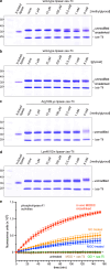
Unmodified lipase domain and ∆ss-Tli were mixed in vitro to form a complex, which was then treated with methylglyoxal (a) or glyoxal (b) at the indicated concentrations for 7 h at 37 °C. Treated complexes were analyzed by SDS-PAGE. Methylglyoxal treatment of Arg180Lys (c) and Lys461Gln (d) lipase domains in complex with ∆ss-Tli. Molecular mass markers (kDa) are on the left. e In vitro crosslinked His6-lipase domains were isolated from ∆ss-Tli by Ni2+-affinity chromatography under denaturing conditions, then refolded by dialysis for phospholipase A1 activity assays. Data are presented as mean values ± standard deviation for three independent experiments. Source data are provided as a Source Data file.
Fig. 6
Fig. 6. Phospholipase activation with an engineered disulfide bond.
a Structure of R. meihei lipase illustrating the disulfide bond linking the N- and C-termini. b Predicted structure of Disulfide-by-design engineered Tle phospholipase domain. c The indicated His6-lipase variants were produced with ∆ss-Tli in E. coli cells, and the complexes purified by Ni2+-affinity chromatography under non-denaturing conditions. Complexes were analyzed by SDS-PAGE under oxidizing and reducing (β-mercaptoethanol, β-ME) conditions. Molecular mass markers (kDa) are on the left. d The indicated His6-lipase domains were isolated from ∆ss-Tli by Ni2+-affinity chromatography under denaturing conditions, then refolded by dialysis for phospholipase A1 activity assays. Phospholipase activity data are presented as mean values ± standard deviation for three independent experiments. Reactions were supplemented with dithiothreitol (DTT) where indicated. Source data are provided as a Source Data file.
Fig. 7
Fig. 7. Model for Tli-dependent Tle activation and T6SS mediated delivery.
Mature Tli (lipo-Tli) is secreted to the periplasm where it is lipidated and embedded in membranes to protect the cell from incoming Tle effectors. E. cloacae cells also produce cytosolic Tli, which presumably forms a transient complex with Tle to enable methylglyoxal crosslinking. The activated effector is then dissociated from the immunity protein and loaded into the T6SS apparatus for export. Upon delivery into the target-cell periplasm, activated Tle hydrolyzes membrane phospholipids.

References

    1. Ruhe, Z. C., Low, D. A. & Hayes, C. S. Polymorphic toxins and their immunity proteins: diversity, evolution, and mechanisms of delivery. Annu. Rev. Microbiol.74, 497–520 (2020). - PMC - PubMed
    1. Hernandez, R. E., Gallegos-Monterrosa, R. & Coulthurst, S. J. TypeV. I. secretion system effector proteins: effective weapons for bacterial competitiveness. Cell Microbiol22, e13241 (2020). - PubMed
    1. Basler, M., Pilhofer, M., Henderson, G. P., Jensen, G. J. & Mekalanos, J. J. Type VI secretion requires a dynamic contractile phage tail-like structure. Nature483, 182–186 (2012). - PMC - PubMed
    1. Allsopp, L. P., Bernal, P., Nolan, L. M. & Filloux, A. Causalities of war: the connection between type VI secretion system and microbiota. Cell Microbiol.22, e13153 (2020). - PMC - PubMed
    1. Brunet, Y. R., Zoued, A., Boyer, F., Douzi, B. & Cascales, E. The Type VI Secretion TssEFGK-VgrG Phage-Like Baseplate Is Recruited to the TssJLM Membrane Complex via Multiple Contacts and Serves As Assembly Platform for Tail Tube/Sheath Polymerization. PLoS Genet.11, e1005545 (2015). - PMC - PubMed

Publication types

MeSH terms