. 2024 Oct 11;7(1):1296.
doi: 10.1038/s42003-024-06971-3.

Type-H endothelial cell protein Clec14a orchestrates osteoblast activity during trabecular bone formation and patterning

Affiliations

Type-H endothelial cell protein Clec14a orchestrates osteoblast activity during trabecular bone formation and patterning

Georgiana Neag et al. Commun Biol. .

Abstract

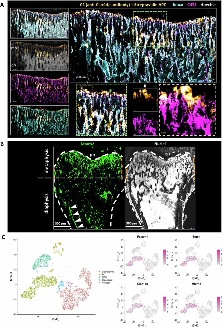
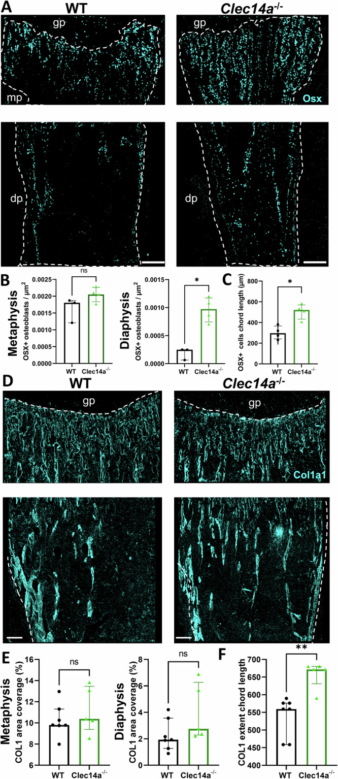
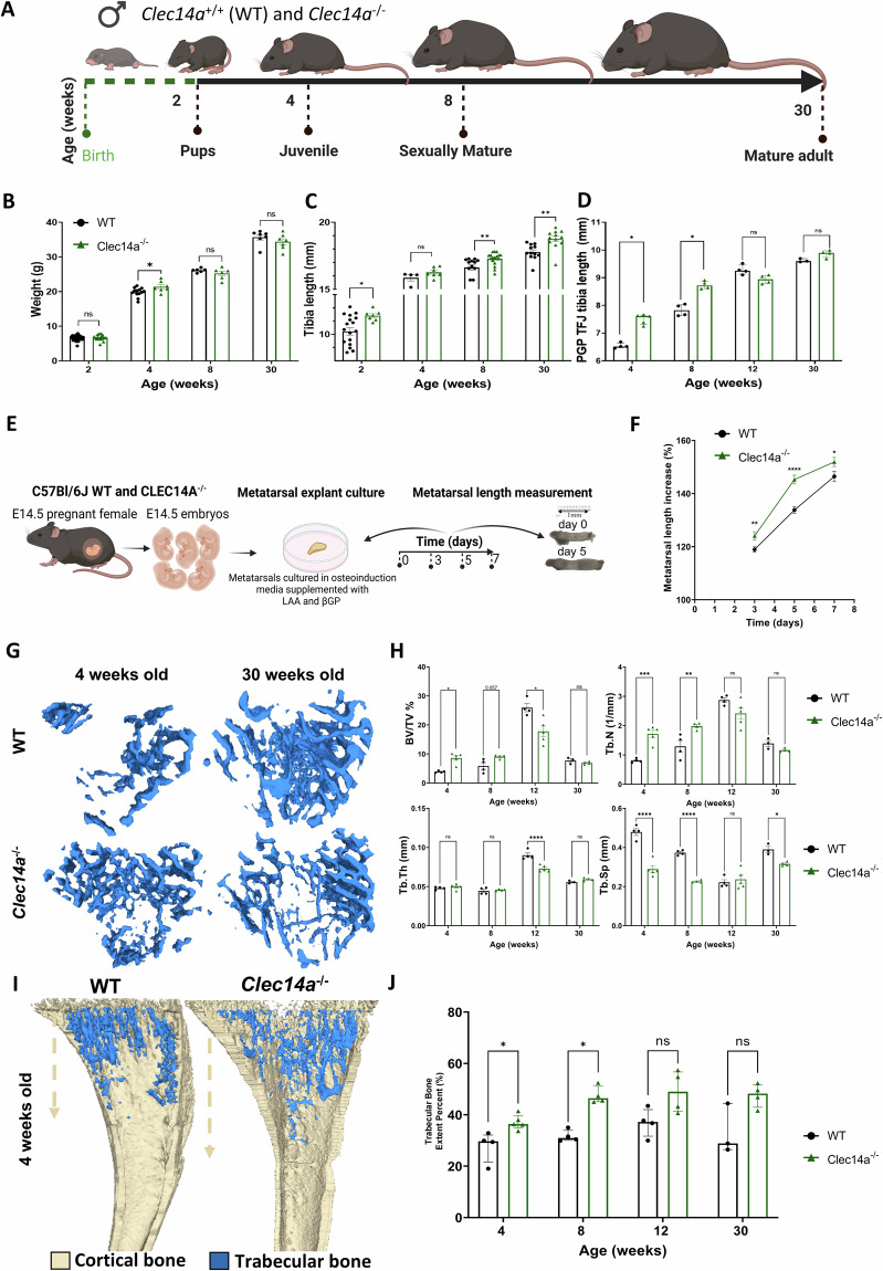
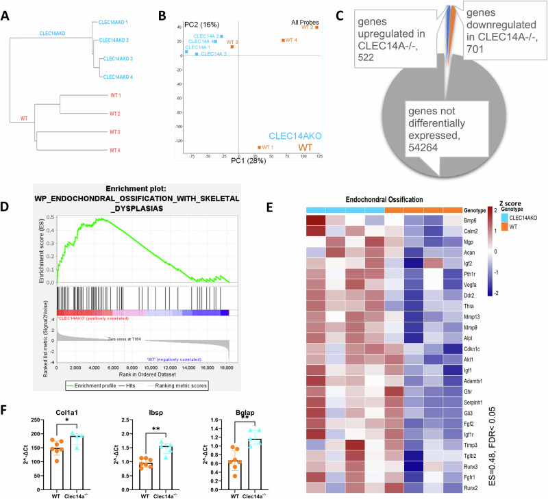
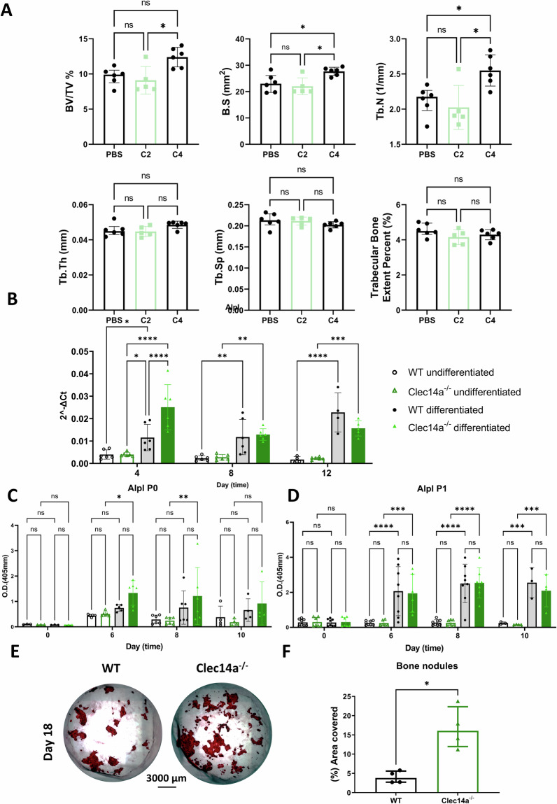
Type-H capillary endothelial cells control bone formation during embryogenesis and postnatal growth but few signalling mechanisms underpinning this influence have been characterised. Here, we identify a highly expressed type-H endothelial cell protein, Clec14a, and explore its role in coordinating osteoblast activity. Expression of Clec14a and its ligand, Mmrn2 are high in murine type-H endothelial cells but absent from osteoblasts. Clec14a-/- mice have premature condensation of the type-H vasculature and expanded distribution of osteoblasts and bone matrix, increased long-bone length and bone density indicative of accelerated skeletal development, and enhanced osteoblast maturation. Antibody-mediated blockade of the Clec14a-Mmrn2 interaction recapitulates the Clec14a-/- phenotype. Endothelial cell expression of Clec14a regulates osteoblast maturation and mineralisation activity during postnatal bone development in mice. This finding underscores the importance of type-H capillary control of osteoblast activity in bone formation and identifies a novel mechanism that mediates this vital cellular crosstalk.


Conflict of interest statement

The authors declare no competing interests.

Figures

Fig. 1
Fig. 1. Mmrn2 and Clec14a expression at sites of trabecular bone formation.
A Maximum intensity projection images show immunofluorescent staining of Clec14a (orange), Endomucin (Emcn - cyan), Cd31 (magenta), Hoechst (grey). Clec14a expression was tracked in vivo, a biotin-labelled monoclonal antibody (C2) raised against Clec14a was injected intraperitoneally and tissue was collected 24 h post injection. Scale bar is 200 µm. i. Region of interest with arrowheads (green) indicating areas where co-expression of all markers (white) is evident. ii. High magnification image demonstrating high expression of Clec14a within bud-like vessel protrusions and anastomotic arches. Scale bar is 10 µm. B Maximum intensity projection images show immunostaining of Mmrn2 (left image - green) and nuclei (Hoechst, right image - grey). Mmrn2 is located predominantly in the anatomical area normally covered by type-H vessels in the metaphysis (mp), moderate Mmrn2 expression is also observed in the region where type-L vessels reside (dp). White arrowheads indicate Mmrn2 staining on an arterial vessel (arterial vessel identified with reference to Kusumbe et al. 2014, Fig. 1 and extended data Fig. 2). White dashed line demarcates the growth plate metaphysis border and the periosteum. Abbreviations: gp-growth plate, mp-metaphysis, dp-diaphysis. Scale bars are 300 µm. C Expression of Clec14a and Mmrn2 in the bone stroma of adult mice (GSE128423). Left panel: t-SNE map of the stromal clusters in the bone coloured-coded according to their cluster name. Right panel: t-SNE maps reveal expression of H-type endothelial cell (EC) markers Pecam1 and Emcn alongside Clec14a and Mmrn2.
Fig. 2
Fig. 2. Changes in vascularisation of the long bone in Clec14a−/− mice.
A Representative flow-cytometry heatmap scatter plots showing Cd31high Emcnhigh EC (H-type) and Cd31low Emcnlow (L-type) endothelial cells (EC) in Cd45 Ter119- cells isolated from the long bones of Clec14a+/+ and Clec14a−/− 2-week-old pups. B Relative abundance of H-type compared to L-type EC in 2-week-old mouse tibiae from Clec14a−/− mice compared to Clec14a+/+ (WT). Data was analysed with a Mann–Whitney test, results are presented as median ± IQR, *P < 0.05, ns = not significant. C High resolution 3D confocal microscopy of the juvenile (4 weeks old) Clec14a+/+ (WT) and Clec14a−/− mouse tibia capillary network. The panel shows representative maximum intensity projection images depicting organisation of H-type vessels in Clec14a+/+ (WT) and Clec14a−/− tibia. Vessels in the tibia section have been immunolabelled with Emcn (magenta). White line marks the limits of the type-H vessel front, note the shorter H capillary front in the Clec14a−/− images. Scale bars are 200 µm. D Left graph: quantification of the length of H type columns in the metaphysis, data represents mean values for each biological replicate. Right graph: quantitative evaluation of the length of the type-H vessels front. In D and E, data is represented as mean ± SD, results were analysed by unpaired T test. **P < 0.01; *P < 0.05; ns = non-significant. E Representative enlarged images of the leading type-H vascular front (Emcn, magenta) in Clec14a+/+ and Clec14a−/− mice. Note dysregulation of the advancing H-type bulges protruding into the growth plate (highlighted by the white dashed ovals in the Clec14a−/− image). Scale bars are 100 µm. F Representative immunofluorescent images of type-H vessels labelled with Emcn (blue) and counterstaining of cells and extracellular matrix in the metaphysis and growth plate with wheat germ agglutinin (WGA - orange). Note blunting of type-H vessel buds in Clec14a−/−. Scale bars are 200 µm (main images) and 50 µm (high magnification insets). G Quantitative evaluation of the fraction of type-H vessels buds showing blunted phenotype in the bone metaphysis. H Maximum intensity projection images show immunostaining of Emcn (fire – colour gradient with high expression in orange and low expression in magenta) and nuclei (Hoechst, grey) in sections collected from 4-week-old Clec14a+/+ and Clec14a−/− male mice. Note absence of filopodia (white arrowhead) in Clec14a−/− images. Scale bars are 20 µm. I, J Immunofluorescent images of the chondro-osseous interface (wheat germ agglutinin WGA - orange, Emcn - blue). Note the normal patterning of the growth plate in both Clec14a+/+ and Clec14a−/−. Scale bar = 200 µm (I) and 50 µm (J).
Fig. 3
Fig. 3. Increased extension of the osteoblast front in the long bone of Clec14a−/− mice.
A Representative images of Osx (cyan) immunolabeled metaphyseal and diaphyseal bone sections from P4 neonatal pups. Dashed line demarcates the bone edge of images and the most distal point of the proximal growth plate. Note the extension of Osx+ cells in Clec14a−/−. Scale bars are 100 µm. B Quantitative measurements of osteoblast density per µm2 in the P4 mouse tibia metaphysis and diaphysis. C Quantification of the length of osteoblast chords in the neonatal (P4) murine tibia. An osteoblast chord was defined as an aggregation of Osx+ cells organised in a column. The length of osteoblast chords from the proximal end of the metaphysis to their most distal point was measured. Individual dots represent the mean length of osteoblast chords in one biological replicate. Data was analysed with a Mann–Whitney test, results are presented as median ± IQR *P < 0.05, ns = not significant. D Representative maximum intensity projections of Col1a1 immunolabeled bone sections in 4-week-old mice. Upper image: white dashed line delineates the boundary between the growth plate (avascular) and the metaphysis. Lower image: dashed line demarcates the area covered by bone including the bone endosteum and periosteum. Scale bars are 100 µm. E Quantification of the percentage (%) area covered by Col1a1 immunolabeling in the tibial metaphysis (left) and diaphysis (right). F Quantification of the length of type I collagen chords in the juvenile murine tibia. Data analysed with a Mann–Whitney test, results are presented as median ± IQR, **P < 0.01, ns = not significant.
Fig. 4
Fig. 4. Bone morphometric parameters in Clec14a−/− male mouse tibia.
A) Diagram illustrating morphometry analyses timepoints (generated in BioRender): Tibiae were collected from C57BL/6J male mice at different skeletal maturation stages: juvenile (2- and 4-weeks-old), sexually mature (8-weeks-old) and mature adult (30-week-old). B Body weight. C Total tibiae length and D tibiae length from the proximal growth plate to the tibiofibular junction - PGP TFJ. Results for graphs B-C presented as mean ± SEM, data analysed with two-way ANOVA, followed by Dunnett’s post hoc. Results for graph D presented as median ± IQR, data analysed with multiple Mann–Whitney tests. E Methodology summary for the in vitro study of metatarsal elongation of E14.5 embryos (generated in BioRender). LAA-L ascorbic acid, βGP- β glycerophosphate. F Quantitation of metatarsal length as percentage increase. Results are presented as median ± IQR, data was analysed with Multiple Mann–Whitney tests. Data was obtained from N = 9, n = 34–40 Clec14a+/+ (WT) and N = 5, n = 13–16 Clec14a−/−, where N = individual biological replicates and n= total number of metatarsals analysed. G Representative μCT analysis images of 4- and 30-week-old Clec14a+/+ (WT) and Clec14a−/− mouse tibias. H Micro-computed tomography (μCT) analysis of trabecular percent bone volume (BV/TV %), trabecular thickness (Tb.Th), trabecular number (Tb.N), trabecular separation (Tb.Sp). For BV/TV %, results are presented as median ± IQR, data was analysed with multiple Mann–Whitney tests. For the other parameters, results are presented as mean ± SEM, data was analysed with two-way ANOVA, followed by Dunnett’s post hoc, compared to WT mice. I Representative μCT images of trabecular bone extent (blue). Note the increased trabecular bone extent in Clec14a−/− samples. J Quantification of trabecular bone extent as a percentage. For all graphs, statistical test results are shown as: *P < 0.05, **P < 0.01; ***P < 0.001; ****P < 0.0001. Sample size varies - each dot represents one biological repeat.
Fig. 5
Fig. 5. RNA sequencing of molecular changes in Clec14a−/− lysates.
AE Clustering and gene ontology analysis of Clec14a+/+ (WT) and Clec14a−/− pup calvaria lysates. A, B Unsupervised hierarchical clustering, (dendrogram – A; principal component analysis – B) of log2 normalised counting reads over exons (RPM-reads per million) for all detected genes, N = 4 for both groups. Clec14a−/− samples (blue), Clec14a+/+ samples (WT - orange). Each sample represents a different individual animal. C) Differentially expressed gene counts. Raw counts were analysed with DESeq2, significantly expressed genes were cut off at log2FC values > 0.5, adjusted p (padj) < 0.05, p value was corrected for multiple comparisons (Benjamini–Hochberg); Orange = genes upregulated in Clec14a−/− samples; Blue = genes downregulated in Clec14a−/− samples. D GSEA analysis enrichment score for pathways related to endochondral ossification. ES-enrichment score, FDR -false discovery rate. Gene expression is presented as a Z score (scale). E GSEA heatmap showing the list of leading-edge genes enriched at the top of the Wikipathways ‘Endochondral Ossification’ gene set. Heatmap presents upregulation of transcripts relevant to endochondral ossification in Clec14a−/− (blue) samples compared to WT control (orange). Gene expression is presented as a Z score. Transcripts presented were identified as statistically significant by GSEA analysis, FDR (q-value) < 0.05, nominal p < 0.01. F Analysis of expression of genes linked to osteoblast maturation and osteogenic activity. P4 pup calvarial lysates were subjected to RT qPCR analysis to dissect expression patterns of genes known to regulate commitment to osteoblast lineage and osteoblast maturation, gene expression was normalised to Gapdh, shown as arbitrary units (2^-ΔCT). Data is presented as median ± IQR, a Mann–Whitney test was used to analyse the results. Plots represent results from two independent experiments (n = 7 Clec14a+/+, n = 4–5 Clec14a−/−), data points in each graph are biological replicates.
Fig. 6
Fig. 6. Increased mineralisation activity of Clec14a−/−-isolated osteoblasts in vitro.
A Bone morphometric parameters in the mouse tibia of C2- and C4-treated mice. Note, C4 blocks and C2 does not block the Clec14a-Mmrn2 interaction, as previously reported. Micro-computed tomography (μCT) analysis of trabecular percent bone volume (BV/TV%), bone surface area (B.S), trabecular number (Tb.N), trabecular thickness (Tb.Th), trabecular separation (Tb.Sp) and trabecular extent. For BV/TV %, results are presented as median ± IQR, data was analysed with multiple Mann–Whitney tests. For the other parameters, results are presented as mean ± SEM, data was analysed with two-way ANOVA, followed by Dunnett’s post hoc, *p < 0.05. B Alpl mRNA expression was analysed by RT qPCR in calvarial osteoblasts. Results were analysed with a two-way ANOVA followed by Dunnett’s correction for multiple testing, data is presented as mean ± SD. C, D Murine calvarial osteoblasts were isolated from Clec14a+/+ (WT) and Clec14a−/− neonatal pups at postnatal day 4 and osteogenesis was induced with osteoinduction media in (C) freshly isolated cells prior to passage (P0) or (D) in cultured cells after passage (P1). Alpl activity was measured by ELISA after 0, 6, 8 and 10 days in culture, in Clec14a+/+ and Clec14a−/− osteoblasts that were incubated with either expansion media (undifferentiated – see key on graph) or osteoinduction media (differentiated – see key on graph). Data is presented as optical density (O.D.). Results were analysed with a two-way ANOVA followed by Dunnett’s post hoc, data is presented as mean ± SD. *p < 0.05, **p < 0.01, ***p < 0.001. E Representative images of alizarin red stained osteoblast-deposited calcium following 18 days in culture. F Alizarin red coverage as percentage. Results were analysed with a Mann–Whitney test, data is presented as median ± IQR, *p < 0.05. For all graphs sample size is represented by each individual dot in the graphs.

References

    1. Carmeliet, P. & Jain, R. K. Angiogenesis in cancer and other diseases. Nature407, 249–257 (2000). - PubMed
    1. Eelen, G., Treps, L., Li, X. & Carmeliet, P. Basic and therapeutic aspects of angiogenesis updated. Circ. Res127, 310–329 (2020). - PubMed
    1. Potente, M., Gerhardt, H. & Carmeliet, P. Basic and therapeutic aspects of angiogenesis. Cell146, 873–887 (2011). - PubMed
    1. Maes, C. et al. Osteoblast precursors, but not mature osteoblasts, move into developing and fractured bones along with invading blood vessels. Dev. Cell19, 329–344 (2010). - PMC - PubMed
    1. Kusumbe, A. P., Ramasamy, S. K. & Adams, R. H. Coupling of angiogenesis and osteogenesis by a specific vessel subtype in bone. Nature507, 323–328 (2014). - PMC - PubMed

Substances

LinkOut - more resources