. 2024 Oct 11;7(1):1310.
doi: 10.1038/s42003-024-07019-2.

Neural activity for complex sounds in the marmoset anterior cingulate cortex

Affiliations

Neural activity for complex sounds in the marmoset anterior cingulate cortex

Rebekah L Gilliland et al. Commun Biol. .

Abstract

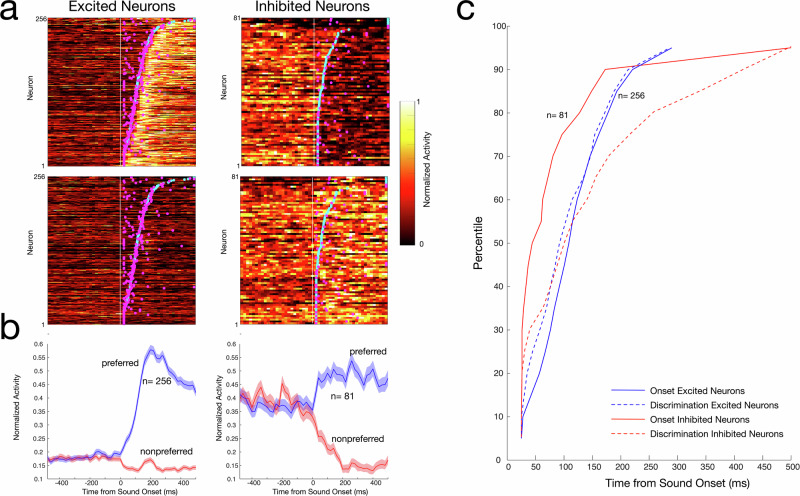
Vocalizations play an important role in the daily life of nonhuman primates and are likely precursors of human language. Recent functional imaging studies in the highly vocal common marmoset (Callithrix jacchus) have suggested that anterior cingulate cortex (ACC) area 32 may be a part of a vocalization-processing network but the response properties of area 32 neurons to auditory stimuli remain unknown. Here we perform electrophysiological recordings in area 32 in marmosets with high-density Neuropixels probes and characterize neuronal responses to a variety of sounds including conspecific vocalizations. Nearly half of the neurons in area 32 respond to conspecific vocalizations and other complex auditory stimuli. These responses exhibit dynamics consisting of an initially non-selective reduction in neural activity, followed by an increase in activity that immediately conveys sound selectivity. Our findings demonstrate that primate ACC area 32 processes species-specific and biologically relevant sounds.


Conflict of interest statement

The authors declare no competing interests.

Figures

Fig. 1
Fig. 1. Recording approach, confirmation of recording sites, and auditory response profiles of example neurons.
a Schematic depiction of Neuropixels probe and trajectory to area 32. b Ex-vivo anatomical MRI images in sagittal plane obtained at 15.2 T showing location of electrode tracts (red arrows) and termination within areas 32 and 32 v of marmoset anterior cingulate cortex in the left hemisphere. c Auditory responses of single area 32 neurons to 11 sound stimuli, recorded simultaneously within a single recording session. Spectrograms of auditory stimuli presented, including marmoset vocalizations, are shown Middle panel, auditory responses of single area 32 neurons aligned on auditory stimulus onset and sorted by depth from probe tip. Each row depicts mean discharge rate within 20 ms bins for a single area 32 neuron, colourmap indicates normalized response magnitude. Lower panel, responses of single neurons to the suite of 11 auditory stimuli. Rasters and spike density functions aligned on stimulus onset. Mean action potential waveforms for each example neuron shown at left.
Fig. 2
Fig. 2. Timing and magnitude of neurons with excited and inhibited responses to auditory stimuli.
a Discharge rates of neurons with excited and inhibited responses to auditory stimuli. Each row depicts activity of a single neuron, heatmap indicates normalized mean discharge rate in 25 ms bins. Neurons are sorted in order of onset times of auditory responses, depicted by cyan dots. Mean spike density functions show discharge rates and standard error of the mean for the subpopulations of area 32 neurons with excited (blue line) and inhibited (red line) responses. b Histograms depicting trough-to-peak waveform durations for neurons with excited and inhibited responses. Proportions of broad and narrow spiking neurons did not differ between response types. c Cumulative onset times of auditory responses for neurons with inhibitory (red line) and excitatory (blue line) responses.
Fig. 3
Fig. 3. Selectivity of excited and inhibited responses of area 32 neurons to auditory stimuli.
a Proportion of selective neurons with excited responses that exhibiting significant activity for each of the 11 sounds, (b) same as (a) for neurons with inhibited responses. c Histogram of selectivity indices computed for the suite of presented sounds for excited neurons. (d) Same as (c) for inhibited neurons.
Fig. 4
Fig. 4. Comparison of responses of area 32 neurons to intact and scrambled sound stimuli.
a Spectrograms of intact and scrambled versions of the 11 auditory stimuli and spike density functions of responses of example neuron to intact (black lines) and scrambled (red lines) stimuli aligned to sound onset. b Scatterplot of selectivity indices for scrambled and intact sounds for neurons with excited responses. c Same as (b) for neurons with inhibited responses.
Fig. 5
Fig. 5. Responses of area 32 neurons to pure tones.
a Responses of 3 neurons to vocalizations and FRA of the same neurons. b Normalized population activity to pure tones for responsive neurons with excited responses, selective neurons with excited responses and responsive neurons with inhibited responses. White asterix indicate significant differences from baseline (Wilconxon signed rank test, p < 0.01).
Fig. 6
Fig. 6. Timecourse of auditory response selectivity of neurons in area 32.
a Top four panels, plots of mean discharge rates of single neurons aligned on auditory stimulus onset, sorted in increasing order of onset times (cyan dots) between their preferred and non-preferred stimuli. Magenta dots depict discrimination times of auditory responses. Colourmap depicts normalized mean discharge rate computed in 25 ms bins. b Mean spike density functions and standard error of the mean for preferred (blue lines) and non-preferred (red lines) auditory stimuli aligned on stimulus onset for excited (bottom left) and inhibited (bottom right) neurons. c Cumulative plot of distribution of onset times (solid lines) and discrimination times (dashed lines) for excited (blue) and inhibited neurons (red). Discrimination times lagged onset times for inhibitory neurons only.
Fig. 7
Fig. 7. Schematic of the proposed model of the hierarchical processing circuit for auditory stimuli involving the amygdala, higher auditory areas, and area 32.
Prior to onset of an auditory stimulus, many area 32 neurons exhibit spontaneous activity. Following onset of an auditory stimulus, input from amygdala neurons silences area 32 neurons, effectively reducing noise in discharge rates. Responses and selectivity of area 32 neurons are enhanced by later input from higher auditory areas (light blue, top right panel). MRI shows pre-processed retrograde tracer injection into area 32 (R01_0095) from BMCR-explorer. Created in BioRender. Everling, S. (2024) BioRender.com/u56u804.

Similar articles

References

    1. Ghazanfar, A. A. & Rendall, D. Evolution of human vocal production. Curr. Biol.18, R457–R460 (2008). - PubMed
    1. Agamaite, J. A., Chang, C.-J., Osmanski, M. S. & Wang, X. A quantitative acoustic analysis of the vocal repertoire of the common marmoset (Callithrix jacchus). J. Acoust. Soc. Am.138, 2906–2928 (2015). - PMC - PubMed
    1. Bezerra, B. M. & Souto, A. Structure and usage of the vocal repertoire of Callithrix jacchus. Int. J. Primatol.29, 671–701 (2008).
    1. Choi, J. Y., Takahashi, D. Y. & Ghazanfar, A. A. Cooperative vocal control in marmoset monkeys via vocal feedback. J. Neurophysiol.114, 274–283 (2015). - PMC - PubMed
    1. Roy, S., Miller, C. T., Gottsch, D. & Wang, X. Vocal control by the common marmoset in the presence of interfering noise. J. Exp. Biol.214, 3619–3629 (2011). - PMC - PubMed

LinkOut - more resources