. 2024 Oct 11;14(1):23816.
doi: 10.1038/s41598-024-74340-5.

Selective activation of PPARα by pemafibrate mitigates peritoneal inflammation and fibrosis through suppression of NLRP3 inflammasome and modulation of inflammation

Affiliations

Selective activation of PPARα by pemafibrate mitigates peritoneal inflammation and fibrosis through suppression of NLRP3 inflammasome and modulation of inflammation

Yutaka Shinkai et al. Sci Rep. .

Abstract

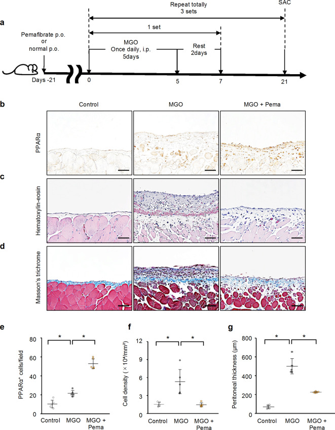
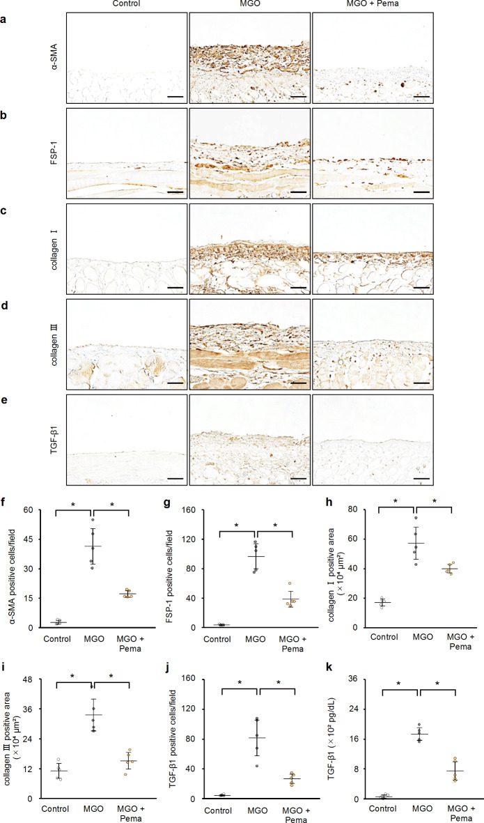
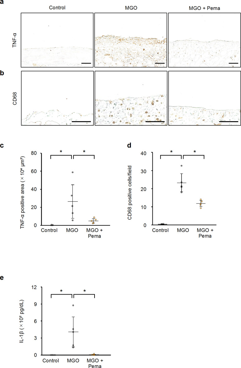
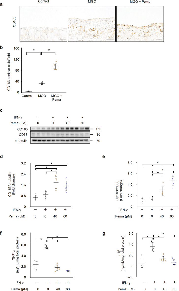
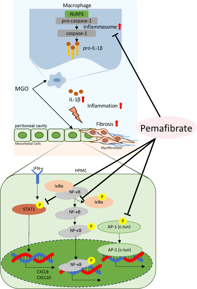
Peritoneal inflammation and fibrosis remain major challenges to the long-term maintenance of peritoneal dialysis. Pemafibrate, a selective peroxisome proliferator-activated receptor α (PPARα) modulator, has been implicated in the management of fibrosis-related disorders. We investigated whether pemafibrate ameliorates peritoneal inflammation and fibrosis and explored the underlying mechanisms in mice with methylglyoxal (MGO)-induced peritoneal fibrosis (MGO mice). MGO mice exhibited peritoneal fibrosis with increased expression of mesenchymal markers, transforming growth factor-β1 (TGF-β1), and substantial deposition of extracellular matrix (ECM) proteins. Additionally, MGO mice exhibited peritoneal inflammation as indicated by elevated tumor necrosis factor-α expression and macrophage infiltration in peritoneal tissue. These effects were mitigated by pemafibrate treatment, which also restored peritoneal membrane function. Furthermore, pemafibrate promoted anti-inflammatory macrophage polarization in both mice and THP-1 cells. In human peritoneal mesothelial cells (HPMCs), pemafibrate effectively inhibited interferon-γ-induced production of TGF-β1 and ECM while suppressing the proinflammatory cytokines nuclear factor-κB (NF-κB) and activator protein 1. The NF-κB inhibitory effect of pemafibrate involved stabilization of the NF-κB inhibitory protein IkBα. Notably, pemafibrate hindered activation of the NLR family pyrin domain containing 3/caspase-1 axis in interferon-γ-stimulated THP-1 cells. These findings suggest that pemafibrate ameliorates peritoneal inflammation and fibrosis, making it a promising candidate for peritoneal fibrosis therapy.

Keywords: Inflammasome; PPARα; Peritoneal dialysis; Peritoneal fibrosis; Peritoneal inflammation.


Conflict of interest statement

The authors declare no competing interests.

Figures

Fig. 1
Fig. 1
Pemafibrate enhanced the expression of PPARα in mice with peritoneal fibrosis. (a) Schematic diagram of the time course of pemafibrate treatment in a mouse model of MGO-induced peritoneal fibrosis. (b) Representative immunohistochemical analyses showing PPARα expression in peritoneal tissues from control mice, MGO-injected mice, and MGO-injected mice with pemafibrate (scale bar, 100 μm). (c) Representative hematoxylin–eosin staining and (d) Masson’s trichrome staining of peritoneal tissues from control mice, MGO-injected mice, and MGO-injected mice with pemafibrate (scale bar, 100 μm). (e–g) Quantitative analysis of (e) PPARα-positive cells, (f) cell density, and (g) peritoneal thickness. n = 5 per group. * indicates P < 0.05 by one-way ANOVA followed by Tukey’s post-hoc test. Pema, pemafibrate; SAC, sacrifice; PO, per os.
Fig. 2
Fig. 2
Pemafibrate ameliorated MGO-induced peritoneal fibrosis in mice. (a-e) Representative immunohistochemical analyses showing (a) α-SMA, (b) FSP-1, (c) collagen I, (d) collagen III, and (e) TGF-β1 expression in peritoneal tissue from control mice, MGO-injected mice, and MGO-injected mice with pemafibrate (scale bar, 100 μm). (f-k) Quantitative analysis of (f) α-SMA-positive cells, (g) FSP-1-positive cells, (h) collagen I-positive area, (i) collagen III-positive area, and (j) TGF-β1-positive cells from control mice, MGO-injected mice, and MGO-injected mice with pemafibrate. n = 5 per group. (k) TGF-β1 protein levels in mouse PD effluent were determined by ELISA. n = 5 per group. * indicates P < 0.05 by one-way ANOVA followed by Tukey’s post-hoc test. Pema, pemafibrate.
Fig. 3
Fig. 3
Pemafibrate reduced inflammatory responses in mice with peritoneal fibrosis. (a, b) Representative immunohistochemical analyses showing (a) TNF-α and (b) CD68 expression in peritoneal tissue from control mice, MGO-injected mice, and MGO-injected mice with pemafibrate (scale bar, 100 μm). (c, d) Quantitative analysis of (c) TNF-α expression and (d) CD68-positive cells from control mice, MGO-injected mice, and MGO-injected mice with pemafibrate. n = 5 per group. (e) IL-1β protein levels in mouse PD effluent were determined by ELISA. n = 5 per group. * indicates P < 0.05 by one-way ANOVA followed by Tukey’s post-hoc test. Pema, pemafibrate.
Fig. 4
Fig. 4
Pemafibrate drove macrophage polarization toward an anti-inflammatory phenotype. (a) Representative immunohistochemical analyses showing CD163 expression in peritoneal tissue from control mice, MGO-injected mice, and MGO-injected mice with pemafibrate (scale bar, 100 μm). (b) Quantitative analysis of CD163-positive cells from control mice, MGO-injected mice, and MGO-injected mice with pemafibrate. n = 5 per group. (c) Representative western blotting analyses showing the expression levels of CD163 and CD68 in THP-1 cells stimulated with IFN-γ. The expression of α-tubulin was utilized as an internal control. (d, e) Quantitative analysis of CD163 (anti-inflammatory macrophage marker) protein expression normalized by (d) CD68 (pan-macrophage marker) and by (e) α-tubulin from control mice, MGO-injected mice, and MGO-injected mice with pemafibrate. n = 5 per group. (f, g) The levels of (f) TNF-α and (g) IL-1β protein expression in THP-1 culture supernatants were determined using ELISA. n = 5 per group. * indicates P < 0.05 by one-way ANOVA followed by Tukey’s post-hoc test. Pema, pemafibrate.
Fig. 5
Fig. 5
Pemafibrate ameliorated functional impairment of peritoneal membrane in mice with peritoneal fibrosis. (a) The dialysate-to-plasma (D/P) ratio of urea nitrogen (UN) and (b) the peritoneal absorption of glucose from dialysate (D/D0) were assessed in control mice, MGO-injected mice, and MGO-injected mice with pemafibrate during a 10-min dialysate dwell time (4.25% dialysis solution). n = 5 per group. * indicates P < 0.05 by one-way ANOVA followed by Tukey’s post-hoc test. Pema, pemafibrate.
Fig. 6
Fig. 6
Pemafibrate inhibited IFN-γ-induced fibrotic and inflammatory markers in HPMCs. (a, b) Representative western blotting analyses showing the expression levels of (a) TGF-β and (b) secreted fibronectin from control HPMCs, and HPMCs with or without pemafibrate (40 or 60 µmol/L) after 48 h of IFN-γ stimulation. (c-h) Representative western blotting analyses showing the expression levels of (c) phospho-NF-κB and NF-κB, (d) phospho-IκBα, (e) phospho-cJun (a component of AP-1), (f) phospho-STAT1, (g) CXCL9, and (h) CXCL10 from control HPMCs, and HPMCs with or without pemafibrate (40 or 60 µmol/L) after 24 h of IFN-γ stimulation. n = 5 per group. * indicates P < 0.05 by one-way ANOVA followed by Tukey’s post-hoc test. Pema, pemafibrate.
Fig. 7
Fig. 7
Pemafibrate reduced IFN-γ-induced NLRP3 inflammasome activity in THP-1 cells. (a, b) Representative western blotting analyses showing the expression levels of (a) NLRP3 and (b) cleaved caspase-1 from control THP-1 cells, and THP-1 cells with or without pemafibrate (40 or 60 µmol/L) after 24 h of IFN-γ stimulation. n = 5 per group. * indicates P < 0.05 by one-way ANOVA followed by Tukey’s post-hoc test. Pema, pemafibrate.
Fig. 8
Fig. 8
Schematic representation of the effect of pemafibrate on peritoneal inflammation and fibrosis.

Similar articles

Cited by

References

    1. Bello, A. K. et al. Epidemiology of peritoneal dialysis outcomes. Nat. Rev. Nephrol.18, 779–793 (2022). - PMC - PubMed
    1. Devuyst, O., Margetts, P. J. & Topley, N. The pathophysiology of the peritoneal membrane. J. Am. Soc. Nephrol.21, 1077–1085 (2010). - PubMed
    1. Zhou, Q., Bajo, M. A., Del Peso, G., Yu, X. & Selgas, R. Preventing peritoneal membrane fibrosis in peritoneal dialysis patients. Kidney Int.90, 515–524 (2016). - PubMed
    1. Kawaguchi, Y. et al. Searching for the reasons for drop-out from peritoneal dialysis: A nationwide survey in Japan. Perit. Dial Int.23 (Suppl 2), S175–177 (2003). - PubMed
    1. Mizuno, M. et al. Peritonitis is still an important factor for withdrawal from peritoneal dialysis therapy in the Tokai area of Japan. Clin. Exp. Nephrol.15, 727–737 (2011). - PubMed

MeSH terms

Substances

LinkOut - more resources