. 2024 Oct 11;19(1):89.
doi: 10.1186/s13062-024-00525-7.

Deciphering the role of SAMHD1 in endometrial cancer progression

Affiliations

Deciphering the role of SAMHD1 in endometrial cancer progression

Ping Qiang et al. Biol Direct. .

Abstract

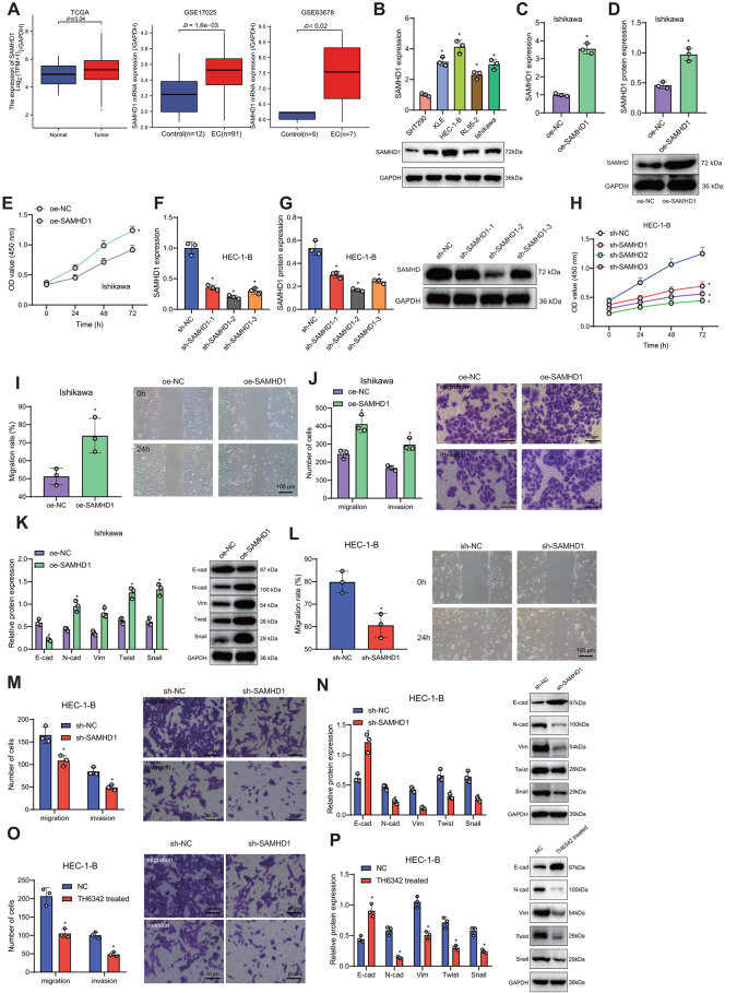
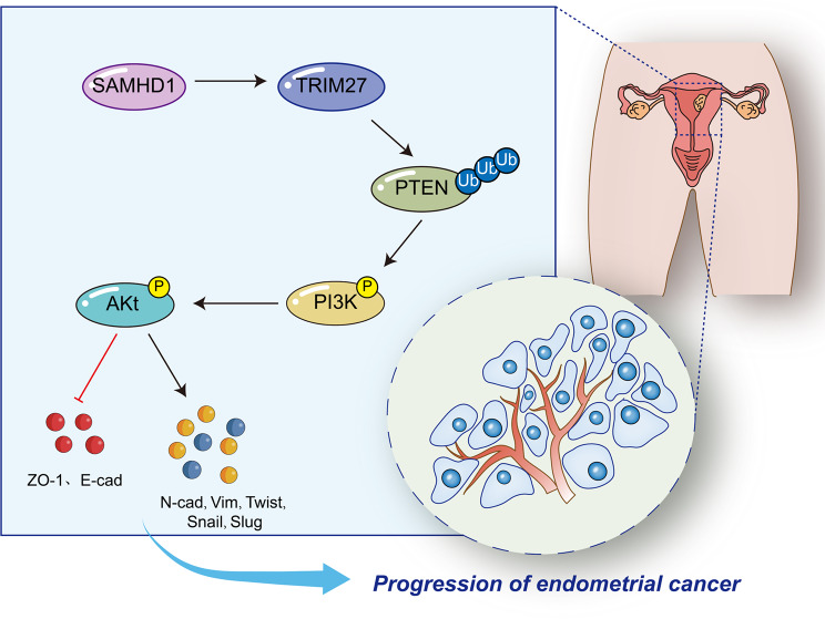
Background: Endometrial cancer (EC) presents significant clinical challenges due to its heterogeneity and complex pathophysiology. SAMHD1, known for its role as a deoxynucleotide triphosphate triphosphohydrolase, has been implicated in the progression of various cancers, including EC. This study focuses on elucidating the role of SAMHD1 in EC through its impact on TRIM27-mediated PTEN ubiquitination.

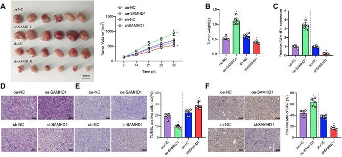
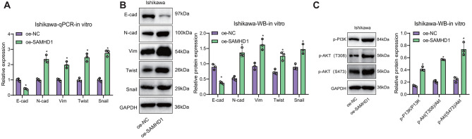
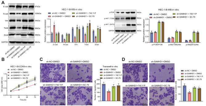
Results: Utilizing a combination of bioinformatics and cellular biology techniques, we investigated the interactions among SAMHD1, TRIM27, and PTEN. Our findings reveal that SAMHD1 modulates PTEN ubiquitination via TRIM27, impacting key pathways involved in EC pathogenesis. These interactions suggest a critical mechanism by which SAMHD1 could influence tumor behavior and progression in EC.

Conclusions: The results from this study underscore the potential of targeting the SAMHD1-TRIM27-PTEN axis as a therapeutic strategy in EC. By providing new insights into the molecular mechanisms underlying EC progression, our research supports the development of novel therapeutic approaches that could contribute to improve treatment strategies for patients with EC.

Keywords: PI3K/AKT signaling pathway; PTEN; SAMHD1; TRIM27; Ubiquitination; dNTP hydrolysis enzyme.


Conflict of interest statement

The authors declare no competing interests.

Figures

Fig. 1
Fig. 1
The effects of overexpression or silence of SAMHD1 on the proliferation, migration, and invasion abilities of endometrial cancer cells Note (A) In the TCGA, GSE17025, and GSE63678 datasets, the expression of SAMHD1 in endometrial cancer samples, ovarian cancer, and SAMHD1 expression in cancer-adjacent samples was examined. The TCGA and GSE17025 data were both normalized using TPM, while the GSE63678 data was normalized using FPKM; (B) Western blot detection of SAMHD1 expression in different endometrial cancer cell lines; (C) qRT-PCR detection of the efficiency of SAMHD1 overexpression; (D) Western blot detection of SAMHD1 expression in stable SAMHD1 overexpressing endometrial cancer cell lines; (E) CCK8 assay to assess the proliferation capacity of SAMHD1 overexpressing cell lines; (F) qRT-PCR detection of the efficiency of SAMHD1 knockdown; (G) Western blot detection of SAMHD1 expression in stable SAMHD1 knockdown endometrial cancer cell lines; (H) CCK8 assay to assess the proliferation capacity of SAMHD1 knockdown cells; (I) Scratch assay to assess the migration ability of Ishikawa cell line overexpressing SAMHD1; (J) Transwell assay to assess the invasion ability of Ishikawa cell line overexpressing SAMHD1; (K) Western blot detection of EMT-related proteins; (L) Scratch assay to assess the migration ability of SAMHD1 knockdown cells; (M) Transwell assay to assess the invasion ability of SAMHD1 knockdown cells (50 µm); (N) Western blot detection of EMT-related proteins; (O) Migration and invasion assay of HEC-1-B cells treated with SAMHD1 inhibitor TH6342; (P) Western blot analysis of EMT-related proteins (N-cadherin, Vimentin, Twist, Snail, E-cadherin) in TH6342-treated HEC-1-B cells. (The statistical analysis employed mean ± standard deviation representation. For comparing two groups, an independent samples t-test was used, while one-way ANOVA was utilized for comparing three or more groups. Data from different time points were analyzed using two-way ANOVA. ‘*’ indicates statistical significance compared to the oe-NC or sh-NC group, where P < 0.05 signifies a significant difference. The experiments were repeated three times.)
Fig. 2
Fig. 2
The effect of overexpression or silencing of SAMHD1 on the tumor growth of endometrial cancer cells in nude mice Note (A) Morphology and volume of subcutaneously transplanted tumors in each group were observed; (B) The weight of subcutaneously transplanted tumors in each group was measured; (C) Expression of SAMHD1 in subcutaneously transplanted tumors of each group was detected by qRT-PCR; (D) Morphology of subcutaneously transplanted tumors in each group was assessed by H&E staining; (E) The rate of cell apoptosis in subcutaneously transplanted tumors of each group was determined by TUNEL staining; (F) Expression of Ki67 protein in subcutaneously transplanted tumors of each group was detected by Ki67 staining (50 μm). Purple * indicates a significant difference compared to the oe-NC group (p < 0.05), blue * indicates a significant difference compared to the sh-NC group (p < 0.05) (the data are presented as mean ± standard deviation. Group comparisons were conducted using one-way analysis of variance, while repeated measures analysis of variance was employed for comparisons across different time points)
Fig. 3
Fig. 3
Regulation of the PI3K/AKT signaling pathway in Ishikawa cells by SAMHD1. (A): Note (A) Expression analysis of EMT-related molecules post SAMHD1 overexpression using qRT-PCR; (B) EMT-related molecule expression analysis post SAMHD1 overexpression using Western blot; (C) Changes in signaling pathways like PI3K, AKT, etc., post SAMHD1 overexpression at the cellular level analyzed through Western blot; data presented as mean ± standard deviation, with analysis conducted using independent sample t-test and cell experiments repeated 3 times). * denotes significance at p < 0.05 compared to the oe-NC group
Fig. 4
Fig. 4
SAMHD1 Regulation of the PI3K/AKT Signaling Pathway in the HEC-1-B Cells and Its Impact on Proliferation, Migration, and Invasion of Endometrial Cancer Cells.Note (A) Western blot analysis of the expression of EMT-related molecules in HEC-1-B cells after treatment with 740 Y-P or SC-79; (B) CCK-8 assay to evaluate the proliferation rate in SAMHD1-silenced HEC-1-B cells upon treatment with 740 Y-P/SC-79. (C-D) Transwell assay to assess the migration and invasion capabilities (50 μm) of SAMHD1-silenced HEC-1-B cells treated with 740 Y-P/SC-79. Multiple comparisons were analyzed using one-way ANOVA. (These data are represented as quantitative data, using mean ± standard deviation. Group comparisons were conducted using one-way analysis of variance, while different time points were analyzed using two-way analysis of variance. Blue * indicates significance compared to the sh-NC + DMSO group (p < 0.05), whereas pink * indicates significance compared to the sh-SAMHD1 + DMSO group (p < 0.05). Cell experiments were repeated three times
Fig. 5
Fig. 5
SAMHD1 regulates PTEN activity to influence the activation of the PI3K/AKT signaling pathway Note (A) Western blot detection of the effect of overexpressed SAMHD1 on PTEN expression; (B) IP to detect the interaction between SAMHD1 and PTEN; (C) Western blot detection of the inhibitory effect of overexpressed SAMHD1 on the PI3K/AKT signaling pathway of PTEN. Purple * indicates a significant difference compared to the oe-NC group or sh-NC + oe-NC group, with p < 0.05. Blue * indicates a significant difference compared to the sh-SAMHD1 + oe-NC group, with p < 0.05.(The data are presented as mean ± standard deviation, and an independent sample t-test was used for statistical analysis. The experiment was repeated 3 times.)
Fig. 6
Fig. 6
Impact of SAMHD1 on the PTEN phosphatase activity mediated by TRIM27-induced PTEN ubiquitination modification Note (A) Western blot detection of the ubiquitination levels of PTEN in the normal immortalized endometrial stromal cell line SHT290 and the endometrial cancer cell line HEC-1-B; (B) Western blot detection of the effect of SAMHD1 on PTEN ubiquitination; (C) Western blot detection of the effect of SAMHD1 on PTEN Ub-k48 and Ub-k63 ubiquitination; .(D) Expression of TRIM27 in TCGA-UCEC; (E) Western blot detection of the regulation of PTEN Ub-k27 ubiquitination by SAMHD1; (F) The correlation between the expression of SAMHD1 and TRIM27 in TCGA-UCEC is depicted by plotting a set of data points and drawing a red trend line to represent the relationship trend between the variables; (G) Western blot detection of the effect of SAMHD1 on TRIM27 expression; (H) Western blot detection of the effect of SAMHD1 on PTEN Ub-k27 ubiquitination mediated by TRIM27; (I) Western blot detection of the efficiency of siTRIM27; (J) CCK8 to detect the cell viability after silencing of TRIM27; (K) Western blot detection of the effect of silencing TRIM27 on SAMHD1-mediated PTEN Ub-k27 ubiquitination; (L) Immunopurification to obtain ubiquitinated PTEN and non-ubiquitinated PTEN; (M) ELISA to detect the effect of SAMHD1 on PTEN phosphatase activity (the data are presented as mean ± standard deviation, and the experiment was repeated three times). Data comparison between the two groups was analyzed using independent samples t-test, while multiple groups were analyzed using one-way analysis of variance. Different time points were analyzed using two-way analysis of variance, with * indicating comparison with the first group in each result, and p < 0.05 signifying statistical significance
Fig. 7
Fig. 7
SAMHD1 knockout inhibits tumor progression by suppressing the PTEN/PI3K/AKT pathway Note (A) Tumor growth curve of HEC-1-B tumor-bearing mice; (B) Tumor size; (C) Tumor weight statistics; (D) Tunel staining for apoptosis level in tumor tissue (scale bar = 50 μm); (E) Ki67 staining for proliferation level in tumor tissue (scale bar = 50 μm); (F-G) Western blot for expression of tumor-related proteins in tumor tissue. N = 6. For multiple group data, one-way analysis of variance was conducted, with pairwise comparisons between groups analyzed using Tukey’s test. Tumor volume data at different time points were analyzed using repeated measures analysis of variance. (* in purple indicates comparison with shNC, p < 0.05; * in blue indicates comparison with shSAMHD1, p < 0.05)
Fig. 8
Fig. 8
SAMHD1 regulates the molecular mechanism of PTEN ubiquitination mediated by TRIM27, affecting the occurrence and development of endometrial cancer

References

    1. Gao S, Zhao T, Meng F, Luo Y, Li Y, Wang Y. Circular RNAs in endometrial carcinoma (review). Oncol Rep. 2022;48(6):212. 10.3892/or.2022.8427. - PMC - PubMed
    1. Fan X, Fan YT, Zeng H, Dong XQ, Lu M, Zhang ZY. Role of ferroptosis in esophageal cancer and corresponding immunotherapy. World J Gastrointest Oncol. 2023;15(7):1105–18. 10.4251/wjgo.v15.i7.1105. - PMC - PubMed
    1. Han P, Cao P, Hu S, et al. Esophageal microenvironment: from Precursor Microenvironment to Premetastatic Niche. Cancer Manag Res. 2020;12:5857–79. 10.2147/CMAR.S258215. Published 2020 Jul 16. - PMC - PubMed
    1. Amant F, Moerman P, Neven P, Timmerman D, Van Limbergen E, Vergote I. Endometrial cancer. Lancet. 2005;366(9484):491–505. 10.1016/S0140-6736(05)67063-8. - PubMed
    1. Cong H, Yang X, Li Z et al. Salvage radiotherapy for locally recurrent cervical and endometrial carcinoma: clinical outcomes and toxicities. BMC Cancer. 2024;24(1):871. Published 2024 Jul 19. 10.1186/s12885-024-12617-8 - PMC - PubMed

MeSH terms

Substances

LinkOut - more resources