. 2024 Oct 11;19(1):90.
doi: 10.1186/s13062-024-00542-6.

miR-210 is essential to retinal homeostasis in fruit flies and mice

Affiliations

miR-210 is essential to retinal homeostasis in fruit flies and mice

Davide Colaianni et al. Biol Direct. .

Abstract

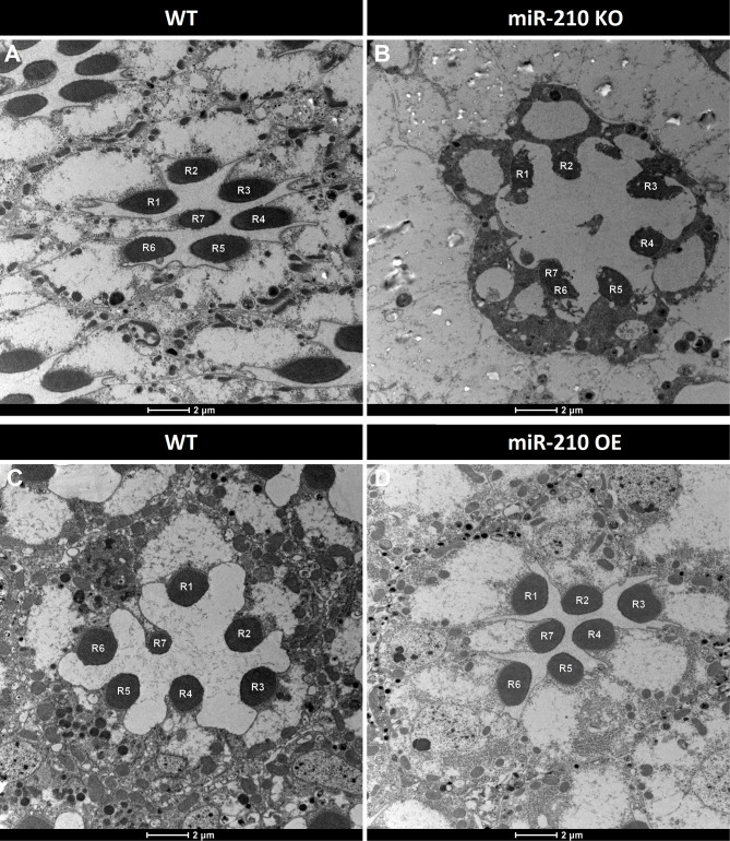
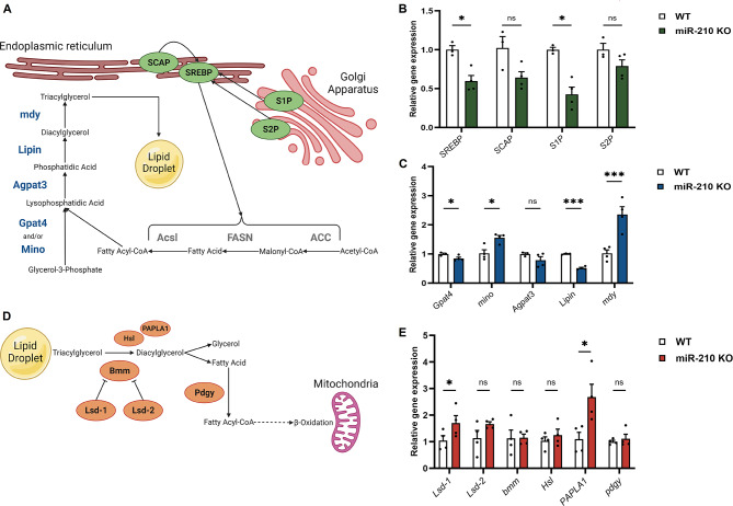
Background: miR-210 is one of the most evolutionarily conserved microRNAs. It is known to be involved in several physiological and pathological processes, including response to hypoxia, angiogenesis, cardiovascular diseases and cancer. Recently, new roles of this microRNA are emerging in the context of eye and visual system homeostasis. Recent studies in Drosophila melanogaster unveiled that the absence of miR-210 leads to a progressive retinal degeneration characterized by the accumulation of lipid droplets and disruptions in lipid metabolism. However, the possible conservation of miR-210 knock-out effect in the mammalian retina has yet to be explored.

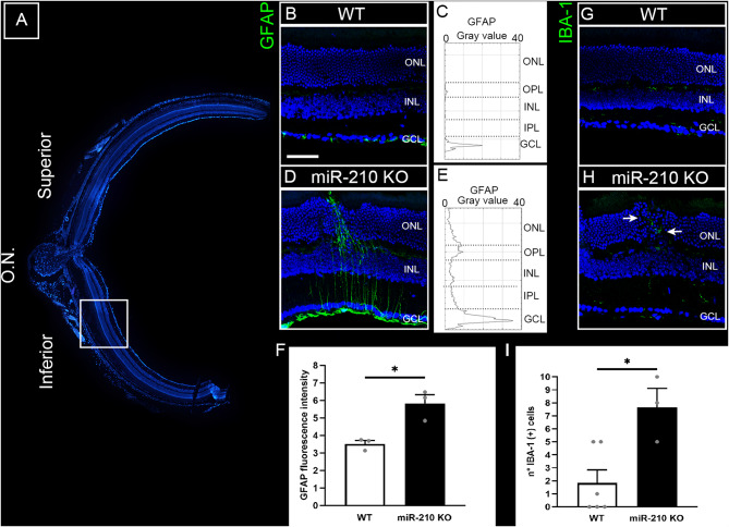
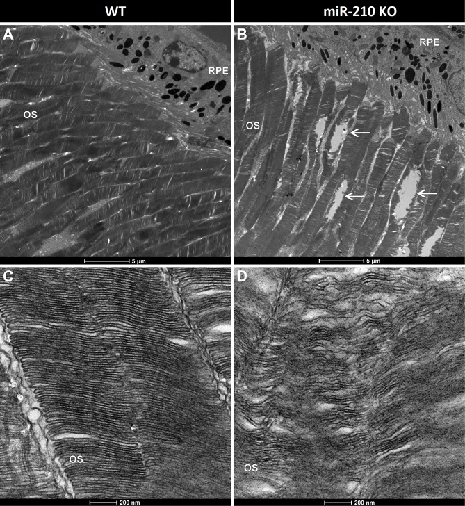
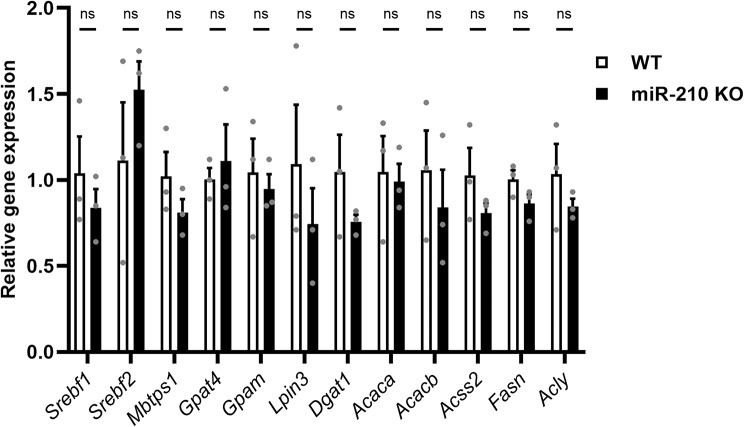
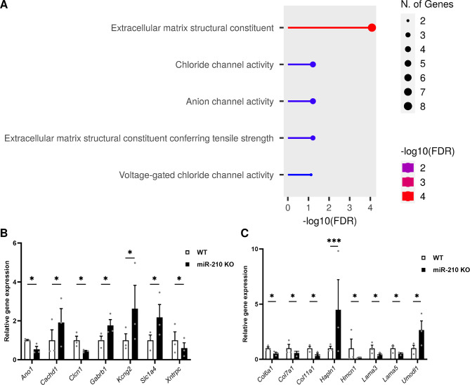
Results: We further investigated lipid anabolism and catabolism in miR-210 knock-out (KO) flies, uncovering significant alterations in gene expression within these pathways. Additionally, we characterized the retinal morphology of flies overexpressing (OE) miR-210, which was not affected by the increased levels of the microRNA. For the first time, we also characterized the retinal morphology of miR-210 KO and OE mice. Similar to flies, miR-210 OE did not affect retinal homeostasis, whereas miR-210 KO mice exhibited photoreceptor degeneration. To explore other potential parallels between miR-210 KO models in flies and mice, we examined lipid metabolism, circadian behaviour, and retinal transcriptome in mice, but found no similarities. Specifically, RNA-seq confirmed the lack of involvement of lipid metabolism in the mice's pathological phenotype, revealing that the differentially expressed genes were predominantly associated with chloride channel activity and extracellular matrix homeostasis. Simultaneously, transcriptome analysis of miR-210 KO fly brains indicated that the observed alterations extend beyond the eye and may be linked to neuronal deficiencies in signal detection and transduction.

Conclusions: We provide the first morphological characterization of the retina of miR-210 KO and OE mice, investigating the role of this microRNA in mammalian retinal physiology and exploring potential parallels with phenotypes observed in fly models. Although the lack of similarities in lipid metabolism, circadian behaviour, and retinal transcriptome in mice suggests divergent mechanisms of retinal degeneration between the two species, transcriptome analysis of miR-210 KO fly brains indicates the potential existence of a shared upstream mechanism contributing to retinal degeneration in both flies and mammals.

Keywords: Drosophila melanogaster; Mus musculus; Chloride channels; Circadian behaviour; Extracellular matrix; Lipid metabolism; Photoreceptor degeneration; Retina; Signal transduction; miR-210.


Conflict of interest statement

The authors declare no competing interests.

Figures

Fig. 1
Fig. 1
Transmission electron microscopy (TEM) analysis of ommatidial structure in the retina of miR-210 KO and OE flies. Ommatidium cross-section of the retina of 5-day-old flies. When compared to controls (A), miR-210 KO flies (B) showed aberrant ommatidial structure, with an altered morphology of the photoreceptor neurons indicating an ongoing retinal degeneration. On the other hand, when compared to their relative controls (C), flies overexpressing miR-210 in retinal cells (D) did not show any ommatidial structural alteration. Scale bar: 2 μm. R1-R7 = photoreceptor neurons. Each image is representative of at least three independent samples
Fig. 2
Fig. 2
Gene expression analysis of the main genes involved in lipid anabolism and catabolism in miR-210 KO flies. (A) A schematic overview of the main proteins involved in lipid anabolism: the SREBP activation pathway, leading to the synthesis of proteins involved in the de novo lipogenesis (reported in grey), is reported in green, while the enzymatic synthesis of triacylglycerols is reported in blue. (Created with BioRender.com) (B) qRT-PCR expression levels of the main genes involved in the SREBP activation pathway in the heads of 5-day-old miR-210 KO flies and controls. (C) qRT-PCR expression levels of the main genes involved in the enzymatic synthesis of triacylglycerols in the heads of 5-day-old miR-210 KO flies and controls. (D) A schematic overview of the main proteins involved in lipid catabolism. (Created with BioRender.com). (E) qRT-PCR expression levels of the main genes involved in triacylglycerols mobilization and lipolysis in the heads of 5-day-old miR-210 KO flies and controls. The results (N = 4) are expressed as mean ± SEM. Student’s t-test was performed to determine significant differences. *p-value < 0.05, ***p-value < 0.005
Fig. 3
Fig. 3
Morphological analysis of the miR-210 KO mouse retina through immunofluorescence (IF) microscopy. (A) Overview of a retinal section crossing the optic nerve (O.N.) stained with bisbenzimide nuclear dye, showing the superior (dorsal) and inferior (ventral) retina. (B-I) Analysis of neuroinflammatory markers GFAP (B-F) and IBA-1 (G-I). (B, D) Representative confocal images of anti-glial fibrillary acidic protein (GFAP) immunostaining acquired at the inferior retina of wild type (WT) (B) and miR-210 KO (D) retinal cryosections obtained from mice aged 10–11 weeks. Bisbenzimide nuclear dye: blue; GFAP: green. Scale bar: 50 μm. Each image is representative of at least three independent samples. (C, E) Profile plots of GFAP fluorescent signals throughout the retinal layers of wild type (WT) (C) and miR-210 KO (E) mice. (F) Column chart of the fluorescence intensity of GFAP fluorescent signal in the ventral retina of wild type (WT) and miR-210 KO mice. The results (N = 3) are expressed as mean ± SEM. Student’s t-test was performed to determine significant differences. *p-value < 0.05. (G, H) Representative anti-ionized calcium binding adaptor molecule 1 (IBA-1) immunostaining acquired at the inferior retina of wild type (WT) (G) and miR-210 KO (H) retinal cryosections. The white arrows indicate IBA-1 (+) cells infiltrating in the outer retina at the site of a structural alteration of the outer nuclear layer (ONL). Bisbenzimide nuclear dye: blue; IBA-1: green. Scale bar: 50 μm. Each image is representative of at least three independent samples. (I) Column chart of microglia quantification in the outer nuclear layer (ONL) through the entire section from superior to inferior of wild type (WT) and miR-210 KO retinal cryosections; the measurement is expressed as number of IBA-1 (+) cells in the outer nuclear layer (ONL). The results (N ≥ 3) are expressed as mean ± SEM. Student’s t-test was performed to determine significant differences
Fig. 4
Fig. 4
Morphological analysis of the miR-210 KO mouse retina through transmission electron microscopy (TEM). (A-D) Transmission electron microscopy (TEM) images showing photoreceptor outer segments (OS) layer (A, B) and photoreceptor ultrastructure (C, D) in the retina of wild type (WT) (A, C) and miR-210 KO (B, D) mice of 10–11 weeks of age. The white arrows indicate the photoreceptor degeneration occurring in the outer segments (OS) of miR-210 KO mice. Scale bars: 5 μm (A, B), 200 nm (C, D). OS = photoreceptor outer segments; RPE = retinal pigment epithelium. Each image is representative of at least three independent samples
Fig. 5
Fig. 5
Representative examples of locomotor activity of miR-210 KO mice under LD cycle and constant darkness. The daily and circadian locomotor activity of WT (A, C) and miR-210 KO mice (B, D) under LD (A, B) and DD conditions (C, D) is reported as actogram. The corresponding mean waves are plotted in (E-H). The comparison between WT and miR-210 KO mice locomotor activity under LD (I) and DD (J) conditions (Student’s t-test), as well as that of the acrophase under LD (K) conditions (two-way RM ANOVA), revealed no differences (p-value > 0.05) between the two experimental groups (N = 7)
Fig. 6
Fig. 6
Gene expression analysis of mouse orthologs of genes involved in lipid metabolism in D. melanogaster. The expression levels of mouse orthologs corresponding to genes implicated in lipid metabolism, which were identified as differentially expressed in miR-210 KO flies, were evaluated via qRT-PCR in the retinas of miR-210 KO and WT mice aged 10–11 weeks. The results (N = 3) are expressed as mean ± SEM. Student’s t-test was performed to determine significant differences
Fig. 7
Fig. 7
RNA-seq and gene ontology (GO) analysis of differentially expressed genes (DEGs) in the retinas of miR-210 KO mice. (A) Gene ontology (GO) analysis was conducted using the ShinyGO annotation tool, focusing on genes exhibiting differential expression between miR-210 KO and WT mice retinas. (B-C) Differentially expressed genes (DEGs) involved in chloride channel activity (B) and in the maintenance and functionality of the extracellular matrix (C), resulting from the RNA-seq analysis. The results (N = 3) are expressed as mean ± SEM. Student’s t-test corrected for multiple testing (Benjiamini-Hochberg method) was performed to determine significant differences
Fig. 8
Fig. 8
Gene ontology (GO) analysis of differentially expressed genes in the brains of miR-210 KO flies. GO analysis performed using ShinyGO annotation tool starting from the genes found to be differentially expressed between miR-210 KO and WT fly brains. The top five significantly enriched biological processes are depicted

References

    1. He L, Hannon GJ. MicroRNAs: small RNAs with a big role in gene regulation. Nat Rev Genet. 2004; 10.1038/nrg1379. - PubMed
    1. Friedman RC, Farh KK, Burge CB, Bartel DP. Most mammalian mRNAs are conserved targets of microRNAs. Genome Res. 2009; 10.1101/gr.082701.108. - PMC - PubMed
    1. Tüfekci KU, Meuwissen RL, Genç S. The role of microRNAs in biological processes. Methods Mol Biol. 2014. 10.1007/978-1-62703-748-8_2. - PubMed
    1. Li Y, Kowdley KV. MicroRNAs in common human diseases. Genomics Proteom Bioinf. 2012. 10.1016/j.gpb.2012.07.005. - PMC - PubMed
    1. Weigelt CM, Hahn O, Arlt K, Gruhn M, Jahn AJ, Eßer J, Werner JA, Klein C, Büschges A, Grönke S, Partridge L. Loss of miR-210 leads to progressive retinal degeneration in Drosophila melanogaster. Life Sci Alliance. 2019. 10.26508/lsa.201800149. - PMC - PubMed

LinkOut - more resources