Review
. 2025 Jan;21(1):3-20.
doi: 10.1080/15548627.2024.2414424. Epub 2024 Oct 23.

Reticulophagy and viral infection

Affiliations
Review

Reticulophagy and viral infection

Alexa Wilson et al. Autophagy. 2025 Jan.

Abstract

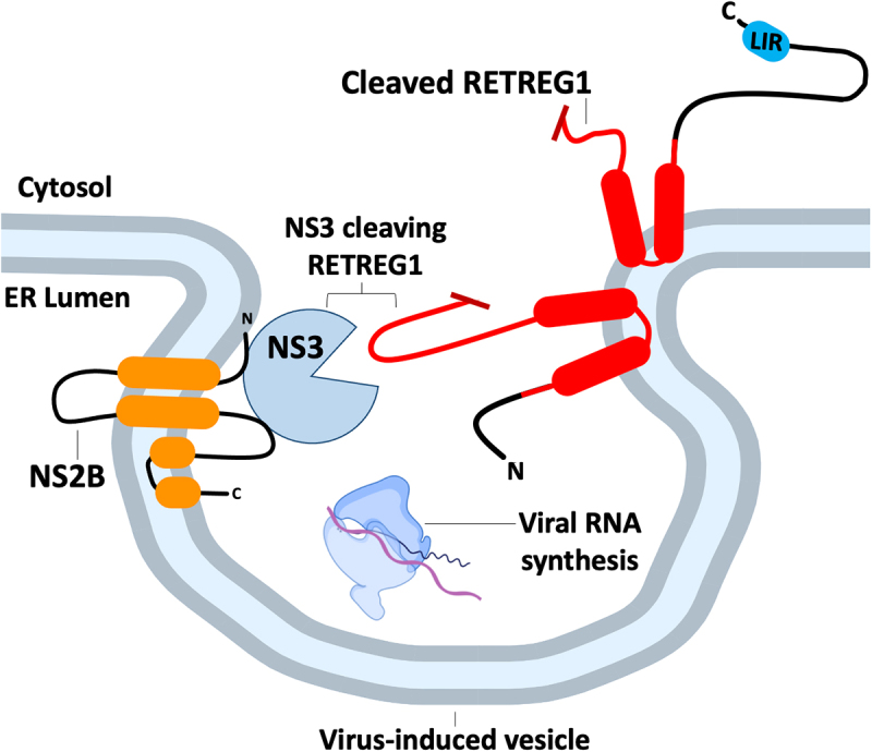
All viruses are obligate intracellular parasites that use host machinery to synthesize viral proteins. In infected eukaryotes, viral secreted and transmembrane proteins are synthesized at the endoplasmic reticulum (ER). Many viruses refashion ER membranes into bespoke factories where viral products accumulate while evading host pattern recognition receptors. ER processes are tightly regulated to maintain cellular homeostasis, so viruses must either conform to ER regulatory mechanisms or subvert them to ensure efficient viral replication. Reticulophagy is a catabolic process that directs lysosomal degradation of ER components. There is accumulating evidence that reticulophagy serves as a form of antiviral defense; we call this defense "xERophagy" to acknowledge its relationship to xenophagy, the catabolic degradation of microorganisms by macroautophagy/autophagy. In turn, viruses can subvert reticulophagy to suppress host antiviral responses and support efficient viral replication. Here, we review the evidence for functional interplay between viruses and the host reticulophagy machinery.Abbreviations: AMFR: autocrine motility factor receptor; ARF4: ADP-ribosylation factor 4; ARL6IP1: ADP-ribosylation factor-like 6 interacting protein 1; ATL3: atlastin GTPase 3; ATF4: activating transcription factor 4; ATF6: activating transcription factor 6; BPIFB3: BPI fold containing family B, member 3; CALCOCO1: calcium binding and coiled coil domain 1; CAMK2B: calcium/calmodulin-dependent protein kinase II, beta; CANX: calnexin; CDV: canine distemper virus; CCPG1: cell cycle progression 1; CDK5RAP3/C53: CDK5 regulatory subunit associated protein 3; CIR: cargo-interacting region; CoV: coronavirus; CSNK2/CK2: casein kinase 2; CVB3: coxsackievirus B3; DAPK1: death associated protein kinase 1; DENV: dengue virus; DMV: double-membrane vesicles; EBOV: Ebola virus; EBV: Epstein-Barr Virus; EIF2AK3/PERK: eukaryotic translation initiation factor 2 alpha kinase 3; EMCV: encephalomyocarditis virus; EMV: extracellular microvesicle; ER: endoplasmic reticulum; ERAD: ER-associated degradation; ERN1/IRE1: endoplasmic reticulum to nucleus signalling 1; EV: extracellular vesicle; EV71: enterovirus 71; FIR: RB1CC1/FIP200-interacting region; FMDV: foot-and-mouth disease virus; HCMV: human cytomegalovirus; HCV: hepatitis C virus; HMGB1: high mobility group box 1; HSPA5/BiP: heat shock protein 5; IFN: interferon; IFNG/IFN-γ: interferon gamma; KSHV: Kaposi's sarcoma-associated herpesvirus; LIR: MAP1LC3/LC3-interacting region; LNP: lunapark, ER junction formation factor; MAP1LC3: microtubule-associated protein 1 light chain 3; MAP3K5/ASK1: mitogen-activated protein kinase kinase kinase 5; MAPK/JNK: mitogen-activated protein kinase; MeV: measles virus; MHV: murine hepatitis virus; NS: non-structural; PDIA3: protein disulfide isomerase associated 3; PRR: pattern recognition receptor; PRRSV: porcine reproductive and respiratory syndrome virus; RB1CC1/FIP200: RB1-inducible coiled-coil 1; RETREG1/FAM134B: reticulophagy regulator 1; RHD: reticulon homology domain; RTN3: reticulon 3; RTN3L: reticulon 3 long; sAIMs: shuffled Atg8-interacting motifs; SARS-CoV: severe acute respiratory syndrome coronavirus; SINV: Sindbis virus; STING1: stimulator of interferon response cGAMP interactor 1; SVV: Seneca Valley virus; SV40: simian virus 40; TEX264: testis expressed gene 264 ER-phagy receptor; TFEB: transcription factor EB; TRAF2: TNF receptor-associated factor 2; UIM: ubiquitin-interacting motif; UFM1: ubiquitin-fold modifier 1; UPR: unfolded protein response; VAPA: vesicle-associated membrane protein, associated protein A; VAPB: vesicle-associated membrane protein, associated protein B and C; VZV: varicella zoster virus; WNV: West Nile virus; XBP1: X-box binding protein 1; XBP1s: XBP1 spliced; xERophagy: xenophagy involving reticulophagy; ZIKV: Zika virus.

Keywords: Autophagy; endoplasmic reticulum; reticulophagy; unfolded protein response; virus.


Conflict of interest statement

No potential conflict of interest was reported by the author(s).

Figures

Figure 1.
Figure 1.
Reticulon homology domain-containing reticulophagy receptors RETREG1 and RTN3L. Reticulon homology domain (rhd)-containing reticulophagy receptors are comprised of transmembrane or membrane-associated RHD domains (in red) connected to domains involved in substrate recognition and LC3 binding. RETREG1 is primarily found in ER sheets where it captures misfolded substrate proteins bound to the ER transmembrane chaperone CANX. ER stress triggers post-translational modifications of the RETREG1 RHD, including CAMK2B-mediated phosphorylation, that contribute to RETREG1 oligomerization and er-scission, while LC3 binding recruits phagophores to initiate local degradation of ER membranes and cargo. RTN3L is primarily associated with the cytoplasmic leaflet of ER tubules, and relatively little is known about substrate recognition mechanisms. Six LIRs on the cytoplasmic amino-terminal domain direct binding to Atg8-like proteins and phagophore recruitment.
Figure 2.
Figure 2.
Transmembrane reticulophagy receptors that lack a reticulon homology domain. There are four known transmembrane reticulophagy receptors that lack reticulon homology domains (RHDs); SEC62 and CCPG1 are predominantly found in ER sheets, whereas TEX264 and ATL3 are predominantly found in ER tubules. SEC62 and CCPG1 have cytoplasmic LIRs; CCPG1 also binds RB1CC1 via a FIR domain. TEX264 has an intrinsically disordered region (IDR), highlighted in red; CSNK2-mediated phosphorylation of residues upstream of the LIR in the IDR aids LC3 binding and phagophore recruitment. ATL3 has two GIMs that confer GABARAP binding. ATL3 associates with ATG13 and ULK1 via a GTPase domain (in black). Through its association with ATG13 and ULK1, ATL3 indirectly interacts with VAPA and VAPB, WIPI2, ATG101 and RB1CC1.
Figure 3.
Figure 3.
Soluble cytosolic reticulophagy receptors. CALCOCO1 is recruited to the cytosolic face of the ER by binding VAPA/B through its C-terminal FFAT-like domain. CALCOCO1 binds GABARAP via multivalent interactions; its amino-terminal CLIR interacts with the GABARAP LIR-docking site (LDS) and its carboxy-terminal UIR interacts with the GABARAP ubiquitin interacting motif docking site (UDS). CDK5RAP3 is a core component of the UFMylation machinery that directs ribosomal protein UFMylation during stalling at the ER translocon; following ribosomal protein UFMylation, CDK5RAP3-DDRGK1/UFBP1 complexes can be released from this role, and bind GABARAP via a sAIM motif in CDK5RAP3, thereby promoting phagophore recruitment for reticulophagy. SRP, signal recognition particle; SRPR, signal recognition particle receptor.
Figure 4.
Figure 4.
The unfolded protein response (UPR) increases reticulophagy receptor levels. ERN1 is an ER localized transmembrane protein that is inactive when bound to the ER lumenal chaperone HSPA5. HSPA5 dissociates from ERN1 in response to the accumulation of unfolded proteins in the ER lumen, which allows ERN1 activation via dimerization and trans-autophosphorylation. ERN1 is an RNA endonuclease that cleaves cytosolic XBP1 mRNA at two proximal hairpin loops; cleavage products are ligated by the RCTB tRNA ligase, which removes a 26-base intron, and shifts the reading frame, allowing synthesis of the XBP1s transcription factor with a carboxy-terminal bZIP transactivation domain. XBP1s translocates to the nucleus and transactivates an array of UPR genes, including genes encoding the RTN3 and CDK5RAP3 reticulophagy receptors, and the MIST transcription factor, which in turn transactivates the gene that encodes the CCPG1 reticulophagy receptor.
Figure 5.
Figure 5.
Flavivirus proteases cleave RETREG1 to prevent xERophagy. Several flaviviruses replicate more efficiently in cells deficient in RETREG1. Flavivirus RNA synthesis takes place in arrays of ER invaginations known as “vesicle packets”, where the viral NS3 protease is tethered to the cytosolic face of the ER by the viral transmembrane adapter protein NS2B. NS3 inactivates RETREG1 by cleaving a loop in the RHD (in red). Preventing RETREG1 cleavage by mutating the protease cleavage site restores xERophagy of viral components.
Figure 6.
Figure 6.
Coronaviruses co-opt reticulophagy receptors for STING1 degradation and then sequester them in protein condensates to prevent xERophagy. In the early stages of coronavirus infection, NSP6 assists in formation of the double-membrane replication organelle, where viral RNA synthesis takes place. NSP6 also assists viral replication by activating EIF2AK3-dependent reticulophagy, involving the RETREG1 and CCPG1 receptors that target STING1 for lysosomal degradation. However, excessive reticulophagy limits replication organelle formation. This is countered by the viral protein ORF8, which binds to RETREG1 or ATL3, along with the selective autophagy receptor SQSTM1, to sequester these reticulophagy receptors in insoluble condensates, thereby inhibiting xERophagy.
Figure 7.
Figure 7.
ATL3 and RTN3L support the formation of SV40 foci during ER escape. SV40 polyomavirus enters cells via a CAV-dependent pathway that delivers virus capsids to the ER lumen. To gain access to the cytosol, SV40 penetrates the ER membrane at foci comprised of multi-tubular ER junctions. Formation of these junctions requires the GTPase activity of ATL3, which assists membrane fusion. ATL3 associates with SV40 VP1 protein, as well as host proteins LNP and RTN4B, to yield a membrane-penetration complex at these sites. RTN3L stabilizes these ER foci and assists membrane penetration by remodeling ER membranes to relieve mechanical stress.

References

    1. Borgese N, Francolini M, Snapp E.. Endoplasmic reticulum architecture: structures in flux. Curr Opin Cell Biol. 2006;18(4):358–364. doi: 10.1016/j.ceb.2006.06.008 - DOI - PMC - PubMed
    1. Shibata Y, Shemesh T, Prinz WA, et al. Mechanisms determining the morphology of the peripheral ER. Cell. 2010;143(5):774–788. doi: 10.1016/j.cell.2010.11.007 - DOI - PMC - PubMed
    1. West M, Zurek N, Hoenger A, et al. A 3D analysis of yeast ER structure reveals how ER domains are organized by membrane curvature. J Cell Biol. 2011;193(2):333–346. doi: 10.1083/jcb.201011039 - DOI - PMC - PubMed
    1. Shibata Y, Voeltz GK, Rapoport TA. Rough sheets and smooth tubules. Cell. 2006;126(3):435–439. doi: 10.1016/j.cell.2006.07.019 - DOI - PubMed
    1. Ron D, Walter P. Signal integration in the endoplasmic reticulum unfolded protein response. Nat Rev Mol Cell Biol. 2007;8(7):519–529. doi: 10.1038/nrm2199 - DOI - PubMed

Publication types