

Classification, angioarchitecture and treatment outcomes of medullary bridging vein-draining dural arteriovenous fistulas in the foramen magnum region: a multicenter study
- PMID: 39395048
- PMCID: PMC11802681
- DOI: 10.1007/s00234-024-03478-w
Classification, angioarchitecture and treatment outcomes of medullary bridging vein-draining dural arteriovenous fistulas in the foramen magnum region: a multicenter study
Abstract
Purpose: This study aimed to classify medullary bridging vein-draining dural arteriovenous fistulas (MBV-DAVFs) located around the foramen magnum (FM) according to their location and characterize their angioarchitecture and treatment outcomes.
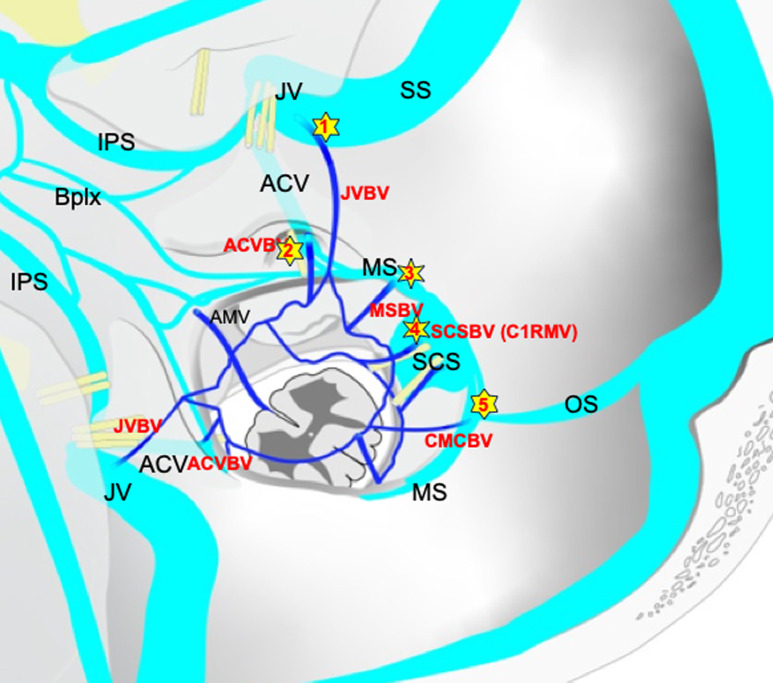
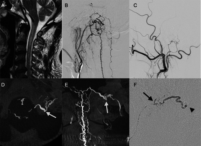
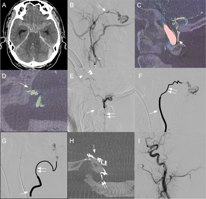
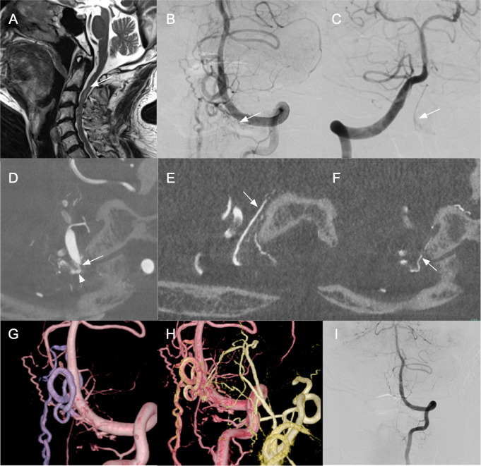
Methods: Patients with MBV-DAVFs diagnosed between January 2013 and October 2022 were included. MBV-DAVFs were classified into four groups. Jugular vein-bridging vein (JV-BV) DAVF: located in proximity to jugular fossa, Anterior condylar vein (ACV)-BV DAVF: proximity to anterior condylar canal, Marginal sinus (MS)-BV DAVF: lateral surface of FM and Suboccipital cavernous sinus (SCS)-BV DAVF: proximity to dural penetration of vertebral artery.
Results: Twenty patients were included, three JV-BV, four ACV-BV, three MS-BV and ten SCS-BV DAVFs, respectively. All groups showed male predominance. There were significant differences in main feeders between JV (jugular branch of ascending pharyngeal artery) and SCS group (C1 dural branch). Pial feeders from anterior spinal artery (ASA) or lateral spinal artery (LSA) were visualized in four SCS and one MS group. Drainage pattern did not differ between groups. Transarterial embolization (TAE) was performed in three, two, one and two cases and complete obliteration was obtained in 100%, 50%, 100% and 0% in JV, ACS, MS and SCS group, respectively. Successful interventions without major complications were finally obtained in 100%, 75%, 100%, and 40% in JV, ACS, MS and SCS group, respectively.
Conclusion: JV-BV DAVFs were successfully treated using TAE alone. SCS-BV DAVFs were mainly fed by small C1 dural branches of vertebral artery often with pial feeders from ASA or LSA, and difficultly treated by TAE alone.
Keywords: Bridging vein; Dural arteriovenous fistula; Foramen Magnum; Surgical obliteration; Transarterial embolization.
© 2024. The Author(s).
Conflict of interest statement
Declarations. Ethical approval: This study protocol was approved by the institutional ethics committee of the participating centers and was performed in accordance with the committees’ guidelines. Informed consent: All patients provided written informed consent. Conflict of interest: The authors report no conflict of interest concerning the materials or methods used in this study or the findings specified in the present paper.
Figures

References
Publication types
MeSH terms
LinkOut - more resources
Full Text Sources
