Review
. 2024 Nov 5;36(4):042003.
doi: 10.1088/1361-6528/ad8626.

Roadmap on magnetic nanoparticles in nanomedicine

Affiliations
Review

Roadmap on magnetic nanoparticles in nanomedicine

Kai Wu et al. Nanotechnology. .

Abstract

Magnetic nanoparticles (MNPs) represent a class of small particles typically with diameters ranging from 1 to 100 nanometers. These nanoparticles are composed of magnetic materials such as iron, cobalt, nickel, or their alloys. The nanoscale size of MNPs gives them unique physicochemical (physical and chemical) properties not found in their bulk counterparts. Their versatile nature and unique magnetic behavior make them valuable in a wide range of scientific, medical, and technological fields. Over the past decade, there has been a significant surge in MNP-based applications spanning biomedical uses, environmental remediation, data storage, energy storage, and catalysis. Given their magnetic nature and small size, MNPs can be manipulated and guided using external magnetic fields. This characteristic is harnessed in biomedical applications, where these nanoparticles can be directed to specific targets in the body for imaging, drug delivery, or hyperthermia treatment. Herein, this roadmap offers an overview of the current status, challenges, and advancements in various facets of MNPs. It covers magnetic properties, synthesis, functionalization, characterization, and biomedical applications such as sample enrichment, bioassays, imaging, hyperthermia, neuromodulation, tissue engineering, and drug/gene delivery. However, as MNPs are increasingly explored forin vivoapplications, concerns have emerged regarding their cytotoxicity, cellular uptake, and degradation, prompting attention from both researchers and clinicians. This roadmap aims to provide a comprehensive perspective on the evolving landscape of MNP research.

Keywords: biomedical application; drug delivery; hyperthermia; magnetic biosensing; magnetic imaging; magnetic nanoparticle; tissue engineering.


Figures

Figure 1.
Figure 1.
One MNP with a core diameter of D. The spin-ordered part of the core has a diameter of Dm, with a surface spin disorder in a spin-canted layer of thickness δ. Original figure prepared by the authors.
Figure 2.
Figure 2.
Schematic drawing representing the zero-field Brownian and Néel relaxations. The dotted line represents the easy axis of the MNP. Original figure prepared by the authors.
Figure 3.
Figure 3.
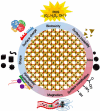
Summary of current applications of MNPs in nanomedicine. Reprinted with permission from [23]. Copyright (2021) American Chemical Society.
Figure 4.
Figure 4.
Types of surface coatings commonly used on MNPs and some examples of each sort. Adapted from [33]. CC BY 4.0.
Figure 5.
Figure 5.
Illustration of functionalized MNPs from various strategies. Original figure prepared by the author.
Figure 6.
Figure 6.
(a) Schematic diagram of the HGMS process. (b) Three possible configurations of the rod matrix in HGMS systems. (c) Effect of the rod diameter on the magnetic field distribution in HGMS matrices. Reprinted from [92], Copyright (2017), with permission from Elsevier.
Figure 7.
Figure 7.
(a) Quadrupole magnetic sorter for magnetic particle separation. (b) Top view of the steel pole pieces (purple) and NdFeB magnets (yellow). (c) Cross-sectional field map. Reprinted from [98], Copyright (2020), with permission from Elsevier.
Figure 8.
Figure 8.
(A) Schematic drawing of various MNP-based bioassays. (A1) Direct assay. (A2) Indirect assay. (A3) Sandwich assay. (A4) Compactivity assay: limited number of probes (left) and limited number of target analytes (right). (A5) DNA assay. (B) Schematic representations of (B1) Néel relaxation and (B2) Brownian relaxation. Original figure prepared by the authors.
Figure 9.
Figure 9.
Point-of-care devices developed utilizing magnetic detection of MNPs using (A) Giant magnetoresistance (GMR) sensor. (B) MPS sensor. (C) NMR sensing implementation. (A) Reprinted from [113], Copyright (2016), with permission from Elsevier. (B) Reprinted with permission from [114]. Copyright (2021) American Chemical Society. (C) From [115]. Reprinted with permission from AAAS.
Figure 10.
Figure 10.
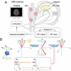
Schematic views of (A) an MRI scanner and (B) T1 and T2 relaxations. (A) Reproduced from [116]. CC BY 4.0. (B) Reproduced from [117] with permission from the Royal Society of Chemistry.
Figure 11.
Figure 11.
Challenges and opportunities of different MRI contrast agents. Original figure prepared by the authors.
Figure 12.
Figure 12.
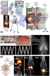
MPI is a new modality that directly & quantitatively images MNPs in vivo. (a) MPI scanning schematic where single-core or multi-core MNPs can be imaged in vivo even in regions such as the lung where MRI or Ultrasound cannot reach. (b) tissue is fully transparent to MPI, thus (c) enabling direct quantitative visualization of perfusion with zero tissue attenuation. (d) Antibodies can be attached to MNPs enabling specific labeling of cancer cells or stem/immune cells. (e) MPI visualizes tumors with high contrast using EPR (or Ab) targeting. (f) MPI tracks the biodistribution of infused stem cells for longitudinal studies lasting up to 89 d. (a) Reproduced with permission from [134]. [insert copyright line, if specified]. (b) Reproduced from [133], Copyright (2012), with permission from Elsevier. (c) Reproduced from [141]. CC BY 4.0. (d) originally prepared by the authors. (e) Reproduced with permission from [142]. Copyright (2017) American Chemical Society. (f) Reproduced from [143]. CC BY 4.0.
Figure 13.
Figure 13.
Superferromagnetic iron oxide nanoparticles (SFMIO) enable >10-fold resolution improvement towards achieving 0.1 mm spatial resolution MPI without a limit-of-detection trade-off (SFMIOs also have >10-fold improved SNR concomitant with the improved resolution). (a) TEM shows SPIOs form chains with strong interparticle interactions when magnetized in a field. (b)/(c) SFMIO theoretical simulations match our ‘chained SPIO’ physics model; (d) 2D SFMIOs show ∼30-fold SNR & resolution boost experimentally in vitro. (a)–(c) are original figures prepared by the authors. (d) Reproduced from [151]. CC BY 4.0.
Figure 14.
Figure 14.
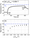
Heating curves and specific loss power (SLP). (a) Heating curves of 0.0326 g of particles with PEG in 500 μl of H2O in a flat-bottomed container, were used for SLP calculations. Each period of temperature increase corresponds to a different AMF amplitude. The frequency ranges from 213–217 kHz depending on AMF amplitude. (b) SLP calculated from the slope dT/dt in the temperature range 22.1–22.9. © [2017] IEEE. Reprinted, with permission, from [152].
Figure 15.
Figure 15.
(a) XRD patterns obtained from fractions. Reference peaks of Gd5Si4 and Gd5Si3 (bottom) match with the patterns. (b) Gadolinium silicide particle sizes obtained from Scanning electron microscopy (SEM) images of size selected gadolinium silicide particles showing the decrease in average particle size with sedimentation time. (c) M-T curve for all fractions and pre-filtered sample (d) Curie temperatures (Tc) for each fraction (S1-S6) Gd5Si4 powder. (e) M-H curve for all fractions and pre-filtered sample; the figure inset shows coercivity (Hc) to fractions. Reproduced from [164]. CC BY 4.0.
Figure 16.
Figure 16.
MNP-mediated neuromodulation demonstrates the activation of cellular ion channels through various techniques, including magneto-mechanical, magneto-thermal, magneto-chemical, magneto-electric, and magneto-catalytic approaches. External magnetic fields can activate MNPs. Specifically, magneto-mechanical forces can activate Piezo1, a mechanosensitive ion channel. The potential for magnetic drug delivery arises, wherein designer G-protein-coupled receptors like DREADDs can be activated under an AMF. Additionally, the magneto-electric effects of MNPs can induce neuronal depolarization under radiofrequency conditions, while the magneto-catalytic effects of MNPs can activate TRPV1 coupled with ferritin [172]. John Wiley & Sons. [© 2023 Wiley Periodicals LLC.].
Figure 17.
Figure 17.
Applications of magnetic fields in biomedical engineering. Low flux density magnetic fields (<50 mT) can align MNP-tagged cells or matrix proteins. Increasing magnetic flux densities (25–250 mT) can initiate magnetic actuation of cells in vitro and in vivo for regenerative medicine applications. Higher magnetic flux densities (>8 T) are often required for magnetic resonance imaging. Created with BioRender.com.
Figure 18.
Figure 18.
Physicochemical characteristics of MNPs for drug delivery systems. Reproduced from [186], with permission from Springer Nature.
Figure 19.
Figure 19.
Overview of the possible endocytosis pathways for MNP entry into cells and MNP intracellular fate. FEME (fast endophilin-mediated endocytosis), CLIC/GEEC (clathrin-independent carrier glycosylphosphatidylinositol-anchored protein-enriched early endocytic compartment endocytosis), EHD2 (Eps15-homology domain-containing protein 2), Cdc42 (cell division cycle 42), ArfI (ADP-ribosylation factor 1), GRAFI (GTPase regulator associated with focal adhesion kinase-1), ROS (Reactive Oxygen Species). Original figure prepared by the authors.

References

    1. Bødker F, Mørup S, Linderoth S. Surface effects in metallic iron nanoparticles. Phys. Rev. Lett. 1994;72:282. doi: 10.1103/PhysRevLett.72.282. - DOI - PubMed
    1. Kittel C. Introduction to Solid State Physics. Wiley; 2018.
    1. Ramos-Guivar J A, Lopez E O, Greneche J-M, Litterst F J, Passamani E C. Effect of EDTA organic coating on the spin canting behavior of maghemite nanoparticles for lead (II) adsorption. Appl. Surf. Sci. 2021;538:148021. doi: 10.1016/j.apsusc.2020.148021. - DOI
    1. Pereira A M, Pereira C, Silva A S, Schmool D S, Freire C, Greneche J-M, Araujo J P. Unravelling the effect of interparticle interactions and surface spin canting in γ-Fe2O3@ SiO2 superparamagnetic nanoparticles. J. Appl. Phys. 2011;109:114319. doi: 10.1063/1.3583652. - DOI
    1. Yari P, Chugh V K, Saha R, Tonini D, Rezaei B, Mostufa S, Xu K, Wang J-P, Wu K. Static and dynamic magnetization models of magnetic nanoparticles: an appraisal. Phys. Scr. 2023;98:082002. doi: 10.1088/1402-4896/ace8d1. - DOI

Substances

LinkOut - more resources