. 2024 Nov;300(11):107886.
doi: 10.1016/j.jbc.2024.107886. Epub 2024 Oct 11.

Site-specific O-GlcNAcylation of progesterone receptor (PR) supports PR attenuation of interferon stimulated genes (ISGs) and tumor growth in breast cancer

Affiliations

Site-specific O-GlcNAcylation of progesterone receptor (PR) supports PR attenuation of interferon stimulated genes (ISGs) and tumor growth in breast cancer

Harmony I Saunders et al. J Biol Chem. 2024 Nov.

Abstract

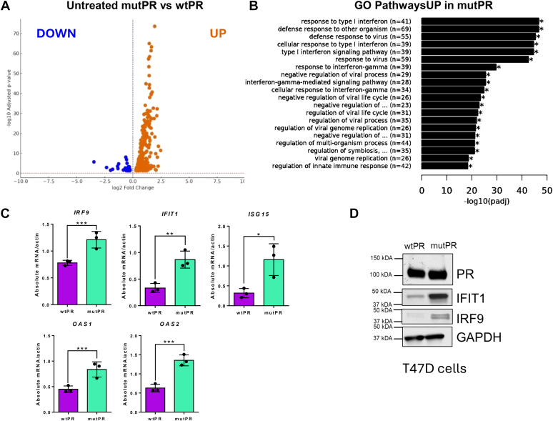
Hormone receptor positive (HR+) breast cancer, defined by expression of estrogen receptor (ER) and/or progesterone receptor (PR), is the most commonly diagnosed type of breast cancer. PR alters the transcriptional landscape to support tumor growth in concert with, or independent of, ER. Understanding the mechanisms regulating PR function is critical to developing new strategies to treat HR+ breast cancer. O-linked β-N-acetylglucosamine (O-GlcNAc) is a posttranslational modification responsible for nutrient sensing that modulates protein function. Although PR is heavily posttranslationally modified, through both phosphorylation and O-GlcNAcylation, specific sites of O-GlcNAcylation on PR and how they regulate PR action have not been investigated. Using established PR-expressing breast cancer cell lines, we mapped several sites of O-GlcNAcylation on PR. RNA-sequencing after PR O-GlcNAc site mutagenesis revealed site-specific O-GlcNAcylation of PR is critical for ligand-independent suppression of interferon signaling, a regulatory function of PR in breast cancer. Furthermore, O-GlcNAcylation of PR enhances PR-driven tumor growth in vivo. Herein, we have delineated one contributing mechanism to PR function in breast cancer that impacts tumor growth and provided additional insight into the mechanism through which PR attenuates interferon signaling.

Keywords: O-GlcNAc; breast; cancer; interferon; progesterone.


Conflict of interest statement

Conflict of interest The authors declare no conflict of interest with the contents of this article.

Figures

Figure 1
Figure 1
Site mapping using LC/MS reveals12PRO-GlcNAc sites.A and B, LC/ESI mass spectral analysis of O-GlcNAcylated peptides. The figure shows the tandem EThcD mass spectra of the triply charged ions at (A) m/z 1290.355 (493DGLPSTSASAAAAGAAPALYPALGLNGLPQLGYQAAVLK531) and (B) 751.4089 (731QLLSVVKWSKSLPGFR746). The b (red), c (pink), y (blue), and z (cyan) fragments are indicated. The parent ion is in green. The full fragmentation patterns are shown in the inserts where the O-GlcNAcylated residues are shown as a subscript next to the modified serine, and “q” represents a deamidated glutamine residue. C, schematic of high confidence O-GlcNAc sites on diagram of full-length PR, shown with previously mapped phosphorylation sites. Activation (AF), DNA-binding (DBD), and ligand-binding (LBD) domains are identified. The translational start sites for the two PR isoforms, PR-B and PR-A, are shown. O-GlcNAc sites are indicated with blue squares. S499, S733, and S735 are shown in red as the chosen sites for further study. O-GlcNAc, O-linked β-N-acetylglucosamine; PR, progesterone receptor.
Figure 2
Figure 2
MutPR (S499A, S733A, S735A) exhibits decreased O-GlcNAcylation. T47D cell lines expressing empty vector control (no PR; vector), wildtype unmodified PR (wtPR), or O-GlcNAc mutant PR (mutPR) were treated with TMG for 18 h, followed by 60 min R5020. PR was immunoprecipitated with a PR-specific antibody (or species-specific IgG control), and membranes were probed with PR or O-GlcNAc antibodies. Densitometry of the ratio between O-GlcNAc/IP PR, as determined using ImageJ, is shown as numerical values. A, representative blot of wtPR and mutPR O-GlcNAc blot is shown. A light and dark exposure is shown for the same PR blot. B, densitometry analysis was performed on three independent experiments as in A and plotted. Error bars represent standard error of the mean. ∗p = 0.04764, as determined using a one Sample t test against the null hypothesis that the ratio is 1. O-GlcNAc, O-linked β-N-acetylglucosamine; PR, progesterone receptor; TMG, Thiamet-G.
Figure 3
Figure 3
Ligand-dependent PR regulated gene expression is unaltered by O-GlcNAc modification on PR. T47D cells with wtPR or mutPR (site specific O-GlcNAc loss) were treated for 6 h with PR ligand, progestin (R5020). Following treatment, RNA was isolated and subjected to RNA-sequencing. A, differential genes expressed in wtPR and mutPR cells with ligand compared to vehicle control. Number of genes increased (up) and decreased (down) relative to untreated controls. B, volcano plot demonstrating the significant differentially expressed genes in vehicle treated cells versus those treated with ligand. wtPR diagram on the left and mutPR on the right. Significant genes are those –log10 p (adjusted) > 1. C, qRT-PCR of T47D variant lines treated with ligand or vehicle control for PR target genes upregulated or repressed in response to ligand. Bar graph represents absolute mRNA expression normalized to housekeeping (B-actin). Significance based on unpaired two tailed t test; ∗∗∗ ≥0.001. mutPR, mutant PR; O-GlcNAc, O-linked β-N-acetylglucosamine; PR, progesterone receptor; wtPR, wildtype unmodified PR.
Figure 4
Figure 4
O-GlcNAcylation of PR impacts ligand independent gene expression. Using T47D cells with wtPR or mutPR (site specific O-GlcNAc loss), RNA was isolated and subjected to RNA-Sequencing. Pathway analysis of differentially expressed genes was performed using gene ontology. A, volcano plot demonstrating the significant differentially expressed genes in vehicle control–treated mutPR and wtPR. Genes increased (up) in the mutPR cells are indicated on the right and decreased (down) on the left. Significant genes are those –log10 p (adjacent) > 1. B, top pathways enriched using genes that were expressed higher in mutPR cells as compared to wtPR. C, qRT-PCR on wtPR and mutPR cells in the absence of ligand on select interferon stimulated genes (ISGs). Bar graphs illustrate absolute mRNA expression normalized to housekeeping gene expression. Significance based on unpaired two tailed t test; ∗ ≥0.05, ∗∗≥0.01, ∗∗∗ ≥0.001. D, immunoblotting was performed on total lysate from untreated T47D variants on the proteins indicated. mutPR, mutant PR; O-GlcNAc, O-linked β-N-acetylglucosamine; PR, progesterone receptor; wtPR, wildtype unmodified PR.
Figure 5
Figure 5
Site specific O-GlcNAcylation affects PR driven mammary tumor growth. T47D cell line variants were injected into the mammary fat pad of NSG/NCG mice [n = 7 (wtPR), n = 5 (mutPR)] supplemented with 0.72 mg estradiol pellets (to support T47D growth in vivo). Tumor growth was followed until humane endpoint. A, tumor growth over time. B, mass of tumors at endpoint. Significance between groups was statistically significant with p value < 0.05 using longitudinal analysis (A) and student’s t test (B). ∗ ≥0.05, ∗∗≥0.01. Error bars indicate SEM (A), and STDEV (B). mutPR, mutant PR; NSG, NOD scid gamma; PR, progesterone receptor; wtPR, wildtype unmodified PR.

References

    1. Andrahennadi S., Sami A., Manna M., Pauls M., Ahmed S. Current landscape of targeted therapy in hormone receptor-positive and HER2-negative breast cancer. Curr. Oncol. 2021;28:1803–1822. - PMC - PubMed
    1. Yersal O., Barutca S. Biological subtypes of breast cancer: prognostic and therapeutic implications. World J. Clin. Oncol. 2014;5:412–424. - PMC - PubMed
    1. Beral V. Breast cancer and hormone-replacement therapy in the million women study. Lancet. 2003;362:419–427. - PubMed
    1. Anderson G.L., Limacher M., Assaf A.R., Bassford T., Beresford S.A., Black H., et al. Effects of conjugated equine estrogen in postmenopausal women with hysterectomy: the Women's Health Initiative randomized controlled trial. JAMA. 2004;291:1701–1712. - PubMed
    1. Scarpin K.M., Graham J.D., Mote P.A., Clarke C.L. Progesterone action in human tissues: regulation by progesterone receptor (PR) isoform expression, nuclear positioning and coregulator expression. Nucl. Recept. Signal. 2009;7 - PMC - PubMed

Publication types