. 2024 Oct 12;15(1):363.
doi: 10.1186/s13287-024-03983-y.

Extracellular vesicles from human cardiac stromal cells up-regulate cardiomyocyte protective responses to hypoxia

Affiliations

Extracellular vesicles from human cardiac stromal cells up-regulate cardiomyocyte protective responses to hypoxia

Andreas Czosseck et al. Stem Cell Res Ther. .

Abstract

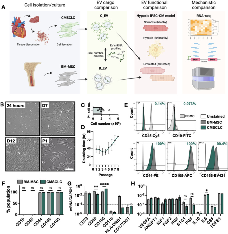
Background: Cell therapy can protect cardiomyocytes from hypoxia, primarily via paracrine secretions, including extracellular vesicles (EVs). Since EVs fulfil specific biological functions based on their cellular origin, we hypothesised that EVs from human cardiac stromal cells (CMSCLCs) obtained from coronary artery bypass surgery may have cardioprotective properties.

Objectives: This study characterises CMSCLC EVs (C_EVs), miRNA cargo, cardioprotective efficacy and transcriptomic modulation of hypoxic human induced pluripotent stem cell-derived cardiomyocytes (iPSC-CMs). C_EVs are compared to bone marrow mesenchymal stromal cell EVs (B_EVs) which are a known therapeutic EV type.

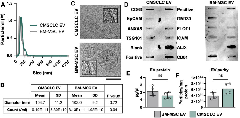
Methods: Cells were characterised for surface markers, gene expression and differentiation potential. EVs were compared for yield, phenotype, and ability to protect hiPSC-CMs from hypoxia/reoxygenation injury. EV dose was normalised by both protein concentration and particle count, allowing direct comparison. C_EV and B_EV miRNA cargo was profiled and RNA-seq was performed on EV-treated hypoxic hiPSC-CMs, then data were integrated by multi-omics. Confirmatory experiments were carried out using miRNA mimics.

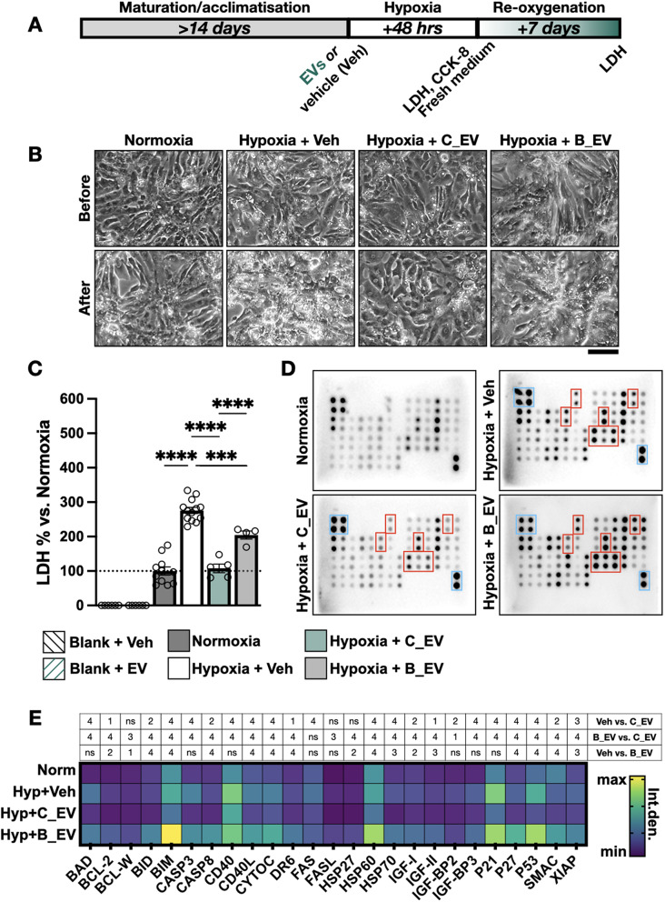
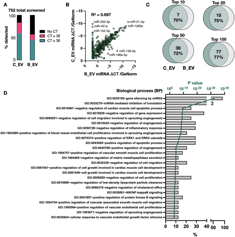
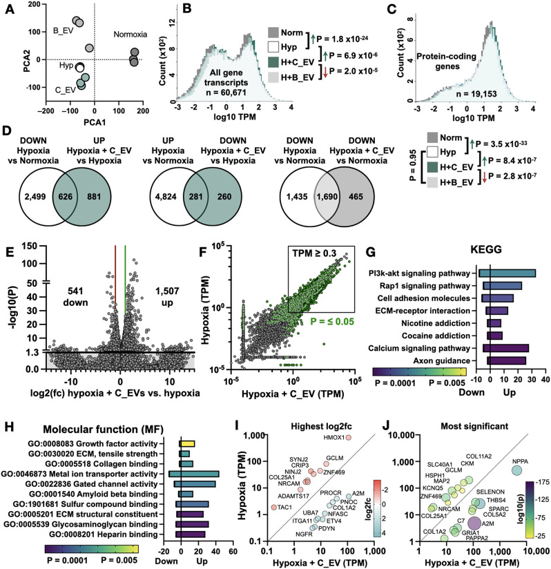
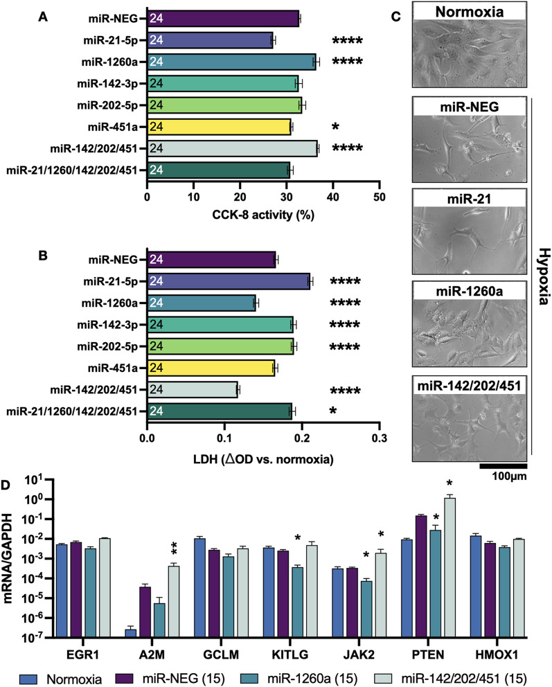
Results: At the same dose, C_EVs were more effective than B_EVs at protecting CM integrity, reducing apoptotic markers, and cell death during hypoxia. While C_EVs and B_EVs shared 70-77% similarity in miRNA content, C_EVs contained unique miRNAs, including miR-202-5p, miR-451a and miR-142-3p. Delivering miRNA mimics confirmed that miR-1260a and miR-202/451a/142 were cardioprotective, and the latter upregulated protective pathways similar to whole C_EVs.

Conclusions: This study demonstrates the potential of cardiac tissues, routinely discarded following surgery, as a valuable source of EVs for myocardial infarction therapy. We also identify miR-1260a as protective of CM hypoxia.

Keywords: Apoptosis; Exosome; Mesenchymal stromal cell; Multi-omics; RNA-sequencing; miRNA.


Conflict of interest statement

On behalf of all authors, the corresponding author states that there is no conflict of interest.

Figures

Fig. 1
Fig. 1
Comparison of cardiac and bone marrow-derived cells. (A) Schematic diagram of experimental design. (B) Images of CMSCLC morphology at 24 h, 7 days and 12 days after isolation, and 24 h after the first passage (P1). Scale bar 100 μm. (C) Number of viable cells obtained at passage 1 for n = 6 donor lines used in this study. (D) Cell doubling time plotted against passage number (average of n = 6 donors). (E) Representative histogram plots of flow cytometric analysis of CMSCLCs (green), BM-MSCs (dark grey) and unstained CMSCLCs (white). Negative markers CD19 and CD45, and positive markers CD44, CD105 and CD166 are shown. PBMCs (light grey) were used as positive controls for CD19 and CD45. (F) Quantification of flow cytometric analysis. n = 3 donors were compared to BM-MSCs by one-way ANOVA. (G) BM-MSC and CMSCLC (n = 3 donors) gene expression levels (as mRNA/GAPDH ratio) of positive and negative MSC markers. A dotted line shows the cut-off for low-expressed genes, which were considered negative. BM-MSC and CMSCLC samples were compared by two-way ANOVA with Sidak’s multiple comparison test. (H) BM-MSC and CMSCLC (n = 3 donors) gene expression of common MSC-associated paracrine factors. A dotted line shows the cut-off for low-expressed markers, which were considered negative. BM-MSC and CMSCLC samples were compared by two-way ANOVA with Sidak’s multiple comparison test. ns = not significant, ** = P ≤ 0.01, **** = P ≤ 0.0001. Pairs without annotations are also not significant (P > 0.05)
Fig. 2
Fig. 2
Extracellular vesicle isolation and characterisation. (A) Representative nanoparticle tracking analysis (NTA) size distribution plots for CMSCLC EVs (green) and BM-MSC EVs (grey). (B) Mean diameter and particle counts of n = 4 separate EV isolations and comparison by unpaired t-test. (C) Representative cryoEM images of isolated EVs. Scale bar 100 nm. A crop showing the lipid bilayer is also shown (inset). (D) Antibody-based membrane array showing human-specific EV surface markers and cargo markers. Two positive controls, a blank, and GM130 (cis-golgi marker) are also included. 50 µg total protein was added per membrane. (E) EV protein concentration (n = 4 per group) (F) Particle to protein ratio. Samples were compared by unpaired t-test. ns = not significant
Fig. 3
Fig. 3
Protection of hypoxic human cardiomyocytes using CMSCLC and BM-MSC EVs (A) Experimental design showing hiPSC-CM seeding and hypoxia treatment. (B) Example images of hiPSC-CMs following 48 h normoxia or hypoxia + vehicle (Veh), or hypoxia with 67 ng/µl CMSCLC EVs (C_EVs) or BM-MSC EVs (B_EVs). Scale bar 100 μm. (C) Culture medium LDH levels after 48 h of hiPSC-CM exposure to each treatment group. Blank samples (without hiPSC-CMs) are also included. Hypoxic hiPSC-CM groups were compared by one-way ANOVA with Tukey’s multiple comparison test. *** = P ≤ 0.001, **** = P ≤ 0.0001 (D) Apoptosis protein arrays from each group (n = 2 per group). Examples of significant differences between samples are highlighted with red boxes. Positive controls are shown as blue boxes in the upper left and lower right corners. (E) Heatmap showing quantification of integrated density of high concentration apoptosis-related proteins (n = 2 per group). All groups were compared using two-way ANOVA with Tukey’s post-test. The table above the heat map describes statistical significance; 1 = P < 0.05, 2 = P ≤ 0.01, 3 = P ≤ 0.001, 4 = P ≤ 0.0001, ns = not significant (P > 0.05)
Fig. 4
Fig. 4
Extracellular vesicle miRNA cargo analysis. (A) Percentage of miRNAs detected in CMSCLC EVs (C_EV) and BM-MSC EVs (B_EVs) for three separate donor samples per group. Those with cycle threshold (CT) values of < 36.0 (green bar) were included in subsequent analyses. (B) Scatter plot of C_EV (Y axis) versus B_EV (X axis) mean miRNA expression levels normalised to reference miRNA (GeNorm) levels. The R-squared correlation is shown in the upper left. (C) Venn diagrams showing degree of overlap between the top 10, 20, 50 and 100 highest expressed C_EV miRNAs compared to B_EV miRNAs. (D) Gene ontology (GO) predictions for biological process (BP) for top 50 expressed C_EV miRNAs. Bars show the % of miRNAs belonging to each GO (lower X axis) and the green line shows the adjusted Fisher P value (upper X axis)
Fig. 5
Fig. 5
RNA sequencing of hypoxic EV-treated human cardiomyocytes. (A) Principal component analysis (PCA) for normoxia, hypoxia + vehicle (Hyp), hypoxia + CMSCLC EV (H + C_EV) and BM-MSC EV groups (H + B_EV). (B) TPM distribution of all gene transcripts or (C) protein-coding gene transcripts for the four experimental groups. Sample distributions were compared by Kolmogorov-Smirnov (KS) test, and the direction of change and P values are shown for each comparison. (D) Venn diagrams showing the number of overlapping genes between the stated comparisons. (E) Volcano plot of hypoxia + vehicle against hypoxia + C_EVs. The Y axis show statistical significance, with the solid line showing P = 0.05. The X axis shows log2 fold change with the red and green lines showing two-fold down- and up-regulation respectively. (F) Scatter plot of hypoxia + vehicle vs. hypoxia + C_EV. Each point represents one gene. Green points indicate P ≤ 0.05 and the box indicates the genes with ≥ 0.3 TPM which were included in subsequent analyses. (G) Pyramid plot of most significantly enriched pathways by KEGG for hypoxia + C_EV vs. hypoxia + vehicle. The X axis shows the number of upregulated and downregulated genes in each group and the bar colours indicate statistical significance. (H) Pyramid plot of molecular function (MF). (I) Scatter plot showing the 10 most differentially-expressed and most statistically significant genes (J) between hypoxia + vehicle vs. hypoxia + C_EV groups
Fig. 6
Fig. 6
Determining hypoxia protection by abundant EV miRNAs. (A) CCK-8 activity of hypoxic AC16 cardiomyocytes incubated with miRNA negative control (miR-NEG), single miRNA mimics or combinations of mimics to a total of 15 nM. Percentages are relative to normoxia. (B) LDH secretion shown as change in absorbance relative to normoxia. Groups were compared to miR-NEG by one-way ANOVA with Dunnett’s multiple comparison test. * = P < 0.05, **** = P ≤ 0.0001. N = 24 samples per group. (C) Representative images from selected conditions. (D) Gene expression of predicted miRNA targets after normoxia, hypoxia + miR-NEG, miR-1260a or miR-142/202/451 at 15 nM. The Y axis shows log scale of gene expression normalised to GAPDH. N = 4 independent samples per bar. Statistical annotations show comparisons against hypoxia + miR-NEG by one-way ANOVA with Dunnett’s multiple comparison test. * = P < 0.05, ** = P ≤ 0.01

Similar articles

Cited by

References

    1. Barile L, Moccetti T, Marbán E, Vassalli G. Roles of exosomes in cardioprotection. Eur Heart J. 2017;38:1372–9. - PubMed
    1. Tariq U, Gupta M, Pathak S, Patil R, Dohare A, Misra SK. Role of Biomaterials in Cardiac Repair and Regeneration: therapeutic intervention for myocardial infarction. ACS Biomater Sci Eng. 2022;8:3271–98. - PubMed
    1. Han MA, Jeon JH, Shin JY, Kim HJ, Lee JS, Seo CW, et al. Intramyocardial delivery of human cardiac stem cell spheroids with enhanced cell engraftment ability and cardiomyogenic potential for myocardial infarct repair. J Controlled Release. 2021;336:499–509. - PubMed
    1. Oldershaw R, Owens WA, Sutherland R, Linney M, Liddle R, Magana L, et al. Human Cardiac-Mesenchymal Stem Cell-Like cells, a Novel Cell Population with therapeutic potential. Stem Cells Dev. 2019;28:593–607. - PMC - PubMed
    1. Nguyen H, Hsu C-C, Meeson A, Oldershaw R, Richardson G, Czosseck A, et al. Differentiation, metabolism, and cardioprotective secretory functions of human cardiac stromal cells from ischemic and Endocarditis patients. Stem Cells Dev. 2024. 10.1089/scd.2024.0103. - PubMed

MeSH terms

LinkOut - more resources