. 2024 Oct 12;15(1):8829.
doi: 10.1038/s41467-024-52966-3.

Principles of paralog-specific targeted protein degradation engaging the C-degron E3 KLHDC2

Affiliations

Principles of paralog-specific targeted protein degradation engaging the C-degron E3 KLHDC2

Daniel C Scott et al. Nat Commun. .

Abstract

PROTAC® (proteolysis-targeting chimera) molecules induce proximity between an E3 ligase and protein-of-interest (POI) to target the POI for ubiquitin-mediated degradation. Cooperative E3-PROTAC-POI complexes have potential to achieve neo-substrate selectivity beyond that established by POI binding to the ligand alone. Here, we extend the collection of ubiquitin ligases employable for cooperative ternary complex formation to include the C-degron E3 KLHDC2. Ligands were identified that engage the C-degron binding site in KLHDC2, subjected to structure-based improvement, and linked to JQ1 for BET-family neo-substrate recruitment. Consideration of the exit vector emanating from the ligand engaged in KLHDC2's U-shaped degron-binding pocket enabled generation of SJ46421, which drives formation of a remarkably cooperative, paralog-selective ternary complex with BRD3BD2. Meanwhile, screening pro-drug variants enabled surmounting cell permeability limitations imposed by acidic moieties resembling the KLHDC2-binding C-degron. Selectivity for BRD3 compared to other BET-family members is further manifested in ubiquitylation in vitro, and prodrug version SJ46420-mediated degradation in cells. Selectivity is also achieved for the ubiquitin ligase, overcoming E3 auto-inhibition to engage KLHDC2, but not the related KLHDC1, KLHDC3, or KLHDC10 E3s. In sum, our study establishes neo-substrate-specific targeted protein degradation via KLHDC2, and provides a framework for developing selective PROTAC protein degraders employing C-degron E3 ligases.


Conflict of interest statement

D.C.S., S.D., E.G., S.C.C., J.R., R.T., C.T.G., H.W.L., J.O., T.C., R.E.L., and B.A.S. are listed as co-inventors in patent filings associated with the technologies described in this manuscript. D.C.S. and B.A.S. are co-inventors of intellectual property that is unrelated to this work (DCN1 inhibitors licensed to Cinsano). J.W.H. is a founder and consultant for Caraway Therapeutics and is a scientific advisory board member for Lyterian Therapeutics. S.J.E. is a founder of, and holds equity in, TScan Therapeutics and Immune ID. S.J.E. is also founder of MAZE Therapeutics, and Mirimus and serves on the scientific advisory board of TSCAN Therapeutics, and MAZE Therapeutics. In accordance with Partners HealthCare’s conflict of interest policies, the Partners Office for Interactions with Industry has reviewed SJE’s financial interest in TSCAN and determined that it creates no significant risk to the welfare of participants in this study or to the integrity of this research. B.A.S. is a member of the scientific advisory boards of Biotheryx and Proxygen. The remaining authors declare no competing interests.

Figures

Fig. 1
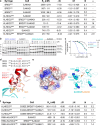
Fig. 1. Discovery of a ligand targeting KLHDC2.
a Thermal shift assay monitoring the denaturation of the KLHDC2SBD in the absence (black) or presence (red) of a diGly substrate peptide from SELK. The change in melting temperature ΔTm is shown. b Summary of workflow for the thermal shift based small molecule screening campaign. c Chemical structure and biophysical characterization of hit SJ6145. Data are the average of n = 2 independent experiments. d Summary of SAR campaign surrounding SJ6145. e Chemical structure and biophysical characterization of lead molecule SJ10278. Data are the average of n = 2 independent experiments. f Crystal structure of KLHDC2SBD (cyan, cartoon) bound to SJ10278 (orange, surface). g Structural superposition of SJ10278-KLHDC2SBD (SJ10278 in orange and KLHDC2SBD in cyan) with the structure of KLHDC2SBD bound to a diGly peptide from EBHB2 (KLHDC2SBD in pale cyan and EBHB2 peptide in brown; 8EBL.pdb). The C-terminal RSR recognition motif from the KLHDC2SBD is shown in sticks. h LigPlot of SJ10278-KLHDC2SBD, hydrogen bonds between SJ10278 and KLHDC2 SBD are shown in dashed black lines with distances indicated. Source data are provided as a Source Data file.
Fig. 2
Fig. 2. Optimization of a KLHDC2 targeting ligand.
a Electrostatic representation of KLHDC2SBD bound to the diGly substrate EPBH2 (chocolate, sticks, 8EBL.pdb). For clarity, only the C-terminal five residues of EPHB2 are shown. b As in (a), but with SJ10278 (orange, sticks). c Summary of SAR campaign surrounding SJ10278. d Chemical structure and biophysical characterization of SJ46411. Data are the average of n = 2 independent experiments. e As in (a) but with SJ46411 shown in surface representation. f LigPlot of SJ46411-KLHDC2SBD, hydrogen bonds between SJ46411 and KLHDC2 are show in black sticks with distances indicated. The water molecule involved in intramolecular hydrogen bonding with SJ46411 is shown as a gray sphere. Source data are provided as a Source Data file.
Fig. 3
Fig. 3. Generation of a PROTAC employing KLHDC2.
a Structural superposition of KLHDC2SBD (cyan, surface) bound to SJ46418 (orange, sticks) with the diGly substrate EPHB2 (chocolate, sticks, 8EBL.pdb). For clarity surface representation is depicted for the KLHDC2SBD bound to SJ46418 only. Probable exit vectors from the ligands are depicted by arrows. Potential neo-substrate interaction surfaces with the KLHDC2SBD U-shaped binding cleft is shown. b Chemical structure and biophysical characterization of SJ46418. Data are the average of n = 2 independent experiments. c Structural superposition of the KLHDC2SBD (electrostatic, surface) bound to SJ46418 (orange, surface) to the diGly substrate EPHB2 (chocolate, surface, 8EBL.pdb) and the KLHDC2 targeting ligand KDRLZK1-KLHDC2SBD (yellow, surface, 8SGE.pdb). For clarity surface electrostatic representation is depicted for KLHDC2SBD bound to SJ46418 only. Probable exit vectors deriving from attachment sites to the ligands is depicted by arrows. d Chemical structure of JQ1 based KLHDC2 PROTAC protein degraders SJ46421 and SJ46423. e Fluorescent scan of gel monitoring SJ46421- and SJ46423-dependent ubiquitylation of the BD2 domain from BRD2, BRD3, or BRD4. Reactions were performed in pulse-chase format, with fluorescent ubiquitin charged-UBE2R2 added to neddylated CRL2KLHDC2 and indicated PROTAC/BRD mixture. Shown is representative panels from n = 2 independent experiments. f Cartoon depiction of the biochemical assay monitoring inhibition of KLHDC2 di-Gly substrate ubiquitylation by PROTAC-mediated ternary complex formation. Briefly, neddylated CRL2KLHDC2 was incubated with BRD3BD2 and the indicated PROTAC prior to adding fluorescent ubiquitin-charged UBE2R2 (i.e. the pre-formed thioester-linked UBE2R2~ubiquitin intermediate) and a di-Gly protein substrate. Loss of di-Gly protein substrate ubiquitylation was quantified. g Morrison fit of data from competition ubiquitylation assay, yielding a Kiapp as a measure of the strength of ternary complexes promoted by SJ46421-BRD3BD2 and SJ46423-BRD3BD2. Data are the average from n = 2 independent experiments. Source data are provided as a Source Data file.
Fig. 4
Fig. 4. SJ46421 promotes selective cooperative ternary complex formation with BRD3BD2.
a Thermodynamic parameters for the indicated binary and ternary complexes. Data are the average +/− 1 s.d. from n = 2 experiments. Values for ΔG and ΔH are expressed in kcal/mol−1. b Biochemical assay monitoring inhibition of KLHDC2 diGly protein substrate ubiquitylation by PROTAC-mediated ternary complex formation, performed as in Fig. 3f. SJ46421-BRD3BD2 or SJ46421 alone (left panel) or SJ46423-BRD3BD2 or SJ46423 alone (right panel) were incubated with neddylated CRL2KLHDC2, prior to adding fluorescent ubiquitin-charged UBE2R2 (i.e. the pre-formed thioester-linked UBE2R2~ubiquitin intermediate) and diGly protein substrate. Effective PROTAC protein degraders promote ubiquitin transfer to BRD3BD2 neo-substrate concomitant with loss of diGly substrate ubiquitylation. Samples were reduced when reaction was quenched by addition of SDS buffer. Loss of substrate ubiquitylation was quantified. Shown is representative panels from n = 2 independent experiments. c Quantification of loss of diGly substrate ubiquitylation from the gels in panel. b Data are the average from n = 2 independent experiments. d Sequence alignment of the second bromodomain from BRD2, BRD3, and BRD4. Residue boundaries of the domains are shown, and E344 from BRD3BD2 is highlighted in red (top panel). Superposition of BRD2BD2 (salmon; 3ONI.pdb) and BRD3BD2 (chocolate; 3S92.pdb) to the structure of the VHL-MZL-BRD4BD2 ternary complex (cyan, orange, and red respectively;5T35.pdb). G382, E344, and G386 from BRD2, BRD3, and BRD4 respectively that mediate cooperative ternary complex formation with VHL are shown in sticks (bottom panel). e Electrostatic surface representation of KLHDC2 highlghting potential basic patches for interaction with BRD3 E344 (left panel). Cartoon representation from (e) with relevant basic residues shown in sphere representation (right panel). f Thermodynamic parameters for the indicated ternary complexes between KLHDC2 and the indicated swap mutant BRD proteins and the R56A mutant of KLHDC2. Experiments were performed n = 2 times. Source data are provided as a Source Data file.
Fig. 5
Fig. 5. KLHDC family selectivity and activity of SJ46421 towards tetrameric KLHDC2.
a Fluorescent scan of gel monitoring SJ46421-dependent BRD3BD2 ubiquitylation by neddylated versions of CUL2KLHDC2, CUL2KLHDC3, CUL2KLHDC10, or CUL5KLHDC1. Assays were performed in pulse-chase format, monitoring PROTAC- and E3-dependent transfer of lysineless (R7) fluorescent ubiquitin from pre-formed thioester-linked UBE2R2~ubiquitin intermediate. Shown is representative panels from n = 2 independent experiments. b Cartoon depiction of the degron-mimic mediated auto-regulation of KLHDC2-EloBC by the inactive tetrameric assembly and active monomer-substrate complexes. c. Fluorescent scan of ubiquitylation assays monitoring the ability of SJ46421-BRD3BD2 to disrupt the KLHDC2-EloBC tetrameric assembly. Assays were performed as in (a). Shown is representative panels from n = 2 independent experiments. d Quantification of the levels ubiquitin conjugated KLHDC2 or BRD3BD2 form gels in panel (c). Data are the average from n = 2 independent experiments. Source data are provided as a Source Data file.
Fig. 6
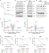
Fig. 6. KLHDC2 PROTAC protein degraders are active in cells and maintain BRD3 selectivity.
a Chemical structure of free-acid and pro-drug PROTAC variants of SJ46421. b Western blot monitoring the levels of the indicated proteins following a 24 h dose of wild-type U2OS or KLHDC2 knockout cells with DMSO, SJ46420, or SJ46421. The asterisk indicates a non-specific reactive band with the KLHDC2 antibody. c Same as (b) but with U2OS KLHDC2 KO/rescue cell line and the indicated pro-drug variants. d Same as (c) but with SJ46420 or SJ48088 in the absence or presence of co-dosing with 1 μM MLN4924. e Volcano plot from TMT proteomics comparing protein levels following a 24 h dose of U2OS KLHDC2 KO/rescue cell line with 1 μM SJ46420. Dashed lines demark proteins stabilized or destabilized > 2-fold with p value < 0.05. Statistical tests used in the volcano plots is two-sided student’s t-test and no correction was made for multiple comparisons. Data are from n = 3 biological replicates. f Same as (e) but with U2OS KLHDC2 knockout cells. g Quantification of dose-response western blot monitoring the levels of BRD2, BRD3, and BRD4 following a 24 h dose of U2OS KLHDC2 KO/rescue with the indicated concentration of SJ46420. DC50 and Dmax values calculated from the fit shown in the table below are the average from n = 2 independent experiments. h Same as (g) but with PC3 cells. i Same as (g) but with 22Rv1 cells. All data in Fig. b-d are representative panels from n = 2 independent experiments. Source data are provided as a Source Data file.

References

    1. Bekes, M., Langley, D. R. & Crews, C. M. PROTAC targeted protein degraders: the past is prologue. Nat. Rev. Drug Discov.21, 181–200 (2022). - PMC - PubMed
    1. Hanzl, A. & Winter, G. E. Targeted protein degradation: current and future challenges. Curr. Opin. Chem. Biol.56, 35–41 (2020). - PMC - PubMed
    1. Verma, R., Mohl, D. & Deshaies, R. J. Harnessing the Power of Proteolysis for Targeted Protein Inactivation. Mol. Cell77, 446–460 (2020). - PubMed
    1. Ramachandran, S. & Ciulli, A. Building ubiquitination machineries: E3 ligase multi-subunit assembly and substrate targeting by PROTACs and molecular glues. Curr. Opin. Struct. Biol.67, 110–119 (2021). - PubMed
    1. Corson, T. W., Aberle, N. & Crews, C. M. Design and Applications of Bifunctional Small Molecules: Why Two Heads Are Better Than One. ACS Chem. Biol.3, 677–692 (2008). - PMC - PubMed

Publication types

MeSH terms